Category Archives: Uncategorized

Bacillus anthracis

by Lauren Mcmannus

Introduction

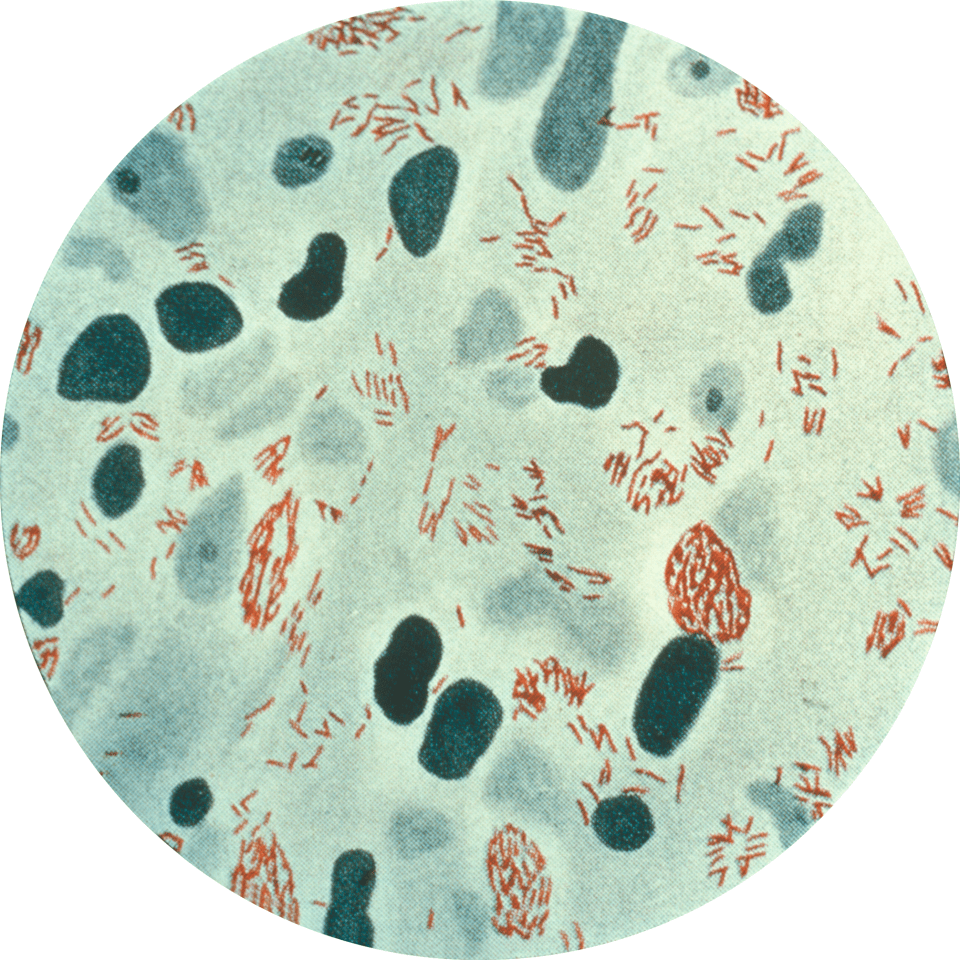
Bacillus anthracis is an endospore-forming bacteria that causes anthrax disease in animals and humans. The endospore (Figure 1) is the inactive, highly-resilient form of a B. anthracis bacterium that can withstand extreme conditions. Anthrax endospores enter its host most commonly through skin wounds, but also by inhalation or ingestion. Human contraction of B. anthracis occurs predominantly through contact with diseased animals or contaminated animal parts.

Figure 1: Image of Bacillus anthracis spores as seen under a microscope using phase contrast. Phase contrast shows endospores as bright white spots where the bacteria is dormant. Source: Public Health Image Library, Center for Disease Control, Larry Stauffer, Oregon State Public Health Laboratory (2002)

Figure 1: Image of Bacillus anthracis spores as seen under a microscope using phase contrast. Phase contrast shows endospores as bright white spots where the bacteria is dormant. Source: Public Health Image Library, Center for Disease Control, Larry Stauffer, Oregon State Public Health Laboratory (2002)

Disease

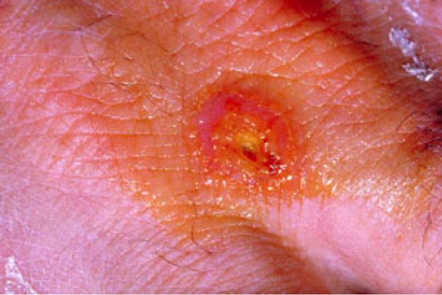
An infection develops in humans after exposure to B. anthracis in specific locations in the body depending on the route of exposure. Cutaneous anthrax occurs when bacteria enter skin through a wound or break in the skin, creating black lesions. Infection can also result from inhaling endospores released into the air from manufacturing wool or hides. Less commonly, anthrax infection can occur in the digestive system from eating contaminated food like undercooked and tainted meat, or through injection of heroin.

Once B. anthracis enters the human, the bacteria encounter macrophages, cells of the immune system that recognize bacterial pathogens and engulf them. Macrophages kill by trapping bacteria and exposing it to a highly acidic environment. These macrophages internalize the B. anthracis, but the endospores (Figure 1) are able to withstand these extreme conditions and survive. From there, B. anthracis can revert back to its non-spore state and replicate within the body, releasing toxins and causing damage.

Epidemiology

Rates of anthrax infection has significantly decreased in the past few decades, largely as a result of vaccine awareness and hygiene standards implemented around the world. Worldwide rates of infections are not well-recorded, but there are only 1 or 2 cases of cutaneous anthrax in the United States each year.

Humans have most commonly contracted anthrax after coming in contact with infected animals or animal products. Veterinarians, farmers, butchers, or industrial workers that handle animal hides or wool are at a higher risk of developing an infection, especially through a wound on the skin. More than 95% of all anthrax cases develop from cutaneous exposure, and this is also the least fatal as the infection is limited to one area.

Cutaneous exposure can have a mortality rate around 20%, while the mortality rates of inhalation and digestive system exposure are 80% and 25 to 75%, respectively. Internal anthrax infection is not as easily treated and is often not recognized until the disease is in its later stages, leading to high mortality rates.

Virulence Factors

When not in endospore form, B. anthracis is more susceptible to immune system defenses when travelling through the body. In order to disguise itself from the host’s immune system cells, like macrophages, B. anthracis surrounds itself in a capsule made up of poly-gamma-D-glutamic acid. When covered in this capsule, the bacteria are less likely to attract attention and can safely multiply and multiply.

The damaging nature of B. anthracis is revealed when large numbers of these bacteria begin to release exotoxins. As seen in Figure 2, bacteria release protective antigen, edema factor, and lethal factor as three separate molecules that are not active by themselves. When the edema factor binds to the protective antigen, the edema factor becomes activated and causes fluid to rush out from cells and collect in the tissue. On the other hand, when the lethal factor binds to the protective antigen, a lethal toxin is created that helps kill macrophages and other cells of the immune system. It also changes the signals cells receive from each other, severely disrupting vital processes that allow for basic functions of cells and leading to cell death.

Figure 2: The secretion of inactive molecules by B. anthracis lead to the production of toxins. The binding of LF and EF alone do not create a toxin. The binding of LF and PA create an active lethal toxin, and the binding of PA and EF create an edema toxin. PA bound with both LF and EF have lethal and edema effects.

Figure 2: The secretion of inactive molecules by B. anthracis lead to the production of toxins. The binding of LF and EF alone do not create a toxin. The binding of LF and PA create an active lethal toxin, and the binding of PA and EF create an edema toxin. PA bound with both LF and EF have lethal and edema effects.

Prevention and Treatment

Though treatments are available, anthrax vaccines are available to humans and animals to offer the best protection against B. anthracis. Human vaccines are not available to the general population, but they are given to people working directly with animals or animal products that are at risk for infection.

Humans

Antibiotic treatments are available and useful when administered early, but they aren’t able to reverse the serious damage done by toxins. Infections can usually be treated with penicillin, an antibiotic that causes cell death by disrupting the links of peptidoglycan on a bacteria’s cell wall. Peptidoglycan is a molecule made up of proteins and sugars that form a protective layer around a bacterial cell.

However, other antibiotics like ciprofloxacin has been used in recent infections in the United States caused by the B. anthracis strain ‘Ames’, as these bacteria produce enzymes called beta-lactamases that can break down antibiotics like penicillin. Ciprofloxacin targets DNA gyrase and DNA topoisomerase IV, which are enzymes that regulate the coiling of bacteria DNA, and this targeting prevents proper DNA replication of B. anthracis.

Animals

Penicillin can also be used to treat animals, like cattle, that have contracted anthrax.

References

Centers for Disease Control and Prevention, National Center for Emerging and Zoonotic Infections Diseases, Division of Foodborne, Waterborne, and Environmental Diseases (2008, August 26 2009). Anthrax.   Retrieved from http://www.cdc.gov/nczved/divisions/dfbmd/diseases/anthrax/technical.html#incidence

Centers for Disease Control and Prevention. (2015, September 1 2015). How People Are Infected. Anthrax.  Retrieved from http://www.cdc.gov/anthrax/basics/how-people-are-infected.html

Drlica, K., & Zhao, X. (1997). DNA gyrase, topoisomerase IV, and the 4-quinolones. Microbiology and Molecular Biology Reviews, 61(3), 377-392.  Retrieved from http://mmbr.asm.org/content/61/3/377

FDA (Food and Drug Administration). (June 17 2015). Anthrax.   Retrieved from http://www.fda.gov/BiologicsBloodVaccines/Vaccines/ucm061751.htm

Jang, J., Cho, M., Chun, J.-H., Cho, M.-H., Park, J., Oh, H.-B., . . . Rhie, G.-e. (2011). The Poly-γ-d-Glutamic Acid Capsule of Bacillus anthracis Enhances Lethal Toxin Activity. Infection and Immunity, 79(9), 3846-3854. doi:10.1128/iai.01145-10

Organization, World Health. (2008). Anthraxin humans and animals (pp. 219).  Retrieved from http://www.who.int/csr/resources/publications/anthrax_webs.pdf

Rogers Yocum, J. R. R., Jack L. Strominger. (1979). The Mechanism of Action of Penicillin. The Journal of Biological Chemistry, Vol. 255(No. 9), 3977-3986. Retrieved from http://www.jbc.org/content/255/9/3977.full.pdf

Schneemann A, Manchester M. Anti-toxin antibodies in prophylaxis and treatment of inhalation anthrax. Future microbiology. 2009;4:35-43. doi:10.2217/17460913.4.1.35.

Spencer, R. C. (2003). Bacillus anthracis. Journal of Clinical Pathology, 56(3), 182-187. doi:10.1136/jcp.56.3.182

Todar, K. (2012). Bacillus anthracis and Anthrax (page 3).   Retrieved from http://textbookofbacteriology.net/Anthrax_3.html

Todar, K. (2012). Structure and Function of Bacterial Cells.   Retrieved from http://www.textbookofbacteriology.net/structure_5.html


Mycobacterium leprae

by Gaëlle-Laurie Dubréus and Emilie Yeh

Introduction

Mycobacterium leprae, a gram positive bacterium, exists as an obligate intracellular pathogen that causes Hansen’s disease, commonly known as leprosy. This disease has been identified as long as 1550 B.C. in Egypt and was first isolated in 1873 by G.A. Hansen (hence the name of the disease). Since then, much has been discovered on its epidemiology, pathogenicity, and treatments –which were successfully created in the 1970s.

Disease

The mode of transmission of M. leprae appears to be from person-to-person either by droplets from sneezing and coughing or by other nasal secretions. Also, the bacteria can use an abrasion in the skin to colonize the superficial site of the epithelium and the peripheral nerves. Families with members who have leprosy have a higher susceptibility of developing this illness due to genetic predisposition. The only organisms known to harvest the infection and develop the disease are humans and armadillos, whom can remain asymptomatic for upto twenty years (long incubation time).

Although patients usually show signs of sores on the skin, leprosy exists in the following two forms: tuberculoid (pauci-auxillary leprosy), which occurs as skin discoloration, and lepromatous (multi-bacillary leprosy), which manifests as skin lesions, plaques, nodules, thickened skin, and nasal complications. The cause for the manifestation of each is based on the T-cell count, where those with a higher count develop the milder form of leprosy. Lepromatous leprosy is the more severe clinical case so it triggers the production of more antibodies against the bacterium, but with a compromised immune cellular response. Moreover, both forms of leprosy can result in collateral damage to the peripheral nervous system. This can permanently affect the arms, legs, eyes, nerves, and the skin.

Epidemiology

Leprosy is present worldwide, especially in warm tropical and subtropical regions. In 2013, based on statistical analyses from five areas of the World Health Organization (WHO), which included 103 countries, there were about 180.6 thousand cases of leprosy reported. Within a community, the emergence of new cases depends on the rate of transmission between the individuals of that society. For instance, this disease seems to be predominant in certain endemic regions; 14 different but specific countries, such as India, Brazil, several areas in Africa, etc., appear to be contributing to approximately 96% of the new cases of leprosy yearly.

Although adults are less likely to acquire the infection, those who do develop the disease have the lepromatous form, which occurs more frequently in men in comparison to women. Conversely, the tuberculoid form of leprosy evolves more quickly in children and its development is equal among both sexes. Since treatment with multi-drug therapies (MDT) debuted, the prevalence of this disease has significantly diminished as in less than 1 per 10,000 individuals. However, in developing countries, there exists a lack of education on this disease as well as low-to-no access to treatment. Consequently, those carrying the bacterium may not be aware of their infection and suffer from severe physical complications and handicaps in the long run– due to late diagnosis.

Figure 1: A biopsy from the skin lesion of a patient infected with M. leprae. Here, the bacteria are stained red. (Source: http://journals.plos.org/plosmedicine/article?id=10.1371/journal.pmed.0020341)

Figure 1: A biopsy from the skin lesion of a patient infected with M. leprae. Here, the bacteria are stained red. (Source: http://journals.plos.org/plosmedicine/article?id=10.1371/journal.pmed.0020341)

Virulence Factors

Leprosy is a difficult infection to study since it cannot be grown in the laboratory. With recent scientific advancements, more of its infection mechanisms have been brought to light, but many details remain unresolved.

 M. leprae begins by infecting Schwann cells found on the exterior of the nerve cells’ axons. The bacterium uses appendages called adhesins to attach itself to the Schwann cells (Figure 2). This results in nerve damage caused by leprosy. In recent years, researchers discovered that M. leprae can accelerate the proliferation of Schwann cell in order to increase the number of cells in which they can infect. Once the host detects a bacterial infection, the immune system initiates the processes that should ultimately lead to the elimination of the invading organism(s). Cells called macrophages are released to phagocytose (or ingest) the bacteria. Normally, the bacteria would be digested within the macrophage, but M. leprae evades cell death using a method that is still not fully understood. M. leprae continues to perpetuate within the macrophage and infects other cells in the host.

Figure 2: The attachment site of M. leprae on Schwann cells (Figure by Emilie Yeh).

Figure 2: The attachment site of M. leprae on Schwann cells (Figure by Emilie Yeh).

Treatment

M. leprae is more difficult to treat than other bacterial infections since it has an abnormally high number of lipid on its cell wall, which acts as a protective barrier against antibiotics from penetrating into the cell. The treatment that is recommended by the World Health Organization are the following multidrug therapies: Dapsone, Rifampin, and Clofazimine. Dapsone inhibits the replication of the cells and all three have bactericidal effects on M. leprae. In the case of tuberculoid leprosy, these antibiotics can reverse heal the patients almost completely; whereas, for lepromatous leprosy, the damage done pre-treatment tend to be irreversible even after the antibiotics have been administered. (Fig. 3) While these drugs have been proven to be extremely effective in curing leprosy, the details on the mechanism of each drug is not yet fully understood.


References

Barker, L. P. (2006). Mycobacterium leprae interactions with the host cell: Recent advances. Indian Journal of Medical Research, 123(6), 748-759.

Hansen’s Disease (Leprosy). Centers for Disease Control and Prevention.

Lastoria J.C., De Abreu M.A.M.M. (2014). Leprosy: Review of the Epidemiological, Clinical, Etiopathogenic Aspects- Part 1. PMC 89(2): 205-218.

Leprosy. World Health Organization. Last updated May 2015.

Leprosy Today. National Institute of Allergies and Infectious Diseases. Last updated February 8, 2011.

Pinheiro R.O., Salles J.D., Samo E.N., Sampaio E.P. (2011). Mycobacterium leprae–host-cell interactions and genetic determinants in leprosy: an overview. NCBI 6(2): 217-230.

Reibel, F., Cambau, E., & Aubry, A. (2015). Update on the epidemiology, diagnosis, and treatment of leprosy. Médecine et Maladies Infectieuses, 45(9), 383-393. DOI: http://dx.doi.org/10.1016/j.medmal.2015.09.002

Clostridium botulinum

By Sara Cantini and Victoria Lee

Introduction

Clostridium botulinum is a rod-shaped bacterium (Figure 1), naturally occurring in the environment, particularly in soil. Under stressful conditions, C. botulinum form resistant spores that can even withstand most standard cooking. In most preservatives and food processing nowadays, acidity and high salt concentrations prevent C. botulinum spores from germinating so that they do not cause damage if ingested. However, if they do develop, growing C. botulinum produces botulinum neurotoxins (BoNT), which are the most toxic substances known to man. Therefore, ingestion of preformed BoNT or growth of the bacterium within a host can be lethal. Interestingly enough, diluted concentrations of BoNT are used in anti-aging Botox.

Figure 1: Microscopic view of gentian violet-stained Clostridium botulinum and their spores. Source: Public Health Image Library, Center for Disease Control (1979).

Figure 1: Microscopic view of gentian violet-stained Clostridium botulinum and their spores. Source: Public Health Image Library, Center for Disease Control (1979).

Botulinum comes from the latin word botulus, meaning sausage. In Europe in the 18th and 19th century, the consumption of blood sausage was sometimes associated with muscle paralysis, breathing problems and even death. This was because sausages provided the perfect environment for C. botulinum to grow and produce toxins at the time. The organism was first isolated and identified in 1897 from contaminated homemade raw ham in Belgium and from the spleen of a man who died from ingesting it.

Disease

C. botulinum can cause a severe neurological disease in humans and animals called botulism. The most common form is food-borne botulism, which often results from the ingestion of poorly preserved foods contaminated with preformed neurotoxins. Moreover, spores can establish themselves in deep wounds, where they can germinate and produce toxins capable of causing wound botulism. Finally, infant botulism occurs in children less than 1 years old and results from the production of BoNTs by established C. botulinum in immature intestines.  Infant botulism has been proposed as a potential cause of sudden death syndrome (SIDS) since both result in a similar sudden respiratory arrest. Botulism is not infectious because the absorption of BoNT in the bloodstream is required for the development of symptoms of the disease.

In all cases, the disease first manifests itself with symptoms such as double vision, inability to focus, difficulty swallowing, slurred speech, dry mouth and muscle weakness. The progression of the disease can lead to paralysis of the muscles responsible for breathing.

Epidemiology

Most incidences of foodborne botulism results from the accidental consumption of poorly preserved foods or home-prepared foods that contain preformed BoNTs. There were many outbreaks in the 20th century due to poor canning processes of foods. In recent years, there have been about 1,000 cases annually worldwide. The incidence of wound botulism is quite low, but almost exclusively occurs in injection drug users.  Most cases of infant botulism are due to the consumption of honey contaminated with C. botulinum spores before the age of 1. Annually, an average of 145 botulism cases are reported in the United States, with the majority of them being cases of infant botulism.

Virulence Systems

C. botulinum produce neurotoxins (BoNTs) which form a large complex with other proteins such as hemagglutinin and other non-toxic neurotoxin associated proteins (NAPs). These proteins protect the neurotoxin in the presence of harsh acidic environments and digestive proteases (enzymes that break down proteins) encountered in the host’s digestive tract. They also facilitate the absorption of the neurotoxin into the general circulation by disrupting the layer of cells which form a protective barrier. BoNT circulates in the bloodstream until it reaches neuromuscular junctions between neuron endings and muscle cells. The domain known as the heavy chain binds to receptors on the cell surface and mediates its internalization via a process called receptor-mediated endocytosis (Figure 3). The heavy chain, now inside a pocket called an endosome, allows the domain known as the light chain to escape. In the nerve endings, there are similar pockets called synaptic vesicles which contain chemicals called acetylcholine. Normally, SNARE proteins are involved in fusing these vesicles to the cell membrane in response to an electrical signal (Figure 2). Acetylcholine is thus released in the synapse (space between neuron endings and muscle cells) so that they can bind to receptors and induce muscle contraction. In the presence of BoNT however, the light chain cleaves these SNARE proteins, which inhibits the release of these neurotransmitters and results in muscle paralysis (Figure 3).

Figure 2: Release of acetylcholine in the synapse. 1. SNARE proteins bringing synaptic vesicles to the cell membrane 2. Fusion of the vesicle and the membrane 3. Release of acetylcholine in the synapse, free to bind receptors on the surface of muscle cells.

Figure 2: Release of acetylcholine in the synapse. 1. SNARE proteins bringing synaptic vesicles to the cell membrane 2. Fusion of the vesicle and the membrane 3. Release of acetylcholine in the synapse, free to bind receptors on the surface of muscle cells.

Figure 3: The inhibition of acetylcholine release in the synapse due to the action of botulinum toxin 1. Engulfment of the BoNT into the nerve cell 2. Translocation of the light chain out of the vesicle 3. Cleavage of SNARE proteins via the light chain 4. Inhibition of the fusion of acetylcholine-containing vesicles to the cell membrane.

Figure 3: The inhibition of acetylcholine release in the synapse due to the action of botulinum toxin: 1. Engulfment of the BoNT into the nerve cell 2. Translocation of the light chain out of the vesicle 3. Cleavage of SNARE proteins via the light chain 4. Inhibition of the fusion of acetylcholine-containing vesicles to the cell membrane.

Treatment

In general, if people are diagnosed and treated early for botulism, most of them will recover muscle strength within weeks to months. Rapid administration of equine botulinum antitoxins and human botulinum immune globulin is used to treat adult and infant botulism respectively. They act by neutralizing the effect of circulating toxins in the bloodstream.  Supportive intensive care such as intubation and mechanical ventilation is often also required. However, failure of rapid diagnosis or proper treatment can result in death within three to ten days due to the rapid development of respiratory failure.


References

Centers for Disease Control and Prevention [CDC]. (2014). Botulism. Retrieved from http://www.cdc.gov/nczved/divisions/dfbmd/diseases/botulism/#treat

Davis, E. L. (2003). Botulism. Current Treatment Options in Neurology, 5(1), 23-31.

Government of Canada. (2013). Botulism (Clostridium botulinum). Retrieved from http://healthycanadians.gc.ca/eating-nutrition/risks-recalls-rappels-risques/poisoning-intoxication/poisoning-intoxication/botulism-botulisme-eng.php

Mukai, Y., & Kaji, R. (2011). [Use of botulinum neurotoxin therapy]. Brain Nerve, 63(7), 775-784.

Peck, M. W. (2010) Clostridium botulinum (pp. 31-52). In Pathogens and toxin in foods: challenges and interventions.  Juneja, V. K. & Sofos, J. N. (eds) Washington, DC: ASM Press.

Rummel, A., & Binz, T. (ed.). (2013). Botulinum neurotoxins. Berlin: Springer.

Shukla, H. D. & Sharma, S. K. (2005). Clostridium botulinum: A Bug with Beauty and Weapon. Critical Reviews in Microbiology, 31(1), 11-18.

Smith, L. D. & Sugiyama, H. (1988). Botulism: The Organism, Its Toxins, The Disease (2nd ed.). Springfield, IL: Charles C. Thomas Publisher.

Sobel J. Botulism. (2005). Clinical Infectious Diseases. 41(8): 1167-1173.

World Health Organization [WHO]. (2013). Botulism. Retrieved from http://www.who.int/mediacentre/factsheets/fs270/en/.


The Outbreak of Tuberculosis in United States during 2002 to 2008

By Chloe (Jin Ying) Liu and Zan Ding

Introduction

Tuberculosis is a disease caused by bacteria called Mycobacterium tuberculosis. This kind of bacteria usually attacks the lungs, but it can also influence other parts of the body such as the brain, kidney or spine. If a person is infected with tuberculosis, the general symptoms include feelings of sickness or weakness, weight loss, fever and night sweats. Besides, the symptoms of tuberculosis on the lungs include coughing, chest pain, and the coughing up of blood. Tuberculosis bacteria can stay in the air for several hours (1) and be easily spread through coughing, sneezing, talking, or singing.

Mycobacterium tuberculosis

Mycobacterium tuberculosis has ropelike structures of peptidoglycans that give it properties of an acid fast bacteria. It is able to form acid-stable complexes when certain arylmethane dyes are added (2). M. tuberculosis has circular chromosomes of about 4,200,000 nucleotides long. Plasmids in M. tuberculosis are important in transferring virulence because genes on the plasmids are more easily transferred than genes located on the chromosomes (2). The tough cell wall of the bacteria with unusual structure and composition prevents passage of nutrients into and out of the cell, therefore M. tuberculosis grows slowly.

Figure 1: How TB Spreads. Source: http://www.cdc.gov/tb/topic/basics/default.htm.

Figure 1: How TB Spreads. Source: http://www.cdc.gov/tb/topic/basics/default.htm.

Tuberculosis outbreak

Most tuberculosis cases do not start outbreaks, however they do occur and they can put tremendous strain on local public health resources. All outbreaks begin with a source case. Recognizing the characteristics of such patients soon after diagnosis could help to interrupt transmission and reduce the risk for an outbreak. There were nonrandom outbreaks of TB in the United States during 2002-2008, which the Centers for Disease Control and Prevention (CDC) assisted in investigation. Outbreaks are defined based on CDC guidelines for contact investigation as detection of tuberculosis disease between more than two people exposed to a person with infectious tuberculosis (5). From this review, the outbreak in the United States had more than 3 culture-confirmed cases that had epidemiologic links and tuberculosis strains with matching genotypes. Linkage by epidemiology means exposure to another outbreak patient by sharing enclosed airspace in the same period. Genotyping methods included spoligotyping and either restriction fragment length polymorphism ore 12-locus mycobacterial interspersed repetitive units. Of the 51 tuberculosis investigations during 2002-2008, a total of 27 met the defining criteria, while 24 cases were excluded because some of these cases include patients with organ transplants and some had insufficient data in CDC reports. Based on the study of this outbreak in the United States, about 84% of patients had pulmonary disease; 25% of patients required hospitalization and 6% died.

Figure 2: Characteristics of source case-patients for 26 investigated tuberculosis outbreaks, United States, 2002–2011. Source: http://wwwnc.cdc.gov/eid/article/21/3/14-1475-t1.

Figure 2: Characteristics of source case-patients for 26 investigated tuberculosis outbreaks, United States, 2002–2011. Source: http://wwwnc.cdc.gov/eid/article/21/3/14-1475-t1.

Causes of outbreaks

Factors contributed to outbreak include delays in seeking medical attention which may cause intense transmission. The infectious period for pulmonary tuberculosis cases was assumed to start three months before tuberculosis symptoms onset and to end with the initiation of tuberculosis (4). Outbreak duration is calculated beginning on the treatment start date for the first reported case and continuing through treatment start date for the last case as noted at the time of the investigation. Prolonged infectious period was defined as more than three months between symptom onset and the date that effective treatment had been administered for two weeks (5). Educating health care providers in order to raise public awareness about tuberculosis is critical. In this way, patients can seek early diagnosis and receive timely treatment  which results in efficient control of potential TB transmission.

Figure 3: Characteristics of source case-patients for 26 investigated tuberculosis outbreaks, United States, 2002–2011. Source: http://wwwnc.cdc.gov/eid/article/21/3/14-1475-t1

Figure 3: Characteristics of source case-patients for 26 investigated tuberculosis outbreaks, United States, 2002–2011. Source: http://wwwnc.cdc.gov/eid/article/21/3/14-1475-t1

When contact investigations are incomplete because of limited resources or hard-to-reach populations, latent tuberculosis infection remains so that TB outbreaks can spread. Identifying high-risk settings is another basic principle of tuberculosis control except applying effective control measures to reduce TB transmission. TB outbreak hotspot was the drug house. According to the data, about 63% of cases were occurred there. Homeless shelters, correctional facility, household, workplace, church, bar, school and public transit were common places for TB outbreak. The most frequently intervention to manage the outbreak is to prioritize contacts based on risk for infection and progression of disease, enabling the highest risk contacts to be completely evaluated.

Tuberculosis  disease can be treated by using antibodies to kill the bacteria M. tuberculosis. Isoniazid (INF) and rifampin (RIF) are most commonly used antibodies in the treatment(6). Effective treatment is difficult and prolonged to achieve because of  unusual mycobacterial cell wall, taking 6 – 9 months. Although therapeutic effects have accomplished and TB incidence has declined in the United States, public health departments still require more efficient strategies to prevent, detect, and treat TB in order to eliminate the disease fundamentally.

Reference:

  1. TB Elimination Tuberculosis: General Information. (n.d.). Retrieved November 3, 2015, from http://www.cdc.gov/tb/publications/factsheets/general/tb.pdf
  2. Uhía, I., Galán, B., Medrano, F. J. & García, J. L. Characterization of the KstR-dependent promoter of the gene for the first step of the cholesterol degradative pathway in Mycobacterium smegmatis. Microbiology 157, 2670 (2011)
  3. Mitruka K, Oeltmann JE, Ijaz K, Haddad MB. Tuberculosis outbreak investigations in the United States, 2002–2008. Emerg Infect Dis. 2011;17:425–31. DOIPubMed
  4. Centers for Disease Control and Prevention. Guidelines for the investigation of contacts of persons with infectious tuberculosis. Recommendations from the National Tuberculosis Controllers Association and CDC. MMWR Recomm Rep. 2005;54(RR-15):1–47 .PubMed
  5. Centers for Disease Control and Prevention Guidelines for the investigation of contacts of persons with infectious tuberculosis: recommendations from the National Tuberculosis Controllers Association and CDC. MMWR Morb Mortal Wkly Rep. 2005;54(RR–15):1–3 [PubMed]
  6. Core Curriculum on Tuberculosis: What the Clinician Should Know (5th ed.). Retrieved June 21, 2013, from http://www.cdc.gov/tb/education/corecurr/pdf/corecurr_all.pdf

Francisella tularensis

by Carle Terrasse and Sarah Stern

Introduction

F.tularensis is an intracellular, nonmotile coccobacillus responsible for the zoonotic (transmissible from animal to human) disease tularemia. It was discovered in 1911 as the cause of a fatal, quickly spreading disease in squirrels in California. It can be divided into 3 main subspecies (figure 1), but not all cause disease in humans. In fact mostly subspecies tularensis and holarctica cause human tularemia, while mediasiatic does not. In North America, F. tularensis tularensis is the most common. This bacterium naturally occurs in small mammals, such as rodents and rabbits that mainly acquire it via arthropod bites. As a result, tularemia is primarily observed in rural areas where small rodents and ticks are abundant. As it is easily spread, highly infective, and deadly, F. tularensis is subject of much concern with regards to bioterrorism. In fact, inhalation of only 10 organisms is enough to cause acute respiratory problems and mortality.

Figure 1: Diagram of the evolution of the 3 subspecies holartica, mediasiatica and tularensis (enclosed in light red) of F.tularensis (enclosed in dark red). Adapted from: Birdsell DN, et al. TaqMan Real-Time PCR Assays for Single-Nucleotide Polymorphisms Which Identify Francisella tularensis and Its Subspecies and Subpopulations. PLoS ONE 2014, 9(9):e107964.

Figure 1: Diagram of the evolution of the 3 subspecies holartica, mediasiatica and tularensis (enclosed in light red) of F.tularensis (enclosed in dark red). Adapted from: Birdsell DN, et al. TaqMan Real-Time PCR Assays for Single-Nucleotide Polymorphisms Which Identify Francisella tularensis and Its Subspecies and Subpopulations. PLoS ONE 2014, 9(9):e107964.

Disease

Depending on the subspecies of F. tularensis, the symptoms can be more or less severe. In general, fever, headaches, body aches and malaise develop within the first 3 to 5 days of infection. The different routes of infection lead to more specific symptoms. A human infection can develop from direct contact with rodents, bites from the ticks and mosquitoes, inhalation of aerosolized bacteria and ingestion or contact with contaminated environment but human-to-human transmission has never been seen. These are the common clinical manifestations of the main forms of tularemia:

  • Glandular: vector borne transmitted by ticks. Painful swollen lymph nodes result from the infection. An ulcer may develop at the site of infection (figure 2) in which case it is called an ulceroglandular infection.
  • Oropharyngeal: ingestion of contaminated food and/or water. A sore throat, vomiting, diarrhea and neck glands swelling are characteristic symptoms.
  • Respiratory or pneumonic: acquired via inhalation. It is the most severe form of tularemia.
  • Oculoglandular: direct contamination with the eye may lead to this form of tularemia. It is characterized by pain and swelling of the eye. An ulcer may develop too.
  • Typhoidal: this form results from an unspecified route of infection and patients will develop a systemic fever where fever, fatigue and chills are the main symptoms.
Figure 2: Ulcer formation at the site of infection by F. tularensis. Source: CDC Public Health Image Library (ID #2037).

Figure 2: Ulcer formation at the site of infection by F. tularensis. Source: CDC Public Health Image Library (ID #2037).

Regardless of the initial site of contamination, in humans the bacteria will spread to the lymph nodes, liver and spleen after entering macrophages. Macrophages are immune system cells that recognize and engulf target cells for destruction. F. tularensis is able to evade the mechanisms within the macrophages that would normally digest it and effectively uses these cells to taxi around the body.

Epidemiology

Francisella tularensis is naturally present in North America. In the United States, Eastern cottontail rabbits are the main reservoirs of the disease. These rabbits also share a range with Canada, so the disease crosses country lines. Here in Canada, snowshoe hares also carry F. tularensis. There have been recent outbreaks across North America, Asia and Europe (Russia, Kosovo, Germany, Austria, and Italy). These outbreaks are significant enough that tularemia is now classified as a re-emerging disease worldwide. In 2000 and 2003 Kosovo saw large outbreaks of more than 300 cases of oropharyngeal tularemia each year. Recent outbreaks occurred in Turkey and Italy due to contaminated well water. Keep an eye on tularemia; this disease is sure to make the news in the near future!

Virulence Factors

Once F. tularensis gets inside the body, it uses some tools to evade the immune system. The host works hard to destroy the pathogen. One way the body tries to rid itself of the bacteria is by a set of proteins called the complement system. These proteins target the pathogen and aim to lyse it by poking holes in the membrane of the bacteria. F. tularensis has adaptations to deal with this problem. The first is the production of a capsule to avoid detection and binding by complement proteins. If the complement protein C3 does happen to bind, F. tularensis is able to cleave the protein, which renders it inactive.

Another aspect of the host defense that F. tularensis has to deal with is antimicrobial peptides. Antimicrobial peptides are positively charged molecules that kill bacteria. These molecules are attracted to the negative surface of bacteria. F. tularensis modifies one of its surface glycolipids, called Lipid A, to make its surface less negative. This tricks the antimicrobial peptides, and they won’t go after the bacteria.

As soon as 1 hour after infection, F. tularensis invades phagocytic cells called macrophages. Macrophages essentially eat up the bacterium. Inside these cells, the bacterium is safe from extracellular defenses. But how does F. tularensis keep from being digested? After being eaten by a macrophage, F. tularensis is trapped inside a digestive pocket called a phagosome. The phagosome acidifies and gains enzymes and reactive oxygen species to digest the material inside of it. F. tularensis has virulence genes that code for products to combat both the acidity and reactive oxygen species inside the phagosome. To combat phagosome acidification, F. tularensis prevents the phagosome from receiving certain acid hydrolase enzymes, so the pH does not fall. In addition F. tularensis produces the acid phosphatases AcpA, AcpB, AcpC, and Hap that degrade reactive oxygen species.

The last step for F. tularensis is to escape from the phagosome to the cytosol, where it reproduces. The exact mechanism of how this occurs is still unknown. One thought is that the bacteria creates a “needle” and the pokes through the phagosome. Regardless of the method, scientists know that the genes for this process are encoded in the Francisella pathogenicity island (FPI), which codes for a bunch of virulence factors. These virulence factors are key in the pathogen’s stealthy evasion of the immune system and journey to replication.

Treatment

So you have tularemia… quick, get to the doctor! This disease can be fatal, so early detection is important. As F. tularensis has the potential to be used for bioterrorism, your doctor must report any incident to the government. Doctors commonly use antibiotics to treat tularemia. Steptomycin is the drug of choice, followed by tetracyclines such as doxycycline. Surgery may be needed to remove ulcers if the infection is ulceroglandular.

When it comes to tularemia, prevention is key. While a vaccine that uses live attenuated bacteria exists, it is not safe for public use. Because F. tularensis is endemic to North America, take care when hiking or working outdoors. Wear protective clothing, and use insect repellent. Do not handle wild animal carcasses without gloves. Take these precautions and you will be saved from tularemia!

References

CDC: Tularemia[Internet]. Atlanta (GA): Centers for Disease Control and Prevention: c2015 [cited 2015 Nov 15]. Available from: http://www.cdc.gov/tularemia/

D’Alessandro D, Napoli C, Nusca A, Bella A , Funari E. Human tularemia in Italy. Is it a re-emerging disease?. Epidemiology and Infection 2015. 143: 2161-2169. doi:10.1017/S0950268814002799.

Dankova V, Balonova L, Straskova A, Spidlova P, Putzova D, Kijek T, Bozue J, Cote C, Mou S, Worsham P et al: Characterization of Tetratricopeptide Repeat-Like Proteins in Francisella tularensis and Identification of a Novel Locus Required for Virulence. Infection and Immunity 2014, 82(12):5035-5048.

Francisella Tularensis (Tularemia). (2014, February 26). Retrieved November 20, 2015, from http://www.upmchealthsecurity.org/our-work/publications/2014/francisella-tularensis-fact-sheet

Jones CL, Napier BA, Sampson TR, Llewellyn AC, Schroeder MR, Weiss DS: Subversion of Host Recognition and Defense Systems by Francisella spp. Microbiology and Molecular Biology Reviews : MMBR 2012, 76(2):383-404.

Kim J-e, Seo Y, Jeong Y, Hwang MP, Hwang J, Choo J, Hong JW, Jeon JH, Rhie G-e, Choi J: A novel nanoprobe for the sensitive detection of Francisella tularensis. Journal of Hazardous Materials 2015, 298:188-194.

Maggio S, Takeda K, Stark F, Meierovics AI, Yabe I, Cowley SC: Control of Francisella tularensis Intracellular Growth by Pulmonary Epithelial Cells. PLoS ONE 2015, 10(9):e0138565.

Santic M, Al-Khodor S, Abu Kwaik Y: Cell biology and molecular ecology of Francisella tularensis. Cellular Microbiology 2010, 12(2):129-139.

Straskova A, Spidlova P, Mou S, Worsham P, Putzova D, Pavkova I, Stulik J: Francisella tularensis type B Delta dsbA mutant protects against type A strain and induces strong inflammatory cytokine and Th1-like antibody response in vivo. Pathogens and disease 2015, 73(8).

Wobeser G, Campbell GD, Dallaire A, McBurney S: Tularemia, plague, yersiniosis, and Tyzzer’s disease in wild rodents and lagomorphs in Canada: A review. The Canadian Veterinary Journal 2009, 50(12):1251-1256.


Listeria monocytogenes

by Stephanie McKee and Racha Lakrouf

Introduction:

Listeria monocytogenes is a gram positive bacteria that is responsible for causing listeriosis. L.monocytogenes was first identified in 1926 based on six sudden deaths in rabbits.  However, L.monocytogenes was not identified as a cause of foodborne illness until 1981 when an outbreak of listeriosis in Halifax, Nova Scotia caused 18 deaths and was linked to consumption of contaminated coleslaw. The most common serotypes that cause disease in humans are 1/2a, 1/2b and 4b and they result in 90 % of all cases. L. monocytogenes is a facultative intracellular parasite meaning that it can survive and grow inside host cells, allowing it to easily evade the immune system.  Unlike most bacteria, L. monocytogenes is cold-adapted allowing it to survive and multiply in some foods in the refrigerator.

Disease:

L. monocytogenes is usually transmitted to humans through ingestion of contaminated food. It may also be transmitted to animals through contamination of silage. L. monocytogenes causes an infection called listeriosis. In most healthy individuals, infection will result in no symptoms or only mild flu-like symptoms. whereas others would exhibit symptoms including headache, fever, myalgias, abdominal cramps, vomiting and diarrhea. L. monocytogenes can be especially dangerous for pregnant women due to its ability to cross the placenta and cause infection in the fetus.  This infection could result in premature birth, spontaneous abortions, or stillbirth. The bacterium invades the gastrointestinal epithelium before entering and multiplying in the host’s monocytes and macrophages, two important defence mechanisms that phagocytose and digest the bacterium using a phagolysosome. The most common form of the disease caused by L. monocytogenes is invasive listeriosis where the pathogen travels from the intestines to the blood causing septicemia, to then invade the central nervous system possibly causing meningitis and meningoencephalitis.  Recent cases of febrile gastroenteritis prove that the bacterium is also able to cause foodborne gastroenteritis with symptoms including fever and diarrhea within 24 to 48 hours after ingestion of the pathogen.

Figure 1: Electron micrograph of a Listeria monocytogenes bacterium in tissue. Source: Public Health Image Library, Center for Disease Control, Dr. Balasubr Swaminathan and Peggy Hayes (2002).

Figure 1: Electron micrograph of a Listeria monocytogenes bacterium in tissue. Source: Public Health Image Library, Center for Disease Control, Dr. Balasubr Swaminathan and Peggy Hayes (2002).

Epidemiology:

Listeriosis is uncommon in healthy individuals but incidence is higher in at risk individuals including pregnant women, young infants, patients undergoing organ transplants, elderly, and immunocompromised individuals.  Infection is more common in males. The CDC estimates that approximately 1600 cases and 200 deaths occur each year due to listeriosis in the United States.  The average annual incidence of listeriosis in the U.S was 0.26 cases per 100, 000 people.  However, the rate of listeriosis among pregnant women is higher, around 12 cases per 100,000 and 115 cases per 100,000 people in AIDS patients.  The largest listeriosis outbreak in U.S history occurred in 2011 when 147 people were affected, 33 deaths and 1 miscarriage occurred due to eating contaminated cantaloupe from a single farm.

The main reservoirs for L. monocytogenes are soil and water from agricultural areas, silage, and minimally processed foods or foods that are preserved by refrigeration.  Foods that are common causes of infection include unpasteurized milk and dairy products, some cheeses, undercooked or minimally processed meat such as hot dogs, chicken and seafood.  Healthy humans and animals may also represent a small percent of reservoirs, since 2-6 % of the population will be healthy carriers.

Virulence Factors:

L. monocytogenes usually colonizes the small intestine. The bacterium enters the cells lining the gastrointestinal tract through a mechanism called the “zipper“ mechanism using invasion molecules to disrupt the host’s cell membrane. Macrophages will also phagocytose the bacteria in order to neutralize it in a phagolysosome. A phagolysosome is formed inside phagocytes by first engulfing the bacteria in a vacuole then fusion of this vacuole with a lysosome containing digestive enzymes. Pathogenic strains of L monocytogenes will produce an exotoxin and virulence factors called listeriolysin O, phospholipase A, phospholipase B and phospholipase C in order to destroy the phagolysosome’s membrane and escape. The bacterium will then multiply in the cytoplasm of the host’s cell and uses the cell’s thin filaments called actin as a tail to move towards the membrane. L monocytogenes will then exit from the cell using pseudopods and enters the neighboring cell forming a double membrane vacuole that the pathogen will need to escape from using the same virulence factors. The pathogen will then spread from cell to cell repeating it’s life cycle and hiding from the immune system.

Figure 2: How Listeria monocytogenes uses virulence factors to avoids the host’s immune system while infecting new cells. LLO: Listeriolysin O, PLA: phospholipase A, PLB: phospholipase B, PLC: phospholipase C.

Figure 2: How Listeria monocytogenes uses virulence factors to avoids the host’s immune system while infecting new cells. LLO: Listeriolysin O, PLA: phospholipase A, PLB: phospholipase B, PLC: phospholipase C.

Treatment:

L. monocytogenes is usually susceptible to treatment with most antibiotics such as penicillin. In order to be effective, the antibiotic must enter host cells and bind to the penicillin-binding protein 3 (PBP3) of listeria and remain active. The antibiotics of choice for treatment of listeriosis are ampicillin and penicillin and are often used in combination with gentamicin.  In patients with penicillin allergies, trimethoprim/sulfamethoxazole can also be used.  However, L. monocytogenes is usually resistant to the cephalosporins and tetracycline resistance has also been described.  A main factor in antibiotic resistance is determined by the presence of a signal transduction system called LisRK.

References:

Camejo A, Carvalho F, Reis O, Leitao E, Sousa S and Cabanes D. 2011. The arsenal of virulence factors deployed by Listeria monocytogenes to promote its cell infection cycle. Virulence. 2(5): 379-394.

Dalton CB, Austin CC, Sobel J, Hayes PS, Bibb WF, Graves LM, Swaminathan B, Proctor ME, Griffin PM. 1997. An outbreak of gastroenteritis and fever due to Listeria monocytogenes in milk. The New England Jol of Medicine. 336(2):100-105.

Drevets DA & Bronze MS. 2008. Listeria monocytogenes :epidemiology,human disease,and mechanisms of brain invasion. Immunol Med Microbiol. 53:151-165.

Ireton K. 2013. Molecular mechanisms of cell-cell spread of intracellular bacterial pathogens. Open    Biology. DOI: http://dx.doi.org/10.1098/rsob.130079.

Jackson KA, Iwamoto M, Swerdlow D. 2010. Pregnancy-associated listeriosis. Epidemiology and infection. 138(10):1503-1509.

O’Neil, H. 2006. Listeria monocytogenes flagella are used for motility, not as adhesins, to increase host cell invasion. Infection and immunity. 74(12): 6675-6681.

Ooi ST, Lorber B. 2005. Gastroenteritis due to listeria monocytogenes. Clinical infectious diseases.oxford journals. 40(9):1327-1332.

Painter J & Slusker L. 2007. Listeriosis in humans. In: E.T Ryser & E.H Marth., editor. Listeria, Listeriosis and Food Safety 3rd ed. Boca Raton, Florida: Taylor and Francis Group. 85-110.

Portnoy D, Auerbuch V and Glomski IJ. 2002. The cell biology of Listeria monocytogenes infection: the intersection of bacterial pathogenesis and cell-mediated immunity.journal of cell biology. 158(3):409-414.

Ramaswamy V, Cresence VM, Rejitha JS, Lekshmi MU, Dharsana KS, Prasad SP, Vijila HM. 2007. Listeria-Review of epidemiology and pathogenesis. J Microbiol Immunol Infect. 40:4-13.

Schlech WF. Foodborne Listeriosis. 2000. Clinical Infectious Diseases. 31: 770-775.

Helicobacter pylori

by Leah Assouline and Jessica Breitstein

Introduction

Helicobacter pylori, has infected humans for more than 58000 years but was only discovered and isolated by two australians, Barry J. Marshall and Robin Warren in 1982. This pathogen was proven to cause ulcers, which was previously linked to stress and eating certain food. Interestingly, Barry J. Marshall, desperate to prove the connection between H. pylori and ulcers, ingested H. pylori taken from the gut of a sick patient and, indeed, developed ulcers.

Disease

The method of transmission of Helicobacter pylori is still unknown. It is believed that it transmitted from human to human through oral or fecal matter. In the stomach, H. pylori will infect gastric epithelial cells after passing through the mucosal layer. This species of bacteria has adapted to survive in the acidic environment of the gut (Figure 1). It is able to modify the host cell’s physiology and undermine the immune system to endure inside the host through its lifetime. Without any intervention, the diseases that are caused by this pathogen range from chronic gastritis with no symptoms, to peptic ulcers, to severe gastric cancer. Furthermore, Helicobacter pylori is the first bacteria to be associated with cancer.

Figure 1: Ulcer formation caused by Helicobacter pylori infection in human stomach.

Figure 1: Ulcer formation caused by Helicobacter pylori infection in human stomach.

Epidemiology

It is shown in multiple studies that over half of the world’s population is infected with H. pylori. In developing countries, the prevalence of H. pylori infection is higher due to the lower sanitation standards, and due to contaminated drinking water. Studies show that H. pylori infection causes 90% of all duodenal ulcers and 80% of all stomach ulcers. Indeed, it is seen that each year there are over 800,000 new cases of peptic ulcers. Furthermore, long term infection of H. pylori is known to cause gastric cancer, which is the second most common cancer worldwide.

Virulence factors

Since H. pylori thrives in the host stomach, a highly acidic environment, it needs to protect itself from the acidity. All H. pylori strains do this by expressing a urease enzyme (Figure 2). A urease will break down the urea in the stomach, a byproduct produced in the liver from breakdown of amino acids, into ammonia and carbon dioxide. The ammonia will neutralize the acidity surrounding the bacteria, allowing it to migrate to the mucus layer bordering the gastric epithelial cells. The mucus will usually protect epithelial cells from bacteria but the ammonia will solubilize the mucus, allowing the H. pylori to cross to the epithelial cells.

Figure 2: Function of Urease, adhesion of H. Pylori by BabA/SabA, T4SS secretion of cagA and its function and the function of the autotransporter VacA.

Figure 2: Function of Urease, adhesion of H. Pylori by BabA/SabA, T4SS secretion of cagA and its function and the function of the autotransporter VacA.

Once H. pylori are through the mucus layer, it will adhere to the host’s epithelial cell membrane through adhesins, SabA and BabA, outer membrane proteins, allowing a delivery system for virulence factors to bind to the host cell (Figure 2). The Type 4 secretory system (T4SS) in Helicobacter pylori, has an inner and outer channel forming needle-like projection, to facilitate contact between bacteria and host. After contact with host cell, H. pylori will use many different virulence factors with different functions, allowing it to survive and colonize the host cells (Figure 2).

One of the virulence factors is the CagA protein injected by the T4SS.  Its function is to decrease cell-to-cell adhesion and rearrange the cells cytoskeleton. The T4SS will also inject peptidoglycan into cell, which will, with CagA trigger the release of a chemokine, il-8, a signalling protein that triggers persistent inflammation as the immune response and potentially causes the ulcers. Furthermore, CagA will affect cell survival after infection, which can lead to the development of gastric cancer.

Another important virulence factor is the VacA protein. It will, unlike the CagA, form an autotransporter in H. pylori membrane to secrete itself. VacA binds to receptor on host cells, triggering the cell to engulf it into vacuoles. The vacuole, or storage bubbles, forms channels in its membrane, allowing protons to flow inside, as well as water, leading to increased acidity and swelling. VacA will also insert inside the mitochondria, leading to the cell death. Finally, as seen in Figure 2, VacA will disrupt the barrier of the cells, allowing the escape of nutrients to the H. pylori and improving its survival in the stomach.

Treatment

Multiple antibiotics can be used for the treatment of H. pylori infection. Common antibiotics that are used include clarithromycin, amoxicillin, metronidazole, tetracycline, and fluoroquinolones. Proton pump inhibitors, which lower acidity in the stomach, and bismuth products are also frequently added to the treatment therapy. For effective treatment, infected individuals will take a combination of two or more antibiotics for a two week period. These antibiotics can each clear the infection of H. pylori in a different way. For example, clarithromycin blocks the bacterial protein synthesis and amoxicillin interacts with the bacterial cell wall to weaken it and cause bacterial cell death.

References

Brown, L. M. (2000). Helicobacter Pylori: Epidemiology and Routes of Transmission. Epidemiologic Reviews. 22.2, 283-97. 

Wandler, A., et al. (2010). A Greasy Foothold for Helicobacter pyloriCell Host & Microbe 7.5, 338-339.

Sutton, P., et al. (2010). Helicobacter Pylori in the 21st Century. Wallingford, Oxfordshire.  

Veiga, N., et al. (2015). Oral and Gastric Helicobacter Pylori: Effects and Associations. PLOS ONE, 10.5. 

Mégraud, F. (2005). A Humble Bacterium Sweeps This Year’s Nobel Prize. Cell 123.6, 975-76. 

Testerman, T.L. (2014). Beyond the Stomach: An Updated View of Helicobacter Pylori Pathogenesis, Diagnosis, and Treatment. World Journal of Gastroenterology 20.36, 12781. 

Posselt, G., et al. (2013). The Functional Interplay of Helicobacter Pylori Factors with Gastric Epithelial Cells Induces a Multi-step Process in Pathogenesis. Cell Communication and Signaling Cell Commun Signal 11.1, 77. 

Cellini, L., et al. (2000). Virulence Factors of Helicobacter PyloriMicrobial Ecology in Health & Disease 12.2, 259-62. 

Adler, I., et al. (2014). Helicobacter Pylori and oral pathology: Relationship with the gastric infection. World Journal of Gastroenterology. 20.29, 9922–9935.

Clostridium perfringens

by Vittoria Lipari and Andréanne Breton-Carbonneau

Introduction

Clostridium perfringens is the 3rd most common form of food poisoning, causing 1 million cases of food poisoning per year. It can occasionally lead to lethal inflammation and necrotic enteritis (death of the intestinal tissue). In addition, C. perfringens induces the fatal disease gas gangrene, which causes myonecrosis (death of muscle tissue). This bacteria was recognized in 1898 and was the primary lethal pathogenic agent from wounds in World War I. C. perfringens is an anaerobic bacteria, meaning it grows in the absence of oxygen. This bacteria can normally live in the human intestine and will decay vegetation when in the environment. C. perfringens can thus be found commensally (without bodily harm) in the human intestinal tract, in sewage, and in soil, however it can also cause serious diseases.

Figure 1: An image of Clostridium perfringens under the microscope. They are rod-shaped and stain purple under the gram stain, indicating that this bacteria is Gram positive (has a peptidoglycan layer not surrounded by an outer membrane) . Source: Centers for Disease Control and Prevention 2015.

Figure 1: An image of Clostridium perfringens under the microscope. They are rod-shaped and stain purple under the gram stain, indicating that this bacteria is Gram positive (has a peptidoglycan layer not surrounded by an outer membrane) . Source: Centers for Disease Control and Prevention 2015.

Disease

Soil-borne pathogens like C. perfringens often colonize low nutrient environments. When a person accidentally eats this bacterium, C. perfringens enters a high-nutrient environment and reproduce quickly. They form spores, a protected and resistant form of the bacteria, that spread rapidly through the circulation. These spores reach tissues of the intestine and muscle, where they further replicate. The rapid multiplication causes tissue damage resulting in necrotic (dead cell) lesions, although the majority of tissue damage and cell death comes from the toxins the bacteria secrete. Symptoms of food poisoning manifest 8-18h after infection and include abdominal cramping, nausea, and diarrhea. This gastrointestinal infection can sometimes evolve into Pidbel, the rare form of clostridial necrotic enteritis, if the infective strain produces the right toxin. Signs of Pigbel disease include vomiting, bloody stool, abdominal pain, and occasionally toxemia (blood poisoning with toxins).

Another way C. perfringens can cause disease is when it enters deep, already existing wounds and mostly infects the surrounding muscle. C. perfringens is capable of anaerobic fermentation within the muscle tissue, a process that produces carbon dioxide (CO2) gas and results in further death of the tissue. Infection can be established in as little as 6-8 hours and death of the host can result within 24-48 hours. Gas gangrene can be easily identified by cell necrosis at the wound site, pain and swelling, fever, and a foul-smelling discharge (Figure 2).

Figure 2: Image of patient with gas gangrene exhibiting swelling and necrotic blisters, or bullae, caused by the CO2 production within the muscles. Source: Schröpfer et. al. 2008.

Figure 2: Image of patient with gas gangrene exhibiting swelling and necrotic blisters, or bullae, caused by the CO2 production within the muscles. Source: Schröpfer et. al. 2008.

Epidemiology

People in areas with poor nutrition and food hygiene, such as New Guinea, are more susceptible to the evolved form of C. perfringens food poisoning: clostridial necrotizing enteritis. Contaminated foods, such as meats and gravies, are common causes of C. perfringens food poisoning. Large quantities of food that remain warm over long periods of time are at the highest risk of causing illness, thus hospitals, schools, and prison cafeterias are typical areas of C. perfringens food poisoning outbreaks.

C. perfringens infection resulting in gas gangrene represents 10%-12% of wound deaths during World War I. With improved battlefield surgical practices, the mortality rate of this disease in the Vietnam War decreased to 0.016%. Gas gangrene results from unhygienic surgical practices and suturing contaminated wounds before elimination of the bacteria, which is why it is commonly associated as a military disease.

Individuals with pre-existing conditions that obstruct blood flow, such as diabetes and atherosclerosis, are the most susceptible to gas gangrene. C. perfringens infection is also associated with multiple sclerosis (MS), however, the interactions between the bacteria and the disease are currently not well understood.

Virulence systems

C. perfringens produces a wide variety of toxins that do harm to the body. The different strains of this species are organised into 5 serotypes (groups) based on which toxins they produce. The commensal type of C. perfringens that reside in human intestines is called type A. Only 1-5% of all type A strains can produce the toxin that causes food poisoning. When the bacteria form spores in the small intestine, they start to release this toxin. In cases where the victim cannot produce diarrhea to eliminate some of the toxin, it will move into the blood to get to other tissues and cause organ damage, eventually leading to heart failure and death. Type C strains produce a different toxin that is responsible for Pigbel disease. Both toxins produced by type A and C strains form a hole in the host cell membrane, causing cell death.

Type A strains also produce the toxins responsible for gas gangrene. One of these toxins tells the host cell to call white blood cells to the blood vessels instead of the site of infection. This inhibits white blood cells from reaching the bacterial cells, thus preventing the body from fighting the infection. The other toxin produced by Type A strains creates a pore in the host cell membrane, causing the host cell to lyse (rupture). Without either one of these two toxins, C. perfringens cannot cause gas gangrene.

Treatment

Antibiotics cannot be used as treatment for C. perfringens intestinal infections because they will kill other bacteria that normally live in the host. If the good bacteria of your intestines are gone, C. perfringens has no competition for nutrients and will grow quickly and cause disease. Successful treatment of this pathogen requires rehydration of the patient and rest so that the body’s immune system can fight the bacteria on its own.

For infections originating from a deep wound, the area must be cleaned as best as possible and antibiotics should be used. Penicillin or a clindamycin/metronidazole combination are good choices. Patients must be under intensive care as organ failure is common with gas gangrene. In some studies, scientists were able to isolate one of the toxins produced by Type A C. perfringens and use it as a vaccine in mice, however further research needs to be done to develop a safer vaccine for humans.

References

Centers for Disease Control and Prevention. Clostridium perfringens. 2015.

Lentino JR. Clostridial Necrotizing Enteritis. 2013. Merck Manual of Diagnosis and Therapy.

Nagahama M, Ochi S, Oda M, Miyamoto K, Takehara M, Kobayashi K. Recent Insights into Clostridium perfringens Beta-Toxin. 2015. Toxins (Basel). 7(2):396–406.

Oda M, Shiihara R, Ohmae Y, Kabura M, Takagishi T, Kobayashi K, Nagahama M, Inoue M, Abe T, Setsu K, Sakurai J. Clostridium perfringens alpha-toxin induces the release of IL-8 through a dual pathway via TrkA in A549 cells. 2012. Biochimica et Biophysica Acta (BBA) – Molecular Basis of Disease. 1822(10):1581-1589.

Pailler JL, Labeeu F. Gas gangrene: a military disease? 1986. Acta Chir Belg. 86(2):63-71.

Schröpfer E, Rauthe S, Meyer T. Diagnosis and misdiagnosis of necrotizing soft tissue infections: three case reports. 2008. Cases J. 1(1): 252.

Stevens DL and Bryant AE. The Role of Clostridial Toxins in the Pathogenesis of Gas Gangrene. 2002. Clinical Infectious Diseases. 35(1): 593-600.

United States Food and Drug Administration. Clostridium perfringens in foods. 1982. Washington, D.C. : Dept. of Health and Human Services, Food and Drug Administration.

Uzal FA, Freedman JC, Shrestha A, Theoret JR, Garcia J, Awad MM, Adams V, Moore RJ, Rood JI, McClane BA. Towards an understanding of the role of Clostridium perfringens toxins in human and animal disease. 2014. Future Microbiol. 9(3): 361–377.

2015 Multidrug-resistant Shigellosis Outbreak in the United States

by Véronique CArtier-Archambault and Sandra Daccache

Introduction

Between May 2014 and February 2015 the Center for Disease Control in the United States confirmed 243 cases in 32 states of multidrug-resistant shigellosis. These reported cases can be further broken down to different types of bacteria that cause the disease. Mainly, it was cases of ciprofloxacin-resistant shigellosis picked up by travellers, also known as “Traveller’s Diarrhea/ Montezuma’s Revenge”. However, around 22 cases were of (DAC)shigellosis with decreased susceptibility to azithromycin (a type of antibiotic). Finally, although there were few reports of extremely drug resistant (XDR), this “superbug” strain is the one with the most potential for harm. The rate of reported cases does not seem to be decreasing.

Description of Disease

Shigellosis is an intestinal illness caused by a group of bacteria with the most common being Shigella sonnei. It infects the digestive system causing symptoms ranging from mild to severe diarrhea, bloody stools (dysentery), abdominal pain, and fever. Some humans can be infected for years and not show any signs but still be infecting those around them, thus, aiding in the spread of this highly contagious disease. New strains of the disease being picked up are proving to be resistant to not only to the most commonly prescribed medicines but now resistant to multiple types of antibiotics causing an urgent threat on society because we can longer treat or prevent the disease if someone gets sick. It usually resolves on its own but immuno-compromised people with weaker immune systems and toddlers are now more at risk than ever for developing complications and even death.

Figure 1: A Timeline of the different kinds of multi-drug resistant Shigellosis isolated in the United States. Source: Ciprofloxacin- and Azithromycin-Nonsusceptible Shigellosis in the United States. (2015, June 4). Source: http://emergency.cdc.gov/han/han00379.asp

Figure 1: A Timeline of the different kinds of multi-drug resistant Shigellosis isolated in the United States. Source: Ciprofloxacin- and Azithromycin-Nonsusceptible Shigellosis in the United States. (2015, June 4), http://emergency.cdc.gov/han/han00379.asp

Source of Outbreak

Shigella is commonly acquired when travelling abroad. A study by the CDC on the recent outbreak revealed that half of the 157 cases identified in 32 states were associated with international travel. The countries that the patients had last visited included Dominican Republic (twenty-two cases), Haiti (four), India (eight), Morocco (three), and other destinations in Asia and Europe. The source of this outbreak is impossible to track down to a single place, it rather seems to come from a worldwide increase in ciprofloxacin-resistant Shigella brought back by travelers. In the United States, the consequence of this is a noticeable increase in the resistance of ciprofloxacin among Shigella cases. Abroad, especially in developing countries with poor sanitation, a traveler can easily contract Shigella by eating contaminated food, drinking contaminated water, or by not observing good hand hygiene.

Figure 2:  Shigella sonnei infections suspected resistant to ciprofloxacin among 239 individuals, by isolation date and patient international travel history — United States, May 2014–February 2015. Source: Bowen, A., et al. (2015). Importation and Domestic Transmission of Shigella sonnei Resistant to Ciprofloxacin—United States, May 2014–February 2015. MMWR, 64(12), 318-320.

Figure 2: Shigella sonnei infections suspected resistant to ciprofloxacin among 239 individuals, by isolation date and patient international travel history — United States, May 2014–February 2015. Source: Bowen, A., et al. (2015). Importation and Domestic Transmission of Shigella sonnei Resistant to Ciprofloxacin—United States, May 2014–February 2015. MMWR, 64(12), 318-320.

Cause of Outbreak

After Shigella is imported in the United States by international travelers, it has to be transmitted between individuals to cause an outbreak. The most common transmission mode of Shigella is by fecal-oral contamination. This can happen when an infected person does not maintain a good hand hygiene. Food and water can also be easily contaminated by fecal particles that comes from a diseased individual. It takes a very small dose of this bacteria to cause infection. In fact, only 10 bacterial cells are needed for someone to develop shigellosis. The fact that fecal-oral is a common transmission route and that only a small dose of the bacteria is needed to cause infections are reasons why one or few cases of shigellosis can easily turn into an outbreak.

Certain groups are more at risk concerning the transmission of Shigella. For example, young toddlers, who tend to orally explore their surrounding environment with their tongues and mouth are more at risk of being contaminated. It is estimated that 59% of gastroenteritis in the United States each year is among children under 10 years old. In 2005 there was a large outbreak of multidrug resistant Shigella in Missouri that originated from a daycare center. Again, poor hand hygiene is to blame, especially among toddlers who might not be able to wash themselves properly. In the case of the 2014/15 ciprofloxaxin-resistant Shigella epidemic, the individuals that were the most affected were homeless. Among the 95 cases of ciprofloxacin-resistant Shigella identified in San Francisco, 74 were associated with homelessness or people without a fixed place of residence. The homeless are at high risk of Shigella contamination mainly because of unsanitary conditions as well as poor hygiene and poor diets.

Aftermath

In their official report, the CDC recommended that travelers suffering from diarrhea do not automatically resort to antibiotics, but rather over-the-counter medication that decreases the symptoms of diarrhea, like Immodium. This would reduce the exposure of the bacteria to antibiotics, which would reduce the occurrence of new drug-resistant Shigella. As for the transmission of the disease, no vaccines against Shigella have been developed yet, so good hand hygiene and increased access to hygiene and sanitation infrastructure among vulnerable populations are the key to reduce the intensity of Shigella outbreaks. Also, promising new research is being done on bacteriophage therapy (using “safe/medicinal” viruses that have no harm on the rest of our normal bacteria in our bodies).

Figure 3: An image of Shigellosis inducing bacteria. Source: http://www.cdc.gov/media/releases/2015/p0402-multidrug-resistant-shigellosis.html

Figure 3: An image of Shigellosis inducing bacteria. Source: http://www.cdc.gov/media/releases/2015/p0402-multidrug-resistant-shigellosis.html

References

Arvelo, W., Hinkle, C. J., Nguyen, T. A., Weiser, T., Steinmuller, N., Khan, F., … & Bowen, A. (2009). Transmission risk factors and treatment of pediatric shigellosis during a large daycare center-associated outbreak of multidrug resistant Shigella sonnei: implications for the management of shigellosis outbreaks among children. The Pediatric infectious disease journal, 28(11), 976-980.

Badiaga, S., Raoult, D., & Brouqui, P. (2008). Preventing and controlling emerging and reemerging transmissible diseases in the homeless. Emerging Infectious Diseases, 14(9), 1353.

Bowen, A. (n.d.). Shigellosis. Retrieved November 21, 2015, from http://wwwnc.cdc.gov/travel/yellowbook/2016/infectious-diseases-related-to-travel/shigellosis

Bowen, A., Hurd, J., Hoover, C., Khachadourian, Y., Traphagen, E., Harvey, E., … & Kimura, A. (2015). Importation and Domestic Transmission of Shigella sonnei Resistant to Ciprofloxacin—United States, May 2014–February 2015.MMWR. Morbidity and mortality weekly report, 64(12), 318-320.

Ciprofloxacin- and Azithromycin-Nonsusceptible Shigellosis in the United States. (2015, June 4). Retrieved November 21, 2015, from http://emergency.cdc.gov/han/han00379.asp

Jamal, M., Chaudhry, W. N., Hussain, T., Andleeb, S., Jamal, M., & Das, C. R. (January 01, 2015). Characterization of new Myoviridae bacteriophage WZ1 against multi-drug resistant (MDR) Shigella dysenteriae. Journal of Basic Microbiology.

Clostridium tetani

by Rosalba Lopez and Rodolphe Goutte

Introduction

In 1884, Carle and Rattone transferred pus containing Clostridium tetani, the bacteria which causes tetanus, from a human patient into an animal, to determine its actions. However, it was only 5 years later that Nicolaier discovered this bacteria forms spores. These spores allow the bacteria to resist extreme temperature changes and disinfectants while remaining “dormant” in soil, dust and manure for years before germinating in optimal conditions within a deep wound or laceration.

Disease

Sharp, pointed objects that have been in the ground for a while, (for example nails) can be covered with C. tetani spores. Then, when an individual steps on the object, the bacteria can enter the deep puncture and cause remote infection as it thrives with the poor oxygen supply. it is important to note that tetanus cannot be transmitted between individuals, and in most cases, it takes 14 days for symptoms to appear. During this time, C. tetani releases a toxin called tetanospasmin (TeNT) which spreads through the bloodstream and inhibits the proper functioning of neurons of the central nervous system, keeps muscles contracted and causes muscle spasms strong enough to fracture bones.

Furthermore, in developing countries with poor medical practices, spores can be found on surgical equipment and can cause neonatal tetanus if they infect a newborn’s umbilical stump. This is especially dangerous for the infant as it can cause death.

Virulence system

Once the TeNT toxin is release into the bloodstream by C.tetani, it gets inside a neuron linked to a muscle called a motor neuron. From there, TeNT travels to the synaptic end of the neuron, crosses a small junction, called the synapse, and is transported into its target inhibitory neuron. The toxin then cleaves proteins that mediate vesicular transport inside the cell (called SNAREs), preventing vesicles from releasing glycine, another protein that inhibits muscle contraction. As a result, the muscle remains primed for contraction and cannot be inhibited. Since neurons damaged by the TeNT keep signalling, it is the amplification of the signals that ultimately cause paralysis.

Diagnosis

The symptoms of tetanus include: locked jaw, difficulty swallowing, laryngospasms (muscle contractions of the vocal cords), stiff arms and legs, opisthotonos (Figure 1) or bowed spine, seizures and in 15% of cases (usually patients who are not vaccinated), even death.

Figure 1: Patient suffering from opisthotonos. Severe muscle spasms cause the spine to arch forward while the head and feet arch backward. Source: Public Health Image Library, Centre for Disease Control, 1995.

Figure 1: Patient suffering from opisthotonos. Severe muscle spasms cause the spine to arch forward while the head and feet arch backward. Source: Public Health Image Library, Centre for Disease Control, 1995.

Treatment

Although there are no clinical tests to confirm tetanus,  emergency medical treatment is important to control spasms and relax muscle with curare (an antitoxin). The patient is also immediately administered with human tetanus immune globulin (TIG), an antibody to the tetanus toxin – which provides temporary immune protection until the patient can produce their own antibodies.

Since there is no available cure for tetanus, it is important to prevent it. Clinicians recommended that children under 7 years of age receive 5 doses of the diphtheria, tetanus and pertussis (DTaP) combination vaccine at specific ages. Then, adolescents and adults should receive Tdap booster vaccines every 10 years thereafter. Moreover, irrespective of the booster shots, women should receive Tdap during the third trimester of each pregnancy to maximize the amount of antibodies passed to the infant(s) and prevent neonatal tetanus.

Epidemiology 

The Incidence of tetanus is 0.03 cases per 100,000 individuals. Although tetanus can occur at any age, worldwide, it is rare in industrialized countries like Canada and the U.S. due to effective vaccination programs, good hygiene and medical practices. In fact, according to the incidence report by the World Health Organization (WHO), there were only 40 cases of tetanus in Canada between 2000-2014. On the contrary, in agriculture-based or developing countries such as Asia, Africa and South America, tetanus remains a leading cause of death. For example, the highest incidences of tetanus, for 2014 alone, were reported in India (5017 cases), Uganda (1358 cases), Nepal (883 cases), and the Philippines (839 cases).

However, in 1999, the United Nations Children’s Fund (UNICEF) and WHO partnered to launch the Maternal and Neonatal Tetanus (MNT) Elimination initiative. Their goal was to create awareness about the dangers of tetanus (in general), improve delivery practices and most importantly eliminate MNT by 2015 through vast immunization programs. Although 21 countries have yet to eliminate MNT, as of August 2015, 38 countries were successful in this endeavour (Figure 2).

Figure 2: World map illustrating 38 out of 59 countries who successfully eliminated Maternal and Neonatal Tetanus (MNT) between 2000 and June 2015 as per the  Maternal and Neonatal Tetanus (MNT) Elimination initiative launched by the United Nations Children’s Fund (UNICEF) and the World Health Organization (WHO) in 1999. Source: http://www.unicef.org/health/index_43509.html.

Figure 2: World map illustrating 38 out of 59 countries who successfully eliminated Maternal and Neonatal Tetanus (MNT) between 2000 and June 2015 as per the Maternal and Neonatal Tetanus (MNT) Elimination initiative launched by the United Nations Children’s Fund (UNICEF) and the World Health Organization (WHO) in 1999. Source: http://www.unicef.org/health/index_43509.html.

References

Barlow, A.L. (1949). Curare in the treatment of tetanus. British Medical Journal. 1(4591):31

Brook, I. (2008). Current concepts in the management of Clostridium tetani infection. Expert Review of Anti-Infective Therapy, 6(3):327-36. doi: 10.1586/14787210.6.3.327.

Brüggemann, H., Gottschalk, G. (2004). Insights in metabolism and toxin production from the complete genome sequence of Clostridium tetani. Anaerobe. 10(2):53-68.

National Center for Immunization and Respiratory Disease (2014). Guidelines for vaccinating pregnant women. In Vaccines and Immunizations. Retrieved from Centers for Disease and Control website:  http://www.cdc.gov/vaccines/pubs/preg-guide.htm#tdap

National Center for Immunization and Respiratory Diseases, Division of Bacterial Diseases. (2013). Tetanus. Retrieved from Centers for Disease and Control website: http://www.cdc.gov/tetanus/about/index.html

Truven Health Analytics. (2012). Tetanus Immune Globulin (Injection). Retrieved from U.S. National Library of Medicine website: http://www.ncbi.nlm.nih.gov/pubmedhealth/PMHT0012359/?report=details

World Health Organization. (2015). Tetanus (total) reported cases. Retrieved from the World Health Organization website: http://apps.who.int/immunization_monitoring/globalsummary/timeseries/tsincidencettetanus.html