Category Archives: Uncategorized

Bordetella bronchiseptica

By Manon Desjardins et Paloma Jacquet

Introduction

Bordetella bronchiseptica is a gram-negative, rod-shaped commensal (and possible pathogen) in many wild and domestic animals (figure 1). It colonizes the respiratory tract and is associated with various lung-related infections. Although many B. bronchiseptica strains possess toxins with the potential to destroy tissue, diseases produced by B. bronchiseptica alone are not always severe. Some diseases can however lead to life-threatening pneumonia. Moreover, infection often predisposes an individual to other infections, some of which can have severe clinical consequences. Although it mainly infects animals, there has been infections in immunocompromised humans and the bacteria is considered zoonotic.

Figure 1: Electron microscopy of B. bronchiseptica biofilm on a glass surface. Source: Nicholson, T.L., Conover, M.S., Deora, R., (2012, November 12). Transcriptome Profiling Reveals Stage-Specific Production and Requirements of Flagella during Biofilm Development in Bordetella bronchiseptica (photograph).

Disease

The main route of transmission for B. bronchiseptica is oral-nasal via direct aerosol droplets (mainly coughing). Infection is initiated by the attachment of B. bronchiseptica to the ciliated cells of the lungs (figure 2A). Inside the lungs, it is able to evade the immune system and create ciliary dysfunction. Ciliated cells have hair-like structure on their surface and are responsible for moving inhaled debris and other pathogens away from the lower respiratory tract. By paralyzing the cilia, Bbronchiseptica increases its chance for colonization and allows for other bacteria to colonize as well. Oftentimes, animals infected with B. bronchiseptica are infected with another bacteria or virus at the same time.

B. bronchiseptica infects a broad range of mammals and gives rise to a wide spectrum of diseases. It is a major cause of “kennel cough” in dogs, which is characterized by persistent, forceful cough, and bronchopneumonia in cats. It is commonly associated with atrophic rhinitis in pigs and snuffles in rabbits. Human disease is rare, but has occurred in individuals that are immunocompromised and occasionally occurs following contact with sick animals. Diseases in this case include pneumonia, sinusitis, and nosocomial tracheobronchitis.

Host symptoms varies depending on the species affected and may include coughing, sneezing, nasal discharge, swelling of the lymph nodes in the neck, lethargy, fever, and difficulty breathing. In severe cases, B. bronchiseptica can be life threatening.

Epidemiology

B. bronchisepticais present and affects animals worldwide. Infections are most commonly found where animals are often in proximity such as animal hospitals, shelters, pet stores and boarding facilities. It is also regularly found in agricultural settings (i.e. commercial rabbiteries) where rapid spread and persistent infection make it difficult to control. Consequent respiratory diseases, which most commonly affects dogs, results in low mortality but morbidity is high. In addition, puppies are much more susceptible than adult dogs because they have yet to develop a strong immune system.

In some situations, the bacteria is present in up to 50% of cat’s nasal swabs from shelters. A European study found that when more cats cohabitate together, more bacteria was isolated from these animals. Another study found that almost 50% of household dogs were carriers of B. bronchiseptica and most of them originated from breeders and pet stores.

Virulence factors

All of B. bronchiseptica s genes for virulence mechanisms are encoded at the bvgAS location in the genome (the genetic material in an organism). Organisms alternate between virulent or non virulent states by turning on or off the bvgAS genes in response to various environmental conditions. Virulence is achieved by causing disfunction in the respiratory tract and the ability to evade the immune system.

Adherence to host cells:

B. bronchiseptica is able to attach to the cells of the upper respiratory system by producing fimbrial adhesins on its surface (figure 2A). They are sticky hair-like structures that allow adherence to host cells and begin infection.

Airway colonization:

The mechanism of ciliated cells paralysis involves the tracheal cytotoxin (TCT) produced by B. bronchiseptica. Ciliated cells in the airways are usually responsible for trapping foreign molecules in a mucus layer and they forcing them out of the body by coughing. TCT targets mitochondria, which produces energy required for the cell’s normal functions. Therefore, it induces ciliostasis meaning it prevents the movement of the cilia. It also causes the extrusion of these cells. Thus, when ciliated cells are paralyzed/killed by this toxin, mucus accumulates in the airways (figure 2B).

Figure 2: Colonization of the ciliated cells by B. bronchiseptica. A) Attachment to the cilia. B) Destruction and paralysis of ciliated cells. Note the mucus that is accumulating in the respiratory tract. Source: Manon Desjardins.

Escape from the immune system:

During a normal immune response, specialized cells such as macrophages and neutrophils migrate to the site of infection and engulf bacteria during a process called phagocytosis (figure 3A) . One of the gene from the bvgAS location encodes for a toxin called adenylate cyclase toxin. The toxin enters the macrophage and/or neutrophil and induces the production of a  molecule called cAMP (figure 3B). High cAMP concentration causes metabolic disturbances and the cell is not able to respond to external signals. Therefore, the macrophage/neutrophil cannot phagocytose the bacteria .

B. bronchiseptica also has a type III secretion system. It allows to deliver toxic components from the bacteria directly into host cells by means of a needle-like structure (figure 3C). These molecules, which are called effectors, induce the host cell to commit apoptosis (cell death). Because the molecules are directly deposited into host cells, they avoid being exposed to antibody mediated immune response or to immune cells, hence why this mechanism is important for bacterial survival inside the host.

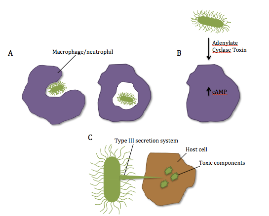
Figure 3: Schematic representation of B. bronchiseptica’s virulence factors. A) Normal mechanism of phagocytosis by neutrophils/macrophages. B) Secretion of Adenylate Cyclase Toxin by the bacteria and inhibition of phagocytosis resulting from elevated cAMP in the cell. C) Direct delivery of toxic components into the host cells with the type III secretion system. Source: Manon Desjardins.

Treatment

In mild cases, the infection can be self-limiting with supportive care. In more severe cases and to treat secondary infections often associated with B. bronchiseptica, antibiotic treatments may be necessary.

Vaccines are available for dogs, cats and swine. Young puppies and kittens may be vaccinated by intranasal, injectable or oral methods. The composition of vaccines depend on the administration method and range from inactivated antigens in injectable vaccines to attenuated avirulent bacteria in intranasal and oral vaccines.

References

Coote, J. G. (2001).  Environmental Sensing Mechanisms in BordetellaAdvances in Microbial Physiology, 44, 141-181. Retrieved from http://www.sciencedirect.com/science/article/pii/S0065291101440136

Ford, R. B. (2014). Vital Vaccination Series: Kennel Cough Revisited. Today’s Veterinary Practice. Retrieved from http://todaysveterinarypractice.navc.com/wp-content/uploads/2016/06/T1407C09.pdf

Huebner, E. S., Christman, B., Dummer, S., Tang, Y.-W., & Goodman, S. (2006). Hospital-Acquired Bordetella bronchiseptica Infection following Hematopoietic Stem Cell Transplantation. Journal of Clinical Microbiology44(7), 2581–2583. http://doi.org/10.1128/JCM.00510-06

Mattoo, S and Cherry, J. D. (2005). Molecular Pathogenesis, Epidemiology, and Clinical Manifestations of Respiratory Infections Due to Bordetella pertussis and Other Bordetella Subspecies. Clinical Microbiology Reviews, 18(2), 326-382. Retrieved from https://www.ncbi.nlm.nih.gov/pmc/articles/PMC1082800/

Nafe, (2014). Diagnostic and Therapeutic Approach: Dogs Infected with Bordetella bronchiseptica & Canine Influenza  Virus (H3N8). Today’s Veterinary Practice. 4(4). Retrieved from http://todaysveterinarypractice.navc.com/wp-content/uploads/2016/06/T1407F03.pdf

Nicholson, T.L., Conover M.S., Deora, R., (2012, November 12). Transcriptome Profiling Reveals Stage-Specific Production and Requirement of Flagella during Biofilm Development in Bordetella bronchiseptica (photograph). Retrieved from http://journals.plos.org/plosone/article?id=10.1371%2Fjournal.pone.0049166

Sykes, J. E. (2013). Bordetellosis. Canine and Feline Infectious Diseases (pp. 372-379). Retrieved from http://www.sciencedirect.com/science/book/9781437707953

Treponema pallidum

by Julia Messina-Pacheco and Lara Montaruli

Introduction

Within the past three decades, an important reemergence of Treponema pallidum infections has been observed worldwide. This infection manifests itself as syphilis and other treponemal diseases such as bejel, pinta and yaws. Most often acquired through close sexual contact, this helically coiled bacterium belongs to the spirochete phylum, which is distinguished by a double membrane. Despite T. pallidum being one of the first successful antibiotic-treated infections, discussion still surrounds the most effective treatment, mainly due to the inability to culture it in vitro and thus study its antimicrobial susceptibility.

Disease

Syphilis is caused by T. pallidum subspecies pallidum, and is divided into four stages of disease progression (primary, secondary, latent and tertiary). The primary stage symptoms are sores at the site of infection, generally around the genitals, anus or mouth and are usually firm, round and painless. Secondary syphilis causes skin rashes, swollen lymph nodes and fever. Once the latent stage is reached, there are no signs or symptoms, as the infection lays ‘dormant’ for multiple years. If left untreated, the disease progresses to the tertiary stage wherein inflammation, apathy, seizures, general paralysis with dementia, and aneurysm formation occur. T. pallidum is generally transmitted through sexual contact, primarily in homosexual men (see Figure 1). It can also be transmitted by transplacental passage during the later stages of pregnancy, giving rise to congenital syphilis. It is highly transmissible as approximately 30% to 60% of those exposed to primary or secondary syphilis will become infected. However, chances of transmission during sex are reduced through the use of condoms to protect the infected area or site of potential exposure.

Figure 1: Number of early syphilis cases by sexual transmission routes between 1992-2008 in Norway. The primary route of transmission overall is by men having sex with men. Source: Biomed Central, BMC Infectious Diseases, Jakopanec et al. (2010) https://doi.org/10.1186/1471-2334-10-105)

Epidemiology

Syphilis rates steadily decreased with the introduction of penicillin in 1947. However, it was not until the 1980s when the trend reversed in concurrence with increased use of intravenous drugs, the exchange of sex for drugs, anonymous sex, and people with multiple sexual partners, reaching its peak of 53.8 cases per 100,000 population in 1990. Still, syphilis rates continue to increase, for example, in the United States, the number of confirmed primary and secondary syphilis cases almost doubled, jumping from 8 724 to 16 663 between the years 2005 and 2013.

The disease primarily affects individuals between 15 and 40 years of age as there is a direct correlation between incidence of T. pallidum infection and increased sexual activity.  Furthermore, there is a noticeable difference in syphilis rates between men and women: males affected with primary and secondary syphilis outweigh females 10 to 1. On an international level, syphilis is distributed worldwide but remains prevalent in developing countries with rates being highest in the Western Pacific region and Southeast Asia.

Virulence Factors

Although T. pallidum may not exhibit the ‘classical’ virulence factors produced by most pathogens, it successfully attaches to, disseminates through and invades host tissues. These bacteria attach to a variety of host cell types by interacting with different host membrane components. In addition, its spiral shape (see Figure 2) allows it to enter through breaches in the skin and easily swim through gel-like substances, such as mucous membranes, to gain access to host blood and lymph systems. Thus, it propels itself by rotating in a corkscrew-like motion. Unlike most motile bacteria whose flagella are extracellular, in this case, the flagella is located in the space between the cytoplasmic and outer membranes, hidden from the host defenses. One of the major components of the host immune system is the generation of specific proteins, called antibodies, that recognize invading pathogens by binding to molecules on their surface. These immunogenic surface molecules are called ‘antigens’ and can be proteins, carbohydrates, or lipids. As such, the flagella of motile bacteria are composed of protein subunits called flagellin, which constitute a group of proteins called the H antigens. The outer membrane thus forms a barrier between the host defenses and T. pallidum‘s flagellum, preventing the binding of host antibodies to H antigens. In addition, T. pallidum is referred to as the ‘stealth pathogen’ due to the sparsity of immunogenic molecules presented on its outer surface, allowing it to avoid triggering an immune response. However, the few proteins that are presented on its surface are highly variable between individual bacteria of the same species. Thus, pathogens such as T. pallidum that can switch the molecular composition of their surface antigens are said to undergo ‘antigenic variation’. This makes it very difficult for the host to identify the bacterial molecules and subsequently raise an appropriate immune response. All in all, this bacterial pathogen effectively bypasses recognition by the host immune system by altering its surface components and by hiding its flagellum, two potential sources of antigenic molecules.

Figure 2: A photomicrograph showing the spiral, corkscrew-like shape of Treponema pallidum. The periplasmic flagella allows for the dissemination of the pathogen through host tissues and viscous substances, while preventing recognition by the host defenses. Source: Public Health Image Library, Center for Disease Control, Susan Lindsley (1972).

Treatment

     To date, there is no vaccine available against T. pallidum due to the sparsity and variability of its surface antigens. Thus, in order to successfully treat syphilis, early detection is crucial and followed by antibiotic treatment of syphilis-infected individuals and their partners. Both the CDC and WHO recommend a 10-day course of penicillin for early syphilis, with longer courses of treatment for those with late syphilis. In fact, penicillin is the only antibiotic shown to be effective in treating syphilis in pregnant women, as macrolides do not cross the placental barrier. However, individuals with penicillin allergies should be given doxycycline (cannot be taken during pregnancy) or ceftriaxone. The commonly used antibiotic azithromycin is not recommended, as resistance has emerged in strains of T. pallidum.

References

Chandrasekar P. H. (2017). Syphilis. Medscape. [2017 November 16] Retrieved from: https://emedicine.medscape.com/article/229461-overview#a2.

Fantry L. E. et al. Treponema Pallidum (Syphilis). Antimicrobe. [2017 November 15] Retrieved from: http://www.antimicrobe.org/b242.asp

LaFond, R. E., & Lukehart, S. A. (2006). Biological Basis for Syphilis. Clin. Microbiol. Rev 19, 29–49.

Liu, J., et al. (2010). Cellular Architecture of Treponema pallidum: Novel Flagellum, Periplasmic Cone, and Cell Envelope as Revealed by Cryo-Electron Tomography. J Mol Biol 403, 546-561.

Peeling, R. W., et al. (2017). Syphilis. Nat Rev Dis Primers 3, 17073.

Penn, C. W., et al. (1985). The outer membrane of Treponema pallidum: biological significance and biochemical properties. Microbiology 13, 2349-2357.

Radolf, J. D. (1996). Treponema. J Med Microbiol. Galveston, Texas: U of Texas Medical Branch.


Yersinia pestis

by Tongzhu Meng and Luyang Yuan

Introduction:

Yersinia pestis, also used to be named as Bacterium pestisBacillus pestis and Pasteurella pestis, is the causative agent of plague, which is a disease primarily affecting rodents via their associated fleas and is able to transmit to humans through infectious fleabites. Y. pestis was thought to be responsible for three devastating pandemics throughout human history, including the Justinian’s plague during the 6th century, the Black Death in Europe and Modern plague in China in the later 19th century. The bacterium was discovered during the epidemic of the plague in Hong Kong and had been used as a biological weapon during the 20th century. Y. pestis can be found exclusively in mammalian hosts and arthropod vectors, such as fleas.

Disease:  

Plague is a serious infectious disease that has three major clinical forms: bubonic plague, septicemic plague and pneumonic plague. If the patient is bitten by an infected flea, Y. pestis bypasses the skin barrier, enters the bloodstream and multiplies inside the lymph nodes. In healthy individuals, macrophages are guards of the immune system that are able to digest invading bacteria. Y. pestis inhibits macrophages from clearing bacteria and causes patients to have fever and one or more swollen lymph nodes. Y. pestis prevents local immune cells from eliminating bacteria and communicating with other remote immune cells that are able to help controlling the infection.

Septicemic plague can exist as the first symptom of plague or as secondary symptom of untreated bubonic plague. This form is the result of infectious fleabites or handling tissues or fluids of an infected animal. Under this kind of scenario, patients’ fingers, toes, the nose and other tissues might get black, due to reduced blood flow. They are also susceptible to develop bleeding into the skin and inner organs. Pneumonic plague is the result of inhaling infectious droplets in the air or from untreated bubonic or septicemic plague after the bacteria have spread to the lungs.  It is the most serious form of the plague and is the only form of the disease that can be spread from person to person through infectious droplets. Infections of lung may lead to pneumonia and cause chest pain, respiratory failure and shock.

Figure 1: Summary of transmission process of Yersinia pestis to human. (by Tongzhu Meng)

Epidemiology:  

From 2010 to 2015, WHO reported 3248 cases worldwide, including 584 deaths. Plague epidemics have occurred in Africa, Asia and South America, but since 1990s, most human cases reported were occurred in Africa. Nowadays, the three most vulnerable countries in the world are Democratic Republic of the Congo, Madagascar and Peru. Bubonic plague has a mortality rate of 30% to 60% while pneumonic plague is always fatal unless treatment started within 20 hours of symptom onset.

Over 80% of cases reported in United States were in bubonic form and there was an average of seven human plague cases reported each year for the past decades. These reported human cases covered people in all ages and in both sexes. Moreover, according to the cases reported in the last 20 years, people living in small towns and villages or agricultural areas are more susceptible than those living in larger towns and cities or urban areas.

Virulence Factor:  

Y. pestis gets into the body by flea bites or by contaminated fluid or by infectious droplets in the air. Phagocytes, such as macrophages, are a type of cell that circulates in the body, which can engulf and digest bacteria and other non-self material. In healthy individuals, these cells would recognize bacteria and then engulf it by a process called phagocytosis. This leads to the formation of a bag of bacteria in the phagocyte, which is called phagosome. Then the enzymes located in the phagocytes are pumped into phagosomes to destroy the bacteria.

However, Y. pestis is resistant to phagocytosis. There are two important virulence factors contributing to this property named F1 (Fraction 1) and LcrV. LcrV may also play a role in suppressing the immune response in an individual, it could inhibit production of signal molecules released by immune cells that would lead to the silence of normal immune response. The bacteria are able to move in the circulation and travel to local lymph nodes. It is resistant to be digested by macrophage and it can replicate within the lymph nodes rapidly, which causes swelling and enlargement of the lymph nodes.

Y. pestis has a needle-like structure (Figure 2), which is necessary to infect the host cells. It injects secretory proteins produced by Yersinia into macrophages and other immune cells. Some of these secreted proteins form pores on the host cell membrane and lead to the destruction of the host cells. Those pores created by the secretory proteins serve as gates for the other secretory proteins to get into the cells. Some of these secretory proteins limit the ability of immune cells to engulf the bacteria and other non-self materials and affect the signaling pathway of the immune system. Moreover, some of them could get into the host cells and lead to the killing of the host.

To sum up, Y. pestis is resistant to phagocytosis. It causes the death of host cells and affects the signals between the immune cells in the host that leads to suppression of normal immune responses.

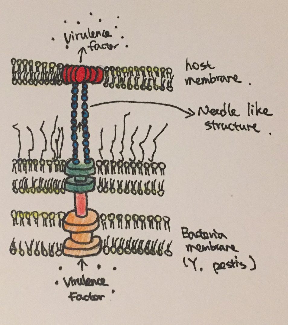
Figure 2: The needle like structure used by Y. pestis to inject virulence factors into host cells.

Prevention:

Contact with dead or infected animals, especially rodents, should be avoided.

Treatment: 

Commonly prescribed antibiotics for enterobacteria can be used to treat Y. pestis infections. CDC recommends that the treatment should begin as soon as plague is suspected. The earlier the treatment starts, the greater chance for patients to survive. Gentamicin and streptomycin are often prescribed as first line treatments due to their ability to stop the bacteria from making proteins it requires and to induce bacterial death. However, patients should not maintain on streptomycin for more than full 10 days in order to avoid the risk of developing endotoxic shock. They should gradually change to other antibiotics to continue the treatment. Treatments can be adjusted depending on the patient’s age, medical history and underlying health conditions as well.

WHO does not recommend vaccination against Y. pestis infections for general populations. However, for those who are often exposed to the risk of contamination and for health care workers, vaccinations should be considered.

Reference:

Auerbach, R. K., Tuanyok, A., Probert, W. S., Kenefic, L., Vogler, A. J., Bruce, D. C., … & Wagner, D. M. (2007). Yersinia pestis evolution on a small timescale: comparison of whole genome sequences from North America. PLoS One2(8), e770.

Galimand, M., Carniel, E., & Courvalin, P. (2006). Resistance of Yersinia pestis to antimicrobial agents. Antimicrobial agents and chemotherapy50(10), 3233-3236.

Li, B., & Yang, R. (2008). Interaction between Yersinia pestis and the host immune system. Infection and immunity76(5), 1804-1811.

Perry, R. D., & Fetherston, J. D. (1997). Yersinia pestis–etiologic agent of plague. Clinical microbiology reviews10(1), 35-66.

Plague.(2015,September 14). Retrieved November 20, 2017, from https://www.cdc.gov/plague/ prevention/index.html

Plague. (n.d.). Retrieved November 20, 2017, from http://www.who.Int/mediacentre/factsheets/ fs267/en/


Acinetobacter baumannii

by Caitlin Deseve and Jessika Marquis-Hrabe

Introduction

Acinetobacter baumannii (see Figure 1) is a multi-drug resistant pathogen that has become of great concern to medical facilities around the world. It causes various infections of the blood, brain, lungs, urinary tracts, and open wounds. The species was first identified in 1911 in soil samples, but became prevalent among returning soldiers who were treated in field hospitals of Iraq – thus earning it the common name ‘Iraqibacter’. Its natural habitat is still unknown.

Figure 1: Computer-generated image of Acinetobacter baumanii based on scanning electron microscopy. Source: Public Health Image Library, Center for Disease Control, James Archer, U.S. Centers for Disease Control and Prevention 2013.

Disease

Acinetobacter baumannii is a nosocomial bacterial pathogen, meaning it originates from hospital settings. The pathogen is capable of growing at various temperatures and pH conditions, which permits A. baumannii to persist in diverse environments. It causes a range of symptoms once it breaches the skin – from skin infections to pneumonia. It is most common in intensive care units, and in patients in hospital stays longer than 90 days with open wounds and invasive devices.

The most common symptoms of A. baumannii are ventilator-associated pneumonia (VAP) and bloodstream infections. VAP develops when pathogens transmit from external equipment and colonize the patient’s airways. Mortality rates due to A. baumannii-associated pneumonia range from 30 to 75%, with VAP responsible for the higher end. The most reported infections are encountered in the respiratory tract, wounds and catheter insertion sites.

Epidemiology

The United States has suffered several outbreaks, mostly associated with the return of military placed in Iraq and Afghanistan. The pathogen is often transmitted by contact to inanimate surface in field hospitals. Improper isolation and disinfecting treatments permitted the transfer of A. baumannii to US hospitals.  In 2004, 83% A. baumannii bloodstream infections were identified in the casualties injured. Infections in military personnel have also been reported in Canada and the United Kingdom.

Additionally, A. baumannii outbreaks have been identified in Europe since the 1980s. In most instances, transmission was caused by transfer of infected patients between hospitals; only one or two epidemic strains were recorded in hospitals. However, the outbreaks were not only limited to the hospitals, but spread internationally through airline travel.

The pathogens` prolonged survival in clinical settings contributes to its continuing outbreaks. Furthermore,  A. baumannii mortality rates are suspected to be associated with warmer temperature conditions, as pathogen colonization appears to be higher in tropical regions.

Virulence Factors

A. baumannii is highly capable of adhering to surfaces using hair-like appendages called pili. Specifically, A. baumannii employs Type IV pili on its surface, which function not only in adherence, but also in motility by extending and retracting. It has become of great concern due to its potential to produce biofilms. Communities of A. baumanni form by sticking together in a slimy extracellular matrix on both skin and hospital equipment. Formation of biofilms (see Figure 2) is a key virulence factor that provides resistance to antibiotics and allows persistence in unfavourable environmental conditions, such as low nutrient availability and desiccation.

Figure 2: Biofilm formation on a surface by Acinetobacter baumannii. 1, Attachment of free cells; 2, cell-cell adhesion in extracellular matrix; 3, dividing cells (proliferation) and cell growth; 4, cells spread and colonize new surfaces.

The presence of competence genes comFECB and comQLONM allow A. baumannii to uptake DNA from the environment and integrate it into its genome through a process called transformation. This factor enables A. baumannii to develop new traits rapidly, such as developing lower susceptibility to antibiotics. Moreover, the pathogen can produce antimicrobial-inactivating enzymes, such as Beta-lactamases. These enzymes destroy antibiotics like penicillins, to render them ineffective.  Finally, A. baumannii has a capsule that surrounds the outer membrane of the bacteria to serve as a protective barrier. Its presence helps the pathogen to evade the human immune response, ultimately making it more persistent in infection sites.

Treatment

A. baumannii is resistant to disinfectants and known antibiotics. Therefore, sufficient control is the current aim in hospitals. To prevent infection, environmental samples, especially from medical equipment, should be cultured and sources of contamination discarded. Additionally, hygiene control could prevent contamination between surface contact or direct person-person interaction. Should patients be infected, they should be isolated until infection has passed. Antibiotic selection is strenuous due to varying strains acquiring different antibiotic resistance. Discerning application of antibiotics in hospitalized patients is important to minimize drug resistance through pathogen evolution. Therefore, the treatment considered should be determined case-by-case.

References

Eliopoulos GM, Maragakis LL, Perl ™. Acinetobacter baumannii: Epidemiology, Antimicrobial Resistance, and Treatment Options. Clinical Infectious Diseases. 46(8): 1254–1263. Available from: https://academic.oup.com/cid/article-lookup/doi/10.1086/529198

Gaddy JA, Actis LA. 2009. Regulation of Acinetobacter baumannii biofilm formation. Future Microbiology. 4: 273-278. Available from: https://www.ncbi.nlm.nih.gov/pmc/articles/PMC2724675/

Geisinger E, Isberg RR. 2015. Antibiotic Modulation of Capsular Exopolysaccharide and Virulence in Acinetobacter baumannii. PLoS Pathog. 11(2): e1004691. Available from: https://www.ncbi.nlm.nih.gov/pmc/articles/PMC4334535/

Piepenbrink KH, Lillehoj E, Harding CM, Labonte JW, Zuo X, Rapp CA, Munson RS, Goldblum SE, Feldman MF, Gray JJ, Sundberg EJ. 2016. Structural Diversity in the Type IV Pili of Multidrug-resistant Acinetobacter. Journal of Biological Chemistry. 291: 22924-22935. Available from: http://www.jbc.org/content/291/44/22924.full

Peleg AY, Seifert H, Paterson DL. 2008. Acinetobacter baumannii: Emergence of a Successful Pathogen. Clinical Microbiology Reviews. 21(3): 538-582. Available from: https://www.ncbi.nlm.nih.gov/pmc/articles/PMC2493088/

Proteus mirabilis

by Rhea Suvarna

Introduction

Proteus mirabilis is commonly the causative agent of complicated urinary tract infections (UTIs), UTIs associated with components that compromises the urinary tract or host defense, especially in individuals with functional or structural abnormalities or with long-term catheterization for patients whose bladders will not empty fully or empty at inappropriate times. This bacterium is frequently found in soil and water, and is also part of the normal bacterial community residing in the human gastrointestinal tract.

Disease

P. mirabilis causes symptomatic infections of the urinary tract by either ascending from the gastrointestinal tract or by person-to-person transmission usually in healthcare settings.

Individuals with P. mirabilis infection may present with urethritis, cystitis, prostatitis, or pyelonephritis. A history of frequent renal stones can be an indication of chronic P. mirabilis infection. Symptoms of urethritis include painful urination, urine containing white blood cells, and increased frequency of urination. In males, symptoms are usually mild and could include urethral discharge. The symptoms of cystitis are more prominent than urethritis. They include painful urination, increased frequency and urgency of urination, small volume urine, dark urine, urine containing red blood cells, suprapubic pain, and back pain. Prostatitis affects men more acutely than cystitis with the same set of symptoms, though sometimes along with fever and chills. Pyelonephritis results from a complication of either of the conditions mentioned above and therefore, the patient presents symptoms of urethritis or cystitis along with flank pain, costovertebral angle tenderness, nausea and vomiting, fever, and sometimes an enlarged kidney.

The interaction between the bacterium and the host immune system determines the level of infection. Adherence followed by colonization is the basis of UTI pathogenesis. P. mirabilis UTIs occur in an ascending manner. The bacteria first contaminate the tissues surrounding the urethra, then enter the bladder through the urethra and form an initial colony. Following initial colonization, P. mirabilis ascends the ureters thanks to its swarming ability and interacts with epithelial cells of the renal pelvis, allowing for colonization of the kidney. Occasionally, the bacteria break through the renal tubular epithelial barrier and enter the bloodstream.

Epidemiology

P. mirabilis causes 1-10% of all UTIs. More specifically, this bacterium accounts for 1-2% of all UTIs in healthy women and in the case of hospital-acquired UTIs and complicated UTIs, P. mirabilis is responsible for 5% and 20-45%, respectively. Recent studies suggest that this species causes 4% of almost 3,000 UTIs in North America.

Factors that may increase the risk for acquiring P. mirabilis UTIs include female sex, longer duration of catheterization, inadequate catheter cleaning or care, underlying illness, and lack of availability of systemic antibiotics.

Virulence Factors

P. mirabilis utilizes a variety of virulence factors that allow it to access and colonize the host urinary tract, including fimbriae and other adhesions, urease and stone formation, iron and zinc acquisition, proteases and toxins, swarming growth, biofilm formation, and regulation of pathogenesis through DNA binding proteins.

The activity of the tiny projections on the bacterium surface, termed fimbriae (or pili), mediate the attachment of the bacterium to host tissue. The tips of these fimbriae contain specific compounds and polysaccharides that permit the bacterium to attach to specific sites in the host. Successful attachment initiates a cascade of events that leads to release of compounds that promote an alkaline pH, making it a suitable environment for survival and propagation. Alkaline pH in the

Urease facilitates virulence via the production of urinary stones. This enzymatic activity results in an increase in local pH, producing an alkaline environment in the urinary tract. The alkaline environment causes the formation of crystalline biofilms and the precipitation of calcium and magnesium ions to form urinary stones. These cause tissue damage and can block urinary flow.

During swarming, the expression of several virulence genes is increased and allows the bacterium to move through ascending urinary tract. The swarming ability of P. mirabilis is especially applicable to catheterized patients, as this bacterium is able to swarm across catheters made of silicon (see Figure 1) or latex. The flagella of P. mirabilis are responsible for the bacterium’s swarming motility, which is fueled by the proton motive force (See Figure 2). Flagellin, the structural component of flagella, is sensed by the host immune system through Toll and NOD-like receptors, which elicits a pro-inflammatory response. However, P. mirabilis flagella encode two flagellins, which allows for antigenic variation. This means that the bacterium is capable of altering its surface proteins to escape recognition by the host immune response.

Figure 1: Episcopic differential interference contrast (EDIC) microscopic image of 24-hour exposure showing multi-layered appearance and highly reflective, motile P. mirabilis (‘grey lines’) on silicone catheter section. Source: PLoS ONE, Wilks et al. (2015).à

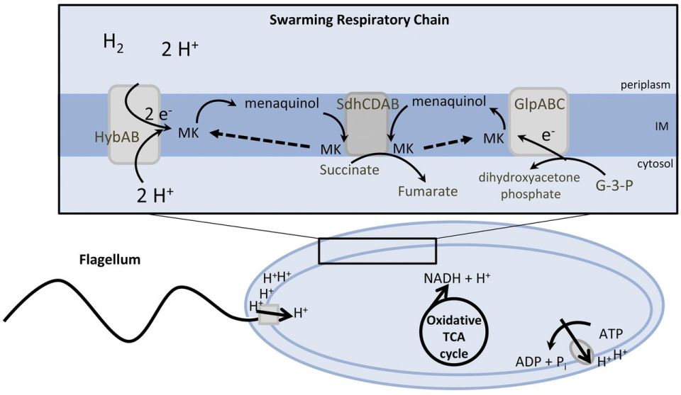
Figure 2: Suggested model for the energy metabolism of P. mirabilis during swarming. The proton gradient is generated by membrane respiration to oxidize NADH to NAD+, which powers flagellum rotation and oxidative phosphorylation. Source: mBio, Alteri et. al (2012).

Treatment

Patients diagnosed with uncomplicated P. mirabilis UTI, patients that are otherwise healthy and have no structural or neurological urinary tract abnormalities, are treated with antibiotics of either a 3-day course of trimethoprim/sulfamethoxazole (TMP/SMZ) or an oral fluoroquinolone. Acute, uncomplicated pyelonephritis may be treated with fluoroquinolone for 7-14 days or a one-time dose of ceftriaxone or gentamycin after either TMP/SMZ, an oral fluoroquinolone, or cephalosporin for 7-14 days. Antibiotic therapy is suggested for patients with more severe P. mirabilis infections.

Those suffering from a complicated P. mirabilis UTI can be treated with oral antibiotics for 10-21 days along with necessary follow-up.

References

Alteri, C.J., Himpsl, S.D., Engstrom, M.D., and Mobley, H.L.T. 2012. Anaerobic respiration using a complete oxidative TCA cycle drives multicellular swarming in Proteus mirabilis. MBio 3(6): e00365-12. American Society for Microbiology. doi:10.1128/mBio.00365-12.

Burall, L.S., Harro, J.M., Li, X., Lockatell, C.V., Himpsl, S.D., Hebel, J.R., Johnson, D.E., and Mobley, H.L.T. 2004. Proteus mirabilis genes that contribute to pathogenesis of urinary tract infection: identification of 25 signature-tagged mutants attenuated at least 100-fold. Infect. Immun. 72(5): 2922–38. American Society for Microbiology (ASM). doi:10.1128/IAI.72.5.2922-2938.2004.

Chen, C., Chen, Y., Lu, P., Lin, W., Chen, T., and Lin, C. 2012. Proteus mirabilis urinary tract infection and bacteremia: Risk factors, clinical presentation, and outcomes. J. Microbiol. Immunol. Infect. 45(3): 228–236. Elsevier. doi:10.1016/J.JMII.2011.11.007.

Foris, L.A., and Snowden, J. 2017. Proteus Mirabilis Infections. In StatPearls. StatPearls Publishing. Available from http://www.ncbi.nlm.nih.gov/pubmed/28723046 [accessed 19 November 2017].

Schaffer, J.N., and Pearson, M.M. 2015. Proteus mirabilis and Urinary Tract Infections. Microbiol. Spectr. 3(5). NIH Public Access. doi:10.1128/microbiolspec.UTI-0017-2013.

Umpiérrez, A., Scavone, P., Romanin, D., Marqués, J.M., Chabalgoity, J.A., Rumbo, M., and Zunino, P. 2013. Innate immune responses to Proteus mirabilis flagellin in the urinary tract. Microbes Infect. 15(10–11): 688–696. doi:10.1016/j.micinf.2013.06.007.

Wilks, S.A., Fader, M.J., and Keevil, C.W. 2015. Novel Insights into the Proteus mirabilis Crystalline Biofilm Using Real-Time Imaging. PLoS One 10(10): e0141711. Public Library of Science. doi:10.1371/journal.pone.0141711.

Escherichia coli Outbreak in Europe (May-June 2011)

by Julianne Audette and Coralie Reymond

Introduction

May 19, 2011, the Robert Koch Institute (RKI) in Germany was notified of a number of cases of bloody diarrhea in northern Germany. The RKI is Germany’s public health institute that investigates all types of outbreaks. These cases were soon identified to be caused by the O104:H4 strain of Shiga toxin-producing Escherichia coli (STEC) bacteria and they rapidly evolved into a full-blown outbreak. Ultimately, almost 4,000 people in 16 countries were affected, but the Northern part of Germany was the most affected, as you can see from Figure 1. The outbreak lasted for approximately two months, from the beginning of May to the beginning of July, with a peak during the month of May.

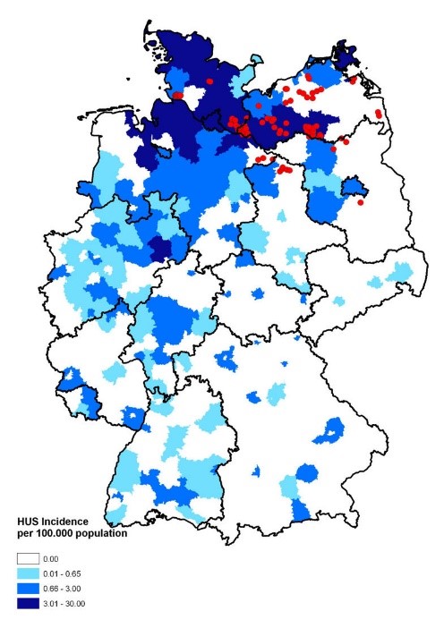
Figure 1: Map of Germany displaying the incidence of HUS [hemolytic uremic syndrome] cases. Source: Wieler LH, Torsten S, Eichhorn I, Antao EM, Kinneman B, Geue L, Karch H, Guenther S, Bethe A. 2011. No evidence of the Shiga toxin-producing E. coli O104:H4 outbreak strain or enteroaggregative E. coli (EAEC) found in cattle faeces in northern Germany, the hotspot of the 2011 HUS outbreak area. Gut Pathogens. [updated 3 November 2011; accessed 17 November 2017]; https://gutpathogens.biomedcentral.com/articles/10.1186/1757-4749-3-17. doi: 10.1186/1757-4749-3-17

Disease description

Escherichia coli is a gram negative, rod-shaped bacteria, see Figure 2, which is also a facultative anaerobe. Most of this bacteria’s strains are commensals and reside in gastrointestinal tract as part of the normal microbiota without causing harm to the host. However, different strains of Escherichia coli have acquired virulence factors that render them pathogenic, capable of causing disease. They acquired virulence factors through mutation or horizontal gene transfer, genes from other bacteria. Escherichia coli is one of the most important food-borne pathogens. Enterohemorrhagic Escherichia coli (EHEC) produce the Shiga toxin which causes hemorrhagic colitis (HC) and hemolytic uremic syndrome (HUS). HC and HUS can cause failure of essential organs, such as kidney and heart, leading to death if left untreated. Those bacteria are highly infectious because they survive for a long period of time outside of the host which increases the time products stay contaminated and increases chances of contracting the infection. Also, they can survive extreme conditions, such as the acidic environment of the stomach. Cases related to Escherichia coli are lower than other bacteria types (Salmonella or Campylobacter spp.), however much higher mortality and hospitalization rates are related to them. Escherichia coli’s natural reservoir is in cattle and the farm environment. Cattle are asymptomatic carriers and shed this bacteria in their feces.

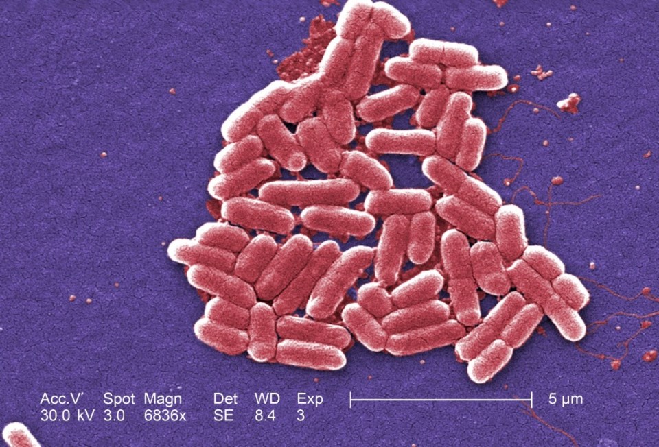
Figure 2: Under a high magnification of 6836X, this digitally-colorized, scanning electron microscopic (SEM) image depicted a growing cluster of Gram-negative, rod-shaped, Escherichia coli bacteria of the strain O157:H7, which is a pathogenic strain of E. coli. Source: Public Health Image Library, Center for Disease Control, Dr. Janice Carr (2006).

Discussion of the source of the outbreak

The outbreak was mainly centered in northern Germany and spread throughout Europe, but very little in the United States, as only 6 cases of people having traveled to this area in Germany were recorded. The common factor of all the people infected was that they ate raw vegetables grown locally in northern Germany. The Robert Koch Institute was able to narrow down the source of the outbreak to raw sprouts. Other cases showed up near Bordeaux, France where the infected people had eaten raw sprouts grown locally. This information led the European Food Safety Authority to discover the real source was from fenugreek seeds imported from Egypt into France and Germany. The origin of the outbreak strain and how the seeds were contaminated remains unclear to this day.

Cause of the outbreak

This outbreak was caused by the O104:H4 strain of Escherichia coli rather than the most common strain O157:H7. The high number of HUS cases and deaths indicated that the O104:H4 strain had an augmented virulence compared to the common strain. The exact reason remains unknown. However, scientists have offered various hypotheses. The outbreak strain combines virulence potentials of STEC and enteroaggregative E. coli (EAEC). The distinguishing feature of EAEC is their ability to attach to tissue cells.  It has been hypothesized that O104:H4 is a typical EAEC strain that acquired the stx2a gene that encodes for the toxin (mutant).  This toxin enters the epithelium cells that line the gastrointestinal tract and lyses the cells which causes bloody diarrhea and HUS. This outbreak pathogen had a longer incubation period (8 days) compared to the O157:H7 strain (3-4 days) which means that there was a longer period between the exposition to the pathogen and the onset of the symptoms, making it more difficult to find the source. The O104:H4 strain also had an increased adherence to intestinal epithelial cells which might facilitate systemic absorption of the toxin. This could explain the high number of patients developing HUS. Antibiotic resistance could also have played a role in this outbreak. This strain is resistant to β-lactam drugs and if this type of antibiotic was used to treat the infection, it would only suppress the competing microbiota making it easier for the O104:H4 strain of Escherichia coli to proliferate.

Measure taken to end the outbreak

To counteract the disease, the infected patients required hospitalization to maintain fluid and electrolyte levels, to monitor and support kidney function and dialysis in case of renal failure.  During the outbreak, public health reviewed the potential therapies going above and beyond symptomatic treatment. Two avenues were explored: antibiotic treatment and passive immunity (toxin neutralizing antibodies). Antibiotic therapy was considered only in very severe cases due to increased release of toxin by bacteria lysis. The antibody treatment was not feasible as they were not yet approved meaning that they were still in the clinical research phase.

Authorities were able to identify the origin at the same time as the epidemic curve was starting to decrease. This decline is due to self-limitation, meaning that there was limited exposure to contaminated food in circulation (Figure 3). Raw sprouts were identified as the outbreak source in less than 3 weeks which is relatively rapid and comparable to other outbreak investigations in other countries (incl. US). On July 5th, the seeds from Egypt were banned which definitively prevented further cases.

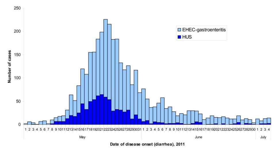
Figure 3: Epidemic curve of the 2011 outbreak of O104:H4 strain of Escherichia coli in Europe.  Source: Werber D, Krause G, Frank C, Fruth A, Flieger A, Mielke M, Schaade L, Stark K. 2012. Outbreaks of virulent diarrheagenic Escherichia coli- are we in control? BMC Medicine. [accessed 2017 Nov 17]; https://bmcmedicine.biomedcentral.com/articles/10.1186/1741-7015-10-11. doi: 10.1186/1741-7015-10-11.

Aftermath

The outbreak had enormous economical consequences because the sale of salads and other vegetables decreased. Spanish vegetable producers were especially affected because Spanish cucumbers and tomatoes had been discussed as a potential source of pathogen.  The European Union compensated farmers from several vegetable-exporting countries for a total of 220 million Euros.

Many criticized how the German government dealt with the outbreak. The notification time from the local health department to the regional and national agencies could have been shortened by a few days. Since then, Germany has put in place a centralized database shared by all health authorities to allow a faster parallel communication between all agencies.

References

Altmann M, Wadl M, Altmann D, Benzler J, Eckmanns T, Krause G, Spode A, An der Heiden M. 2011. Timeliness of Surveillance during Outbreak of Shiga Toxin–producing Escherichia coli Infection, Germany, 2011. Emerging Infectious Diseases. [accessed 2017 Nov 17]; 17(10): 1906-1909. https://www.ncbi.nlm.nih.gov/pmc/articles/PMC3310688/. doi : 10.3201/eid1710.111027.

Bielaszewska M, Mellmann A, Zhang W, Köck R, Fruth A, Bauwens A, Peters G, Karch H. 2011. Characterisation of the Escherichia coli strain associated with an outbreak of haemolytic uraemic syndrome in Germany, 2011: a microbiological study. The Lancet Infectious Diseases. [accessed 2017 Nov 17]; 11(9): 671-676. http://www.sciencedirect.com/science/article/pii/S1473309911701657. doi: 10.1016/S1473-3099(11)70165-7.

Burger R. 2012. EHEC O104:H4 in Germany 2011: Large Outbreak of BloodyDiarrhea and Haemolytic Uraemic Syndrome by Shiga Toxin-Producing E. Coli via Contaminated Food. In: Improving Food Safety Through a One Health Approach: Workshop Summary. Washington (DC): National Academies Press; [accessed 2017 Nov 17]. https://www.ncbi.nlm.nih.gov/books/NBK114499/.

Centers for Disease Control and Prevention (CDC). 2013. Outbreak of Escherichia coli O104:H4 Infections Associated with Sprout Consumption — Europe and North America, May–July 2011. Morbidity and Mortality Weekly Report (MMWR). [updated 20 December 2013; accessed 17 November 2017]; 62(50);1029-1031. https://www.cdc.gov/mmwr/preview/mmwrhtml/mm6250a3.htm.

Frank C, Werber D, Cramer JP, Askar M, Faber M, An der Heiden M, Bernard H, Fruth A, Prager R, Spode A, et al. 2011. Epidemic Profile of Shiga-Toxin–Producing Escherichia coli O104:H4 Outbreak in Germany. The New England Journal of Medicine. [accessed 2017 Nov 17]; 365: 1771-1780. http://www.nejm.org/doi/full/10.1056/NEJMoa1106483#t=article. doi : 10.1056/NEJMoa1106483.

Karch H, Denamur E, Dobrindt U, Finlay BB, Hengge R, Johannes L, Ron EZ, Tonjum T, Sansonetti J, Viencente M. 2012. The enemy within us: lessons from the 2011 European Escherichia coli O104:H4 outbreak. EMBO Molecular Medicine. [accessed 2017 Nov 17]; 4(9): 841-848. https://www.ncbi.nlm.nih.gov/pmc/articles/PMC3491817/. doi : 10.1002/emmm.201201662.

Youn Lim J, Yoon JW, Hovde CJ. 2010. A Brief Overview of Escherichia coli O157:H7 and Its Plasmid O157. Journal of Microbiology Biotechnology. [updated 20 January 2010; accessed 17 November 2017]; 20(1):5–14. https://www.ncbi.nlm.nih.gov/pmc/articles/PMC3645889/. doi: PubMed Central PMCID: PMC3645889.


Staphylococcus saprophyticus

Introduction

Staphylococcus saprophyticus is a frequent causative agent of uncomplicated lower urinary tract infections (UTI) in humans. Due to its high prevalence in slaughtered animal carcasses, abattoir workers’ gloves and animal products, the bacterial pathogen is thought to have a zoonotic origin.

To differentiate between the various species of staphylococci, the bacteria are evaluated on their ability to produce coagulase. As S. saprophyticus is among the coagulase-negative staphylococci (CoNS), it lacks the enzyme that catalyzes the formation of fibrin from fibrinogen. Before 1960, CoNS observed in urine samples were assumed to be urinary contaminants. However, in 1962, Toress-Pereira established the first connection between CoNS and human disease when he isolated CoNS from the urine of 40 women with acute UTIs. S. saprophyticus officially became acknowledged as a common cause of UTIs in the early 1970s.

Disease

S. saprophyticus is predominantly transmitted to humans through the consumption of contaminated animal products. The microorganisms first colonize the gastrointestinal tract. Then, activities such as sexual intercourse facilitate the transfer of the bacteria from the anus to the periurethral area, where they can ascend into the urethra and eventually the bladder. An infection in the lower urinary tract, also known as honeymoon cystitis, is established when S. saprophyticus circumvents the host’s defences, colonizes, invades and replicates within the uroepithelium. Symptoms of S. saprophyticus lower UTIs include: red and white blood cells in urine (hematuria and pyuria), painful urination (dysuria), frequent urination and back pain. If the bacteria continue to rise to the kidneys, a potentially life-threatening infection in the upper urinary tract can result called pyelonephritis.

Epidemiology

Women are 15 times more prone to developing UTIs than men due to their shorter urethras. Over 150 million women worldwide suffer from UTIs each year. Though the infections are primarily caused by E. coli, S. saprophyticus is responsible for 10 to 15% of cases. The actual rate of infection by S. saprophyticus is likely higher, however, as the bacteria tend to be overlooked when dominant uropathogens are also present in urine.

S. saprophyticus colonization is especially prevalent in late summer and fall. The most susceptible individuals to cystitis caused by S. saprophyticus are sexually-active young women between the ages of 16 and 25. Among this group, S. saprophyticus accounts for 42.3% of all cases. Infected women are also more likely to have recently had a menstrual period, experienced a symptomatic UTI within the previous year, swam in a pool, had sexual intercourse while suffering from vaginal candidiasis or used vaginal spermicides that disrupt the normal vaginal flora. Though less common, the infection can also target young boys, male homosexuals and elderly men with urinary catheters.

Virulence systems

Once the periurethral region becomes contaminated with S. saprophyticus from the gut, the uropathogen first colonizes the urethra before traveling up to the bladder. To eliminate the bacteria, the host executes defences such as the secretion of mucus to prevent bacterial adherence to the bladder mucosa. Poorly attached bacteria are then expelled from the body with urine, which encompasses antibacterial properties (e.g. acidic, high osmolality, high concentration of urea and organic acids). Despite the host’s best efforts, the properties of S. saprophyticus allow it to colonize the bladder. To attach to the uroepithelial cells, S. saprophyticus expresses several adhesins: extracellular slime, the collagen- and fibronectin-binding protein Sdrl, the surface-associated fibrillar lipase Ssp and the autolytic/adhesive protein Aas. In addition, the adhesin and hemagglutinin UafA binds to both fibronectin and membrane proteins on red blood cells, causing the red blood cells to agglutinate (clump together). S. saprophyticus can also produce urease (Figure 1). The enzyme hydrolyzes the urea in urine to ammonia and carbon dioxide (Figure 1). Ammonia supports the bacteria’s survival by increasing the pH of urine (Figure 1) and triggering the formation of urinary stones. Furthermore, ammonia is toxic to uroepithelial cells and causes direct tissue damage.

Figure 1: The production of urease by S. saprophyticus; As the pathogen cannot survive in low pH environments such as that found in urine, it produces the enzyme that hydrolyzes the urea in urine to ammonia and carbon dioxide. Ammonia raises the pH around S. saprophyticus, permitting its existence (Figure by Liz Brenhouse).

Once the host senses a bacterial infection, immune system cells (macrophages and neutrophils) are called in to kill the invading pathogen in a process called phagocytosis (Figure 2). Normally in phagocytosis, the macrophages and neutrophils surround the bacteria and engulf them. The bacteria are then trapped inside a pocket, the phagosome, within the immune system cells. Digestion of the pathogen occurs when the phagosome fuses with a specialized vesicle that contains degradative enzymes. However, S. saprophyticus bacteria can escape phagocytosis by embedding themselves within the urinary stones. Additionally, in coating themselves with the host’s proteins (collagen and fibronectin), the bacteria can disguise themselves to prevent being identified by antibodies, blood proteins that recognize and bind to foreign substances. Once bound, antibodies act as flags for macrophages to come and destroy their targets.

Figure 2: The immune response to S. saprophyticus urinary tract infection; Neutrophils attempt to engulf S. saprophyticus but are impeded by the bacteria’s virulence factors (Figure by Liz Brenhouse).

Treatment

Individuals suffering from UTIs caused by S. saprophyticus are prescribed antibiotics for one week or less. Though the uropathogen is resistant to nalidixic acid, a common treatment for UTIs, it is generally susceptible to other antibiotics including ampicillin, cephalosporins, nitrofurantoin, tetracyclines, sulfonamides and trimethoprim. While the medications differ in their modes of action, frequently-prescribed trimethoprim-sulfamethoxazole works by blocking the production of tetrahydrofolic acid (THF) which bacteria require for growth. Preventative measures can also be taken such as staying hydrated, wiping from front to back after using the bathroom and urinating before and after sexual intercourse.

References

Elizabeth, T. & Jones, B. (2008). Introduction to Pathogenic Bacteria. In: Zourob, M., Elwary, S. & Turner, A., editors. Principles of Bacterial Detection: Biosensors, Recognition Receptors and Microsystems. Berlin (DE): Springer. 3-13.

Flores-Mireles, A.L., Walker, J.N., Caparon, M. & Hultgren, S.J. (2015). Urinary tract infections: epidemiology, mechanisms of infection and treatment options. Nat Rev Microbiol 13(5), 269-284.

Gatermann, S. & Crossley, K.B. (2009). Urinary Tract Infections. In: Crossley, K.B., Jefferson, K.K., Archer, G.L. & Fowler, V.G., editors. Staphylococci in Human Disease. Hoboken (NJ): Blackwell Publishing Ltd. 455-469.

Hedman, P., Ringertz, O., Lindström, M. & Olsson, K. (1993). The origin of Staphylococcus saprophyticus from cattle and pigs. Scand J Infect Dis 25(1), 57-60.

Hitchings, G.H. (1973). Mechanism of Action of Trimethoprim-Sulfamethoxazole—I. The Journal of Infectious Diseases 128(3), S433–S436.

Hovelius, B. & Mårdh, P. (1984). Staphylococcus saprophyticus as a Common Cause of Urinary Tract Infections. Reviews of Infectious Diseases 6(3), 328-337.

Kline, K.A., et al. (2010). Characterization of a Novel Murine Model of Staphylococcus saprophyticus Urinary Tract Infection Reveals Roles for Ssp and SdrI in Virulence. Infect Immune 78(5), 1943-1951.

Koneman, E.W. (2006). Color Atlas and Textbook of Diagnostic Microbiology (6th ed.) Philadelphia (PA): Lippincott Williams & Wilkins.

Lo, J., Choi, W.H., Chan, J.Y.H. & Lange, D. (2015). Overview of Urinary Tract Infections. In: Lange, D. & Chew, B., editors. The Role of Bacteria in Urology. Berlin (DE): Springer. 7-20.

Raz, R., Colodner, R. & Kunin, C.M. (2005). Who Are You — Staphylococcus Saprophyticus? Clinical Infectious Diseases 40(6), 896-898.

Zachary, J.F. (2016). Pathologic Basis of Veterinary Disease Expert Consult (6th ed.) St. Louis (MO): Elsevier Health Sciences.


Brucella canis

by Baylie Daghofer-Hawes

Introduction

Brucella canis is a gram-negative coccobacilli (figure 1) animal pathogen and is the causative agent of canine brucellosis, which affects the reproductive organs of both male and female dogs. It is a zoonotic pathogen, meaning that although it normally exists in animals, it can be transmitted from animals to infect humans. B. canis was first isolated from dogs in the United States in 1966 after a widespread outbreak occurred in Beagles. Transmission of B. canis is caused by contact with fluids and secretions from infected dogs making this a widespread problem in many dog kennels.

Figure 1: Microscopic image of an extraction of Brucella canis, which is a gram-negative coccobacilli found in dogs, and appears as fine sand under microscope when stained. Source: Public Health Image Library, Centers for Disease Control and Prevention, Larry Stauffer (2002).

Disease

Infection by B. canis is mainly acquired by penetration of mucous membranes by the organism. B. canis usually resides in fluids such as genitourinary secretions, vaginal discharge and semen or milk from the mother and is transmitted through sexual or oral contact with infected fluids. It can also be transmitted in utero (from infected mothers to babies) or by fluids from infected aborted fetuses and placentas, as well as by inhalation or ingestion. Once the organism enters the dog it will get phagocytosed and sent to the lymphatic tissue to multiply and develop. After being incubated the bacteria will then travel to other organs in the dog by hematogenic (blood) routes. This infection can lead to reproductive failure, abortion, prolonged vaginal discharge and stillbirths in female dogs and infertility, testicle atrophy and epididymitis in male dogs. Other symptoms may include puppies that are born live but weak, that will often die soon later, as well as later development of lymphadenitis which is an enlargement of the lymph nodes. Although B. canis is a zoonotic disease, its effect on humans is not as severe as it is on animals and is not yet very well understood.

Epidemiology

The rate of infection from B. canis is still not very well-understood.  In 1970, the prevalence of the disease was around 6% worldwide. However, this varies by location.  In Canada 1.6% of dogs were infected in 1970 but in Asia 11% of dogs were infected in 1976-1977. Rates of infection are a lot higher in low income countries and the incidence of B. canis infection is still on the rise. There have been reported cases of the disease worldwide, especially in dog kennels and dog breeding facilities where many dogs are in close contact with each other. All breeds of dogs are susceptible to canine brucellosis however, they are more vulnerable if they are in confined populations and they are breeding or during abortions as the disease can spread faster. Stray dogs are also more susceptible to this disease than pet dogs, and dogs who have been spayed or neutered present a much lower risk at acquiring the disease. Canine brucellosis can occur in dogs of any age or sex but is most common in newborns and female dogs. The mortality rate from this disease is low except for in fetus or newborn dogs. In such cases, the risk of death is around 75% during outbreaks. Most outbreaks are due to contaminated fluids or introduction of contaminated dogs into kennels or breeding facilities as it facilitates the transmission of the disease. In this case outbreaks occur rapidly since even contaminated saliva or urine can carry the organism. Infected mothers can have infected offspring which can also spread the disease.

Virulence

B. canis will start infection by penetrating through the membrane of fluids, which will then enter the dog in order to get to the lymphatic and genital tissues. When B. canis enters the dog, it can then invade both phagocytic and non-phagocytic cells in the bloodstream.

In the case of phagocytic cells, the organism is phagocytosed by cells in the dog. These cells are called phagocytes and they pick up the organism to kill it. However, this bacterium is able to circumvent the killing action of the phagocytes in order to replicate within the host. When in the bloodstream, B. canis quickly develops into intracellular pathogens contained within circular polymorphonuclear cells (PMNs) and macrophages. This means they can easily survive the killing action of these cells. The main reason that they can circumvent the killing action is because of their rough lipopolysaccharide (LPS) coat. The adenine and guanine monophosphate production of the coat inhibits phagosomal fusion and oxidative burst activity. This prevents killing of the organism by phagocytes.

In the case of non-phagocytic cells, the organism hides in there to avoid the immune system. It is easily able to overcome innate immunity at very early times of infection. Since B. canis has low virulence in the host, it a poor activator of cytokines such as tumor necrosis factor (TNF). These are cells that promote inflammation in the host in response to a pathogen. As a result, B. canis is able to avoid activation of the complement system of the immune system.

The surviving brucella are then transported to the lymphatic and genital tissues hematogenically, which means it gets to these tissues by blood routes. Once in these tissues, the bacteria will multiply and develop and a leukocyte-associated bacteremia (bacteria that is in the blood) will distribute the bacteria to other organs and systems within the dog (figure 2). The spread of the bacteria within the dog will cause the development of clinical lesions in the dog and hyperplasia of the lymphoid tissue, which is enlargement of the lymphoid tissue due to the concentration of bacteria in localized regions. After the lymphoid tissue expands it can cause the production of Peyer’s patches which are small masses of lymphatic tissue and there is also an increase in lymphocytes, macrophages and plasma cells. This increase in cells will promote chronic infection in the dog and lead to reproductive failure after a few months incubation. Symptoms only show after a few months incubation because as part of the bacteria’s strategy it remains asymptomatic for a while in order to spread throughout the dog.

Figure 2: Pathway of infection and damage of Brucella canis in dogs and zoonotic potential of the virus in humans. Source: Baylie Daghofer-Hawes.

Prevention and Treatment

In order to prevent further spread of canine brucellosis, isolation of infected dogs is first required. Spaying or neutering dogs has also been shown effective in prevention of this disease in many cases. Treatment of canine brucellosis can be accomplished rather easily due to the fact that B. canis is short-lived outside of the body. It is rather sensitive to many disinfectants and degreasers as they are able to effectively remove the biofilm that builds up in the population. Long-term treatment with antibiotics has also been shown to successfully treat dogs with canine brucellosis. Since B. canis has a long incubation time inside the animal, the antibiotics needs to be given consistently on the long-term. A combination of minocycline and streptomycin have been proven the most effective antibiotics as they sequester the bacteria to inhibit its growth.

References

Al-Nassir, Wafa. “Brucellosis.” Medscape, https://emedicine.medscape.com/article/213430-overview#a2.

Bramlage, Donald J, et al. “Best Practices for Brucella Canis Prevention and Control in Dog Breeding Facilities .” United States Department of Agriculture, www.aphis.usda.gov/animal_welfare/downloads/brucella_canis_prevention.pdf.

“Canine Brucellosis: Brucella Canis.” The Center for Food Security and Public Health, http://www.cfsph.iastate.edu/Factsheets/pdfs/brucellosis_canis.pdf.

Chacón-Díaz, Carlos, et al. “Brucella Canis Is an Intracellular Pathogen That Induces a Lower Proinflammatory Response than Smooth Zoonotic Counterparts.” Infection and Immunity, vol. 83, no. 12, May 2015, pp. 4861–4870., doi:10.1128/iai.00995-15

Ledbetter, Eric C., et al. “Brucella Canisendophthalmitis in 3 Dogs: Clinical Features, Diagnosis, and Treatment.” Veterinary Ophthalmology, vol. 12, no. 3, 2009, pp. 183–191., doi:10.1111/j.1463-5224.2009.00690.x

Morisset, R. “Epidemic Canine Brucellosis Due To A New Species, Brucella Canis.” The Lancet, vol. 294, no. 7628, 1969, pp. 1000–1002., doi:10.1016/s0140-6736(69)90551-0.

Quah, Stella R., and William C. Cockerham. “Brucellosis .” International Encyclopedia of Public Health, 2nd ed., vol. 1, Elsevier/AP, 2017, pp. 281–285.

2014 Plague Outbreak in Madagascar

by Emma Rollins and Celeste Welch

Introduction

August 31, 2014: A man in Soamahatmana village, Madagascar, was diagnosed with the plague. The World Health Organisation (WHO) was not notified until November 4. By November 14, a total of 119 plague cases had been confirmed, with 40 deaths. By the end of the outbreak, there had been a total of 308 human plague cases, and 81 deaths. Out of the total cases, 23 (7%) were pneumonic plague, while the rest were bubonic plague.

Description of the disease

Yersinia pestis, the pathogen responsible for the plague, is a gram negative, zoonotic bacteria. It is most commonly transmitted from fleas to humans, but can also be transmitted through interpersonal contact, or indirectly through biting or scratching of a rodent intermediate. The flea is an essential vector for transmission as it provides the pathogen with the ability to penetrate the skin barrier when the flea bites humans to take a blood meal. Rats are used as reservoir hosts for Y. pestis as they also serve as a food source for fleas.

In humans, the infection begins as bubonic plague, characterized by swelling of the lymph nodes in the armpits and groin. Bacterial cells invade and replicate in macrophages and proliferate in lymph nodes resulting in painful, swollen lymph nodes called buboes (Fig. 1). In this case, the infection cannot be transmitted from person to person. Bubonic plague can be successfully treated with antibiotics such as Streptomycin if detected soon enough.

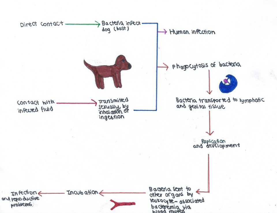
Figure 1: The characteristic buboes of the bubonic plague, seen here in a man’s armpit. The bubo is circled in red. Source: Centre for Disease Control and Prevention. 2017. https://phil.cdc.gov/Details.aspx?pid=2045. Accessed 2017 November 19.

Infrequently, Y. pestis can reach the lungs, which characterizes the pneumonic plague (Fig. 2). Droplets containing Y. pestis are released into the air when an infected host coughs and are then breathed in by another person, allowing for person to person transmission. Pneumonic plague is far more deadly: patients can die within 24 hours of infection. If the plague is left untreated, Y. pestis can infect the bloodstream, causing septicemic plague (Fig. 3). This is characterized by inflammation throughout the entire body and septic shock, which leads to low blood flow to the extremities and death. As seen in Fig. 3, the low blood flow causes necrosis of the extremities, causing them to turn black.

Figure 2: A chest X-ray showing Y. pestis in a person’s lungs, causing pneumonic plague. Source: Centers for Disease Control and Prevention. Plague Symptom. 2015. https://phil.cdc.gov/Details.aspx?pid=1955

Figure 3: Septicemic plague, caused by an infection of Y. pestis in the blood stream. Necrosis of the extremities is seen here, caused by low blood flow. Source: Centers for Disease Control and Prevention. Plague Symptom. 2015. https://www.cdc.gov/plague/symptoms/index.html

The Outbreak

Y. pestis was initially introduced to Madagascar in 1898 from a steamship containing fleas and rats from an infested area. Since then, there have been almost yearly outbreaks of Y. pestis. The first case in the 2014 epidemic was reported in Soamahatamana village. Y. pestis continued to spread, reaching a total of 16 districts. As of November 16, 2014, two cases of plague were reported in Antananarivo, the capital of Madagascar. The city’s dense population and weak healthcare system promoted rapid spread of Y. pestis.

It is primarily socioeconomic factors that led to the spread of Y. pestis on the island of Madagascar. Much of the population is impoverished with limited access to healthcare. This, in combination with poor living conditions and increased exposure to bacterial vectors, makes it much harder for people to receive the immediate treatment needed to eradicate Y. pestis. Without treatment, the plague continues to spread.

Plague season occurs during the rainy season in Madagascar, between November and April. During this time, rats take shelter in populated areas, often in the straw roofs of homes, increasing spread of infection by bringing the fleas they carry into close proximity with humans. While pesticides such as deltamethrin can be used to eliminate fleas, increased resistance has led to persistence of Y. pestis.

Response

The outbreak was mediated by targeting the primary vector of transmission, fleas, and administering antibiotics to infected patients. Pesticides were applied in order to eradicate flea populations and prevent spread of the bacteria. The WHO brought antibiotics to Madagascar to treat individuals infected with the bubonic plague, and individuals with pneumonic plague were isolated to prevent person-to-person transmission.  

Aftermath

Unfortunately, these measures did not fully eliminate the disease vector population: further re-emergence of Y. pestis occured in subsequent years, including a massive outbreak in 2017. As of November 10th, there have been a total of 2119 confirmed cases, and 171 deaths. This outbreak is increasingly of concern due to the high number of pneumonic plague cases, totaling at 76% of reported cases, compared to 7% in the 2014 outbreak. More than half of the 2017 cases have been recorded in the capital of Antananarivo and the main port of Toamasina, the two most densely populated areas in Madagascar. Death tolls will continue to climb as the plague ravages Madagascar’s cities until effective public health and sanitation measures are put into place.

Recent cases of Y. pestis infection have spread from Madagascar due to travelers contracting the disease and bringing it to other countries such as Seychelles and South Africa, where it spreads among local populations. The WHO warns that it is likely that Y. pestis will spread to numerous countries in Africa and the Indian Ocean due to high contact with Madagascar for trade and travel.

More effective precautions must be taken in the future to prevent re-emergence of infection; long term solutions include improved health care, wastewater treatment, and rodent and vector control. Enhanced case finding and monitoring must also be implemented with strengthened epidemiological surveillance in affected districts, early investigation, isolation and treatment of all pneumonic cases, as well as provision of free prophylactic antibiotics. The public and health care workers should also be educated on the prevention of the plague and infection control measures in burial practices.

References

Bertherat EG. 2015. Plague in Madagascar: overview of the 2014-2015 epidemic season. Weekly Epidemiological Record [accessed 2017 November 17]; 90(20): 250-252. http://web.a.ebscohost.com.proxy3.library.mcgill.ca/ehost/pdfviewer/pdfviewer?vid=1&sid=018af9c5-74c2-4386-978f-2333ec7fcaa3%40sessionmgr4010.

Plague – Madagascar. 6 September 2015. World Health Organization; [accessed 2017 November 17]. http://www.who.int/csr/don/06-september-2015-plague/en/

Plague – Madagascar. 15 November 2017. World Health Organization; [accessed 2017 November 18]. http://www.who.int/csr/don/15-november-2017-plague-madagascar/en/

Plague – Madagascar. 21 November 2014. World Health Organization; [accessed 2017 November 16]. http://www.who.int/csr/don/21-november-2014-plague/en/

Ratovonjato J, Rajerison M, Rahelinirina S, Boyer S. 2014. Yersinia pestis in Pulex irritans Fleas during Plague Outbreak, Madagascar. Emerging Infectious Diseases [accessed 2017 November 16]; 20(8): 1414-1415. https://www.ncbi.nlm.nih.gov/pmc/articles/PMC4111190/. Doi: 10.3201/eid2008.130629

Actinobacillus pleuropneumoniae

by An Bui

Introduction

Actinobacillus pleuropneumoniae, formerly known as Haemophilis pleuropneumoniae, is a member of the Pasteurellaceae family. This bacterium exists as a nonmotile, gram-negative coccobacillus and is the causative agent of pleuropneumoniae in pigs. A. pleuropneumoniaie is prevalently found in the porcine respiratory tract and does not survive long outside this environment.  It was first described in 1957 in the United States and has since then caused great economic losses worldwide in the pig industry.

Disease

A. pleuropneumoniae is a highly contagious and severe respiratory disease. It is often transmitted through aerosols or direct contact via nasal secretions between pigs. A. pleuropneumoniae, migrates to the swine lower respiratory system where it comes into contact with leukocytes inside the lungs. Leukocytes are cells belonging to the immune system that are used to protect the host from pathogens. This is usually done by phagocytosis; a process in which leukocytes engulf and digest pathogens. However, A. pleuropneumoniae can attack the immune cell by releasing toxins on the surface of its membrane causing the rupture of the leukocyte’s cell wall. When this occurs, lysosome, a membrane organelle of the immune cell, are released. The release of the lysosome content causes damage to the surrounding lung tissue, because of its low pH. Lesions and abscesses begin to form in the lungs (Figure 1) and symptoms such as respiratory distress, fever, diarrhea, bloody discharge from the mouth and lethargy appear. Finally, the lungs begin to hemorrhage and, due to cyanosis, lung tissue progressively dies, leading to the death of the animal. The porcine lung becomes dark, swollen and generally ooze bloody fluid (Figure 2).

Figure 1: Lungs infected with A. pleuropneumoniae after 21 days after initial infection. White arrows indicate abscess-like nodules between connective tissue. Source: BioMed Central, BMV Veterinary Research, Brauer, C., et al. (2012).

Figure 2: Lung infected by A. pleuropneumoniae showing pathological symptoms (a). Microscopic view of the lung tissue with fibrinous exudates between alveolar spaces and connective tissue (b). Source: BioMed Central, BMV Veterinary Research, Sassu, E.L., et al. (2017).

Infection, however, can also be asymptomatic depending on the strain of the bacteria. Pigs who survive the infection also become asymptomatic carriers. These asymptomatic carriers become an important reservoir for the pathogen as it continue to spread from one individual to another.

Epidemiology

A. pleuropneumoniae is found worldwide. It is more prevalent in pigpens where overstocking and bad ventilation are an issue. Unusual stress can also increase the spreading of the disease. It causes up to 20% of all bacterial pneumonia in swine. Although it is more commonly seen in growing pigs, it can affect swine of all ages. Mortality is higher for pigs between the ages of 12 to 16 weeks. For these growing pigs, death rate can reach up to 80% during outbreaks. Outbreaks are generally characterized by a large number of sudden deaths in a short period of time.

A. pleuropneumoniae continues to be a problem in various parts of the world but outbreaks have decreased in North America.

Virulence Factors

A. pleuropneumoniae has a number of virulence factors that make it a successful pathogen.

First of all, the bacterium is enveloped in a capsule which gives it protection from phagocytosis. The capsule is a large structure made of polysaccharide outside the cell envelope. The capsule is a virulence factor, because it makes the bacteria harder to kill by the host, and more resistant to bacterial viruses or desiccation. The pathogen also secretes lipopolysaccharides (LPS) and proteases. LPS is found on the surface of A. pleuropneumoniae’s surface and acts as a toxin that causes tissue damage. Proteases can cleave hemoglobin to facilitate the acquisition of the host’s iron reserves.

Finally, the most important virulence factor of this pathogen are the Apx toxins. They are responsible for the hemolytic functions of the bacteria. These exotoxins attack porcine lung immune cells such as macrophages and neutrophils. They provoke cell death by forming pores in leukocytes and by inducing oxidative bursts. Oxidative bursts are caused by the production of reactive oxygen species that lead to lysis of the cells. The release of toxic oxygen radicals also causes damage to the surrounding lung tissue. Apx toxins are also harmful to endothelial cells and red blood cells and are the main cause of the lesions found in the respiratory tract. They play an essential role for the pathogenicity of A. pleuropneumoniae.

Treatment

When infected with A. pleuropneumoniae, pigs must be treated immediately and continuously. Because of how rapid the pathogen is transmitted and its persistence, treatment can be difficult. Treatment usually include the use of antibiotic such as amoxicillin, penicillin or streptomycin. Swine in neighboring pens should also be treated as a form of prevention. It is important to note that these treatment typically only work for acute outbreaks and not chronic outbreaks. In the case of chronic outbreaks, infected pigs are usually asymptomatic.

The main concern are the surviving pigs because they remain carriers of the bacteria. However, some vaccines have proven to be effective in controlling the disease.

The best way to prevent an outbreak is by carefully managing the environment

References

Bossé, Janine T. et al. “Actinobacillus Pleuropneumoniae: Pathobiology and Pathogenesis of Infection.” Microbes and Infection, vol. 4, no. 2, 2002, pp. 225-235, doi:10.1016/s1286-4579(01)01534-9.

Dee, Scott A. “Pleuropneumoniae in Pigs.” Merck Sharp & Dohme Corp Accessed 7 November 2017.

Haesebrouck, F. et al. “Actinobacillus Pleuropneumoniae Infections in Pigs: The Role of Virulence Factors in Pathogenesis and Protection.” Vet Microbiol, vol. 58, no. 2-4, 1997, pp. 239-249, doi:https://doi.org/10.1016/S0378-1135(97)00162-4.

Inzana, Thomas J. “Virulence Properties of Actinobacillus Pleuropneumoniae.” Microbial Pathogenesis, vol. 11, no. 5, 1991, pp. 305-316, doi:10.1016/0882-4010(91)90016-4.

Marsteller, Thomas A. and Brad. Fenwick. “Actinobacillus Pleuropneumoniae Disease and Serology.” Swine Health Prod., vol. 7, no. 4, 1999, pp. 161-165.

Sassu, E. L. et al. “Host-Pathogen Interplay at Primary Infection Sites in Pigs Challenged with Actinobacillus Pleuropneumoniae.” BMC Vet Res, vol. 13, no. 1, 2017, p. 64, doi:10.1186/s12917-017-0979-6.