
by An Bui
Introduction
Actinobacillus pleuropneumoniae, formerly known as Haemophilis pleuropneumoniae, is a member of the Pasteurellaceae family. This bacterium exists as a nonmotile, gram-negative coccobacillus and is the causative agent of pleuropneumoniae in pigs. A. pleuropneumoniaie is prevalently found in the porcine respiratory tract and does not survive long outside this environment. It was first described in 1957 in the United States and has since then caused great economic losses worldwide in the pig industry.
Disease
A. pleuropneumoniae is a highly contagious and severe respiratory disease. It is often transmitted through aerosols or direct contact via nasal secretions between pigs. A. pleuropneumoniae, migrates to the swine lower respiratory system where it comes into contact with leukocytes inside the lungs. Leukocytes are cells belonging to the immune system that are used to protect the host from pathogens. This is usually done by phagocytosis; a process in which leukocytes engulf and digest pathogens. However, A. pleuropneumoniae can attack the immune cell by releasing toxins on the surface of its membrane causing the rupture of the leukocyte’s cell wall. When this occurs, lysosome, a membrane organelle of the immune cell, are released. The release of the lysosome content causes damage to the surrounding lung tissue, because of its low pH. Lesions and abscesses begin to form in the lungs (Figure 1) and symptoms such as respiratory distress, fever, diarrhea, bloody discharge from the mouth and lethargy appear. Finally, the lungs begin to hemorrhage and, due to cyanosis, lung tissue progressively dies, leading to the death of the animal. The porcine lung becomes dark, swollen and generally ooze bloody fluid (Figure 2).

Figure 1: Lungs infected with A. pleuropneumoniae after 21 days after initial infection. White arrows indicate abscess-like nodules between connective tissue. Source: BioMed Central, BMV Veterinary Research, Brauer, C., et al. (2012).
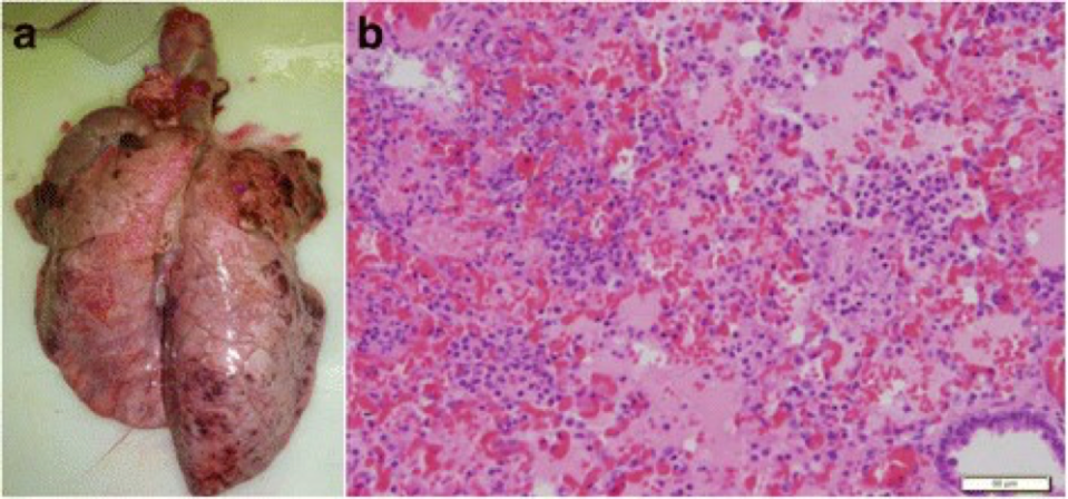
Figure 2: Lung infected by A. pleuropneumoniae showing pathological symptoms (a). Microscopic view of the lung tissue with fibrinous exudates between alveolar spaces and connective tissue (b). Source: BioMed Central, BMV Veterinary Research, Sassu, E.L., et al. (2017).
Infection, however, can also be asymptomatic depending on the strain of the bacteria. Pigs who survive the infection also become asymptomatic carriers. These asymptomatic carriers become an important reservoir for the pathogen as it continue to spread from one individual to another.
Epidemiology
A. pleuropneumoniae is found worldwide. It is more prevalent in pigpens where overstocking and bad ventilation are an issue. Unusual stress can also increase the spreading of the disease. It causes up to 20% of all bacterial pneumonia in swine. Although it is more commonly seen in growing pigs, it can affect swine of all ages. Mortality is higher for pigs between the ages of 12 to 16 weeks. For these growing pigs, death rate can reach up to 80% during outbreaks. Outbreaks are generally characterized by a large number of sudden deaths in a short period of time.
A. pleuropneumoniae continues to be a problem in various parts of the world but outbreaks have decreased in North America.
Virulence Factors
A. pleuropneumoniae has a number of virulence factors that make it a successful pathogen.
First of all, the bacterium is enveloped in a capsule which gives it protection from phagocytosis. The capsule is a large structure made of polysaccharide outside the cell envelope. The capsule is a virulence factor, because it makes the bacteria harder to kill by the host, and more resistant to bacterial viruses or desiccation. The pathogen also secretes lipopolysaccharides (LPS) and proteases. LPS is found on the surface of A. pleuropneumoniae’s surface and acts as a toxin that causes tissue damage. Proteases can cleave hemoglobin to facilitate the acquisition of the host’s iron reserves.
Finally, the most important virulence factor of this pathogen are the Apx toxins. They are responsible for the hemolytic functions of the bacteria. These exotoxins attack porcine lung immune cells such as macrophages and neutrophils. They provoke cell death by forming pores in leukocytes and by inducing oxidative bursts. Oxidative bursts are caused by the production of reactive oxygen species that lead to lysis of the cells. The release of toxic oxygen radicals also causes damage to the surrounding lung tissue. Apx toxins are also harmful to endothelial cells and red blood cells and are the main cause of the lesions found in the respiratory tract. They play an essential role for the pathogenicity of A. pleuropneumoniae.
Treatment
When infected with A. pleuropneumoniae, pigs must be treated immediately and continuously. Because of how rapid the pathogen is transmitted and its persistence, treatment can be difficult. Treatment usually include the use of antibiotic such as amoxicillin, penicillin or streptomycin. Swine in neighboring pens should also be treated as a form of prevention. It is important to note that these treatment typically only work for acute outbreaks and not chronic outbreaks. In the case of chronic outbreaks, infected pigs are usually asymptomatic.
The main concern are the surviving pigs because they remain carriers of the bacteria. However, some vaccines have proven to be effective in controlling the disease.
The best way to prevent an outbreak is by carefully managing the environment
References
Bossé, Janine T. et al. “Actinobacillus Pleuropneumoniae: Pathobiology and Pathogenesis of Infection.” Microbes and Infection, vol. 4, no. 2, 2002, pp. 225-235, doi:10.1016/s1286-4579(01)01534-9.
Dee, Scott A. “Pleuropneumoniae in Pigs.” Merck Sharp & Dohme Corp Accessed 7 November 2017.
Haesebrouck, F. et al. “Actinobacillus Pleuropneumoniae Infections in Pigs: The Role of Virulence Factors in Pathogenesis and Protection.” Vet Microbiol, vol. 58, no. 2-4, 1997, pp. 239-249, doi:https://doi.org/10.1016/S0378-1135(97)00162-4.
Inzana, Thomas J. “Virulence Properties of Actinobacillus Pleuropneumoniae.” Microbial Pathogenesis, vol. 11, no. 5, 1991, pp. 305-316, doi:10.1016/0882-4010(91)90016-4.
Marsteller, Thomas A. and Brad. Fenwick. “Actinobacillus Pleuropneumoniae Disease and Serology.” Swine Health Prod., vol. 7, no. 4, 1999, pp. 161-165.
Sassu, E. L. et al. “Host-Pathogen Interplay at Primary Infection Sites in Pigs Challenged with Actinobacillus Pleuropneumoniae.” BMC Vet Res, vol. 13, no. 1, 2017, p. 64, doi:10.1186/s12917-017-0979-6.
