Category Archives: Uncategorized

Taylorella equigenitalis

By Roxanne Losier and Giuliana Matta

Introduction

Taylorella equigenitalis is the pathogen known to cause contagious equine metritis (CEM), a sexually transmitted disease mostly targeting horses, but also seen in donkeys. This pathogen was first identified in 1977 in the United Kingdom, which then led to diagnosis in many other countries. T. equigenitalis is highly contagious and is considered a notifiable disease by the World Organization for Animal Health (OIE).

Disease

T. equigenitalis can be transmitted a number of ways such as during reproduction between an infected stallion and a mare, by contaminated equipment, or by infected semen used in artificial insemination. The primary site of infection of T. equigenitalis is the mare’s urogenital membranes, and it will not propagate through the mare’s body, since CEM is a nonsystematic infection. The infection can be characterized by mucopurulent vaginal discharge, a discharge containing mucus and pus, that can be observed as early as two days after contamination. Although one of the symptoms observed is an early return to estrus, the female’s menstrual cycle, an infection may result in a temporary loss of fertility for the mare. The symptoms typically disappear within days, however the mare may remain infected for several months. As for the stallion, the bacterium will shelter in the smegma of the prepuce and on the penis’ surface mostly in the urethral surface (Figure 1). Infected stallions will show no symptoms. While most infections will heal with no repercussions, the mare and the stallion can develop a chronic infection. In these cases, the pathogen will infect the clitoral fossa and sinuses of the mare and the stallion will simply remain a carrier (Figure 1). When the infection becomes chronic, both sexes will become asymptomatic, that is they will not show any symptoms of infections, making diagnosis much harder. 

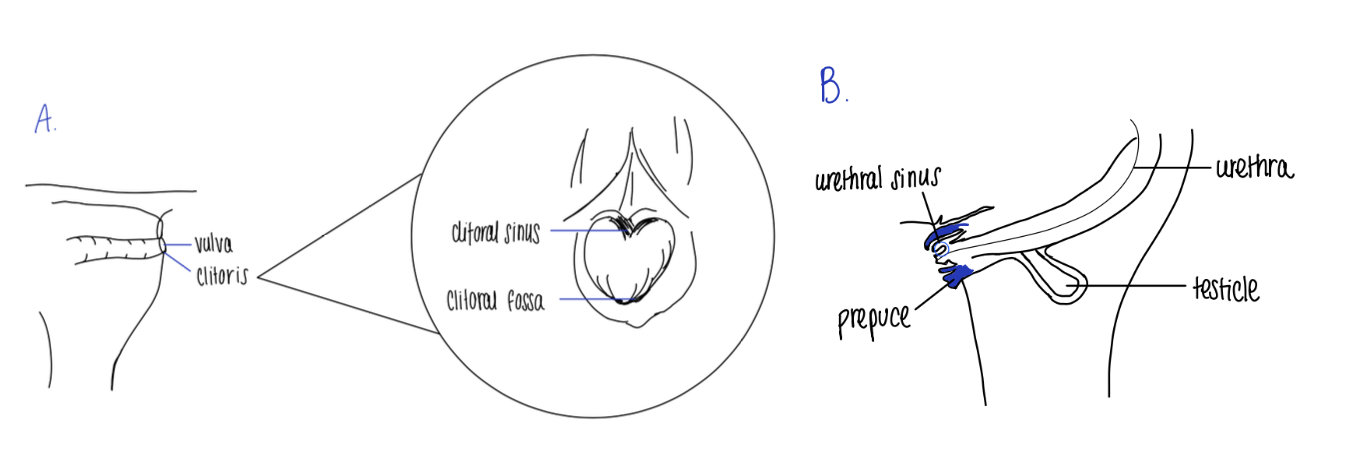
Figure 1: Site of infection of T. equigenitalis for the A. mare and B. stallion. Source: Roxanne Losier.

Epidemiology

Europe is the main continent affected by outbreaks of T. equigenitalis, and, in this area, it is characterized as an endemic disease. However, the presence of T. equigenitalis has never been detected in Canada, and the United States has successfully eradicated the bacterium. Therefore, they, and many other countries, are referred to as CEM-free countries. This status provides them a secure place within the trading market as well as an optimal reproductive efficiency. 

In countries where outbreaks still occur, the main causative agent would be the reproduction of undiagnosed horses. Since T. equigenitalis is highly contagious, it is crucial to take the required prevention against this pathogen. For example, in 2000, an imported horse was an undiagnosed carrier for the disease. In a short period of 8 years, reported cases were found amongst 48 countries and a total of 1100 horses were reportedly exposed. Since the infection caused by T. equigenitalis does not lead to the death of the horse, the major impact of outbreaks would be economic. Indeed, the infected horses can no longer contribute to reproduction of the species for the period of the infection and must be properly treated for it not to become a chronic infection.

In order to be prepared in the case of an outbreak, Canada’s Government has prepared a protocol. This protocol was issued by the Canadian Food Inspection Agency (CFIA) and aims to eradicate the disease as soon as possible in order to re-establish Canada’s CEM-free country status. Some of the disease control methods include tracking of potentially infected horses, strict quarantine and treatment of infected horses and rigorous decontamination to redefine disease-free areas.

Virulence Factor

No virulence factors are known or confirmed since T. equigenitalis has fastidious growth requirements, meaning specific factors and conditions are needed for it to grow and survive in the laboratory. Additionally, the number of bacteria on the external genitalia of stallions is low, resulting in the disease to possibly be missed by culturing alone. Consequently, isolating and studying this disease is hard, thus discovering virulence factors becomes difficult, which can be one of the many reasons why no virulence factors have been documented. T. equigenitalis can also go unnoticed because the bacteria cause no harm to the host cells, essentially resulting in no damage to the external genitalia and the animals being asymptomatic. Given that the only animals concerned with this disease are mostly horses and occasionally donkeys, research is not rushed. Because the infection does not display direct harm to the tissues, the urgency to create a cure is low and therefore virulence factors are not identified.

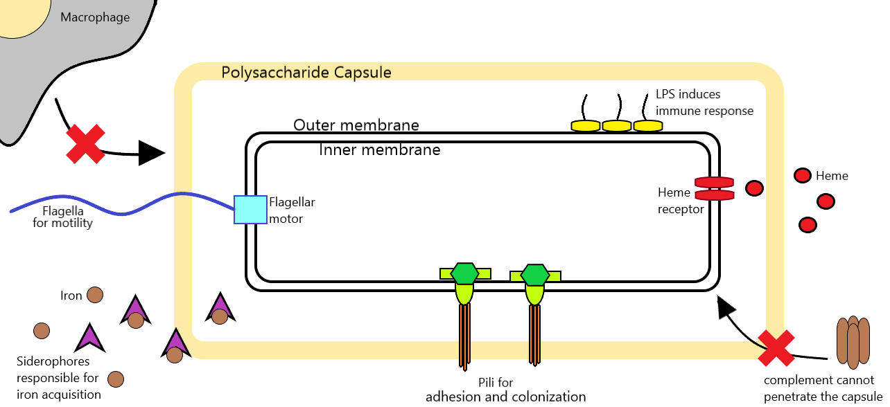
On the other hand, according to theory, we can hypothesize about a virulence factor that T. equigenitalis possesses. Adhesins are proteins located on the bacteria’ surface and aid in attachment to different places on the host cells’ surface by recognizing specific molecules (Figure 2). For T. equigenitalis to colonize the tissue, it must be able to recognize and bind to the cells of the external genitalia. With the adhesin proteins, the bacterium can bind and stay on the genitalia of horses and donkeys. Adhesins do not cause symptoms of the infection, but help the bacteria colonize in different niches, which is critical for the establishment of the infection. Therefore, it is very likely that T. equigenitalis contains adhesin proteins on its surface.

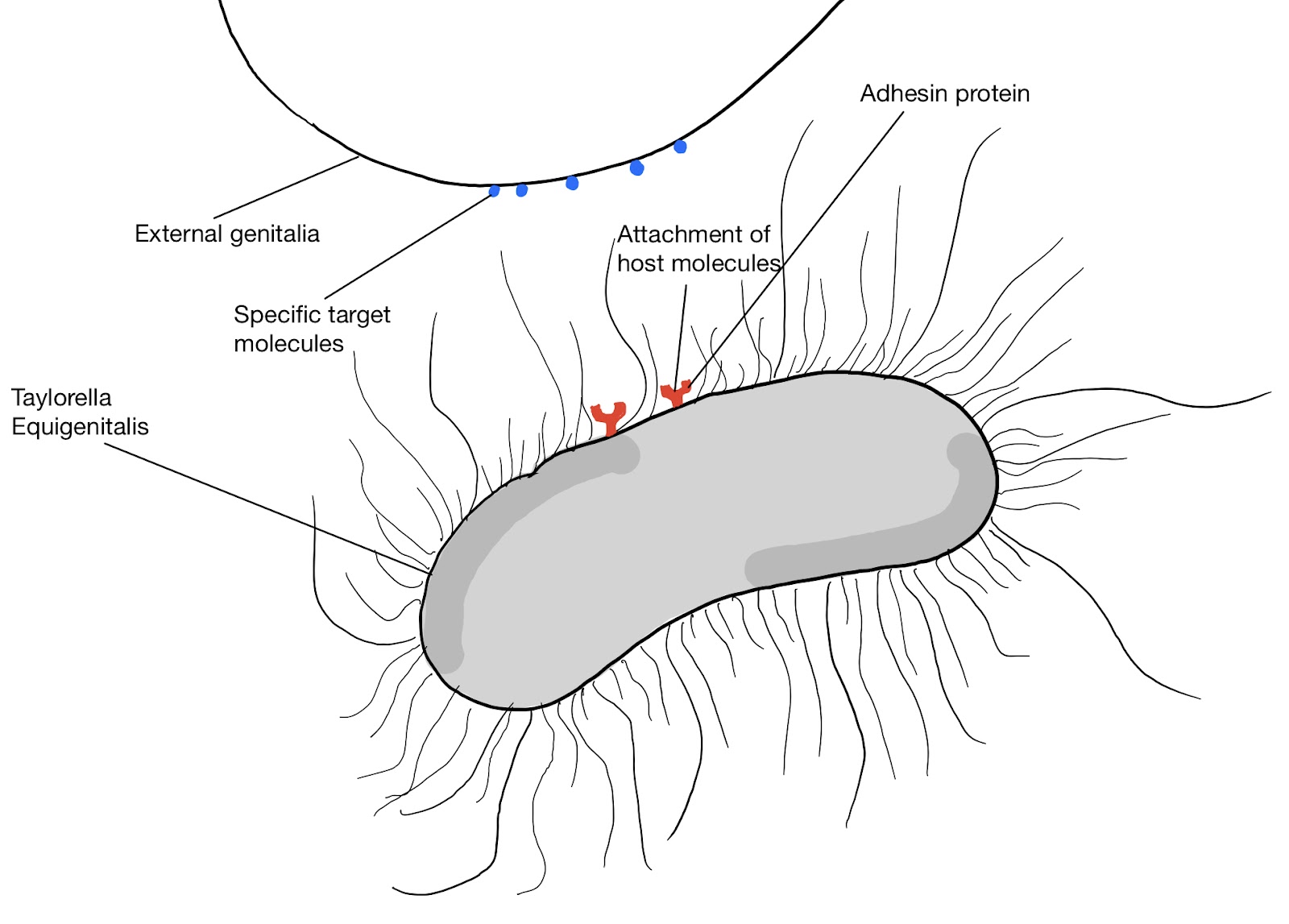
Figure 2: Adhesin proteins on T. equigenitalis. Source: Giuliana Matta

Treatment

As of today, no vaccine exists for T. equigenitalis, but treatments are available like disinfectants and antibiotics, which are provided topically. One antibiotic being ampicillin, which fights against certain Gram-negative organisms like T. equigenitalis. Even though this bacterium is sensitive to many antibiotics, no cure will completely eradicate and rapidly resolve the infection. In mares, the best treatment is performing daily washes for 5 days or more of the external genitalia. A 4% chlorhexidine solution, an antibacterial agent, is applied first, which clears the skin of infection and preps the area for the 0.2% nitrofurazone antibiotic ointment added afterwards, which fights Gram-negative bacteria. For stallions, the same procedures are implemented, but with 2% chlorhexidine solution instead of 4%. The application times for both mares and stallions varies depending on the severity of the disease. If the regimen does not work, they may have to remove the reproductive tract of the stallion or mare. In the end, the best way to prevent the transmission and the appearance of T. equigenitalis is to ensure the external genitalia of the animals are kept clean and to examine them for the disease because the horses or donkeys may be asymptomatic. Additionally, the animals should be assessed before entering any new stable or farm. 

References

Castle, S.S. (2008). Ampicillin. xPharm: The Comprehensive Pharmacology Reference, (pp.1-6) Retrieved November 17, 2021, from https://doi.org/10.1016/B978-008055232-3.61227-9

Hébert, L., Moumen, B., Pons, N., Duquesne, F., Breuil, M. F., Goux, D., Batto, J. M., Laugier, C., Renault, P., & Petry, S. (2012). Genomic characterization of the Taylorella genus. PloS one, 7(1), e29953. Retrieved November 17, 2021 from, https://doi.org/10.1371/journal.pone.0029953

Hitchcock, P. J., Brown, T. M., Corwin, D., Hayes, S. F., Olszewski, A., & Todd, W. J. (1985). Morphology of three strains of contagious equine metritis organism. Infection and immunity, 48(1), 94–108. Retrieved November 17, 2021 from, https://doi.org/10.1128/iai.48.1.94-108.1985

IBM Micromedex. (2021, October 1). Chlorhexidine (topical application route) precautions. Mayo Clinic. Retrieved November 17, 2021, from https://www.mayoclinic.org/drugs-supplements/chlorhexidine-topical-application-route/precautions/drg-20070874?p=1. 

Luddy, S., & Kutzler, M. A. (2010). Contagious equine metritis within the United States: A review of the 2008 outbreak. Journal of Equine Veterinary Science, 30(8), 393–400. Retrieved November 17, 2021 from, https://doi.org/10.1016/j.jevs.2010.07.006 

OIE. (2018). Contagious Equine Metritis. Description and importance of disease – oie. Retrieved November 17, 2021, from https://www.oie.int/fileadmin/Home/eng/Health_standards/tahm/3.05.02_CEM.pdf. 

Parlevliet, J. M., Bleumink-Pluym, N. M. C., Houwers, D. J., Remmen, J. L. A. M., Sluijter, F. J. H., & Colenbrander, B. (1997). Epidemiologic aspects of. Theriogenology, 47(6), 1169–1177. Retrieved November 17, 2021 from, https://doi.org/10.1016/s0093-691x(97)00097-6 

Ricketts, S. W., Barrelet, A., Barrelet, F. E., & Stoneham, S. J. (2006). The stallion and mare reproductive system. The Equine Manual, 2(13), 713–763. Retrieved November 17, 2021 from, https://doi.org/10.1016/b978-0-7020-2769-7.50018-6 

Rugbank. (n.d.). Nitrofural. Uses, Interactions, Mechanism of Action | DrugBank Online. Retrieved November 17, 2021, from https://go.drugbank.com/drugs/DB00336. 

Timoney, P. J. (1996). Contagious equine metritis. Comparative Immunology, Microbiology and Infectious Diseases, 19(3), 199–204. https://doi.org/10.1016/0147-9571(96)00005-7.

Mycobacterium avium

By Karim Al-Itani and Yasamin Nassimi

Introduction

The Mycobacterium avium complex (MAC) is composed of multiple nontuberculous mycobacterial species (NTM); this complex primarily consists of M.avium and Mycobacterium intracellulare. M.avium, is a slow-growing Gram-positive bacteria that is commonly found in soil and water sources. This intracellular pathogen has the ability to resist the immune mechanisms of the host, and the respiratory tract is amongst the most susceptible sites of infection. In addition to the infections within the respiratory tract, M.avium can also invade the bones, joints, lymph nodes and spread systemically. Infection acquisition may occur through ingestion, inhalation and dermal contact with environmental sources or medical devices. 

Disease

M. avium is an opportunistic pathogen, meaning it can cause disease when the host’s immune system is impaired (e.g. HIV). Acquisition of M. avium results in the adhesion of the microorganism to the mucosal epithelial cells in the intestinal lining and infection of the macrophages. Upon the invasion of the submucosal tissue and lymph nodes, the bacterium will be carried to the rest of the body via the lymphatics. Additionally, another risk factor for MAC infections includes the presence of underlying lung diseases. 

There are two major phenotypes associated with MAC pulmonary diseases (MAC-PD): fibrocavitary and nodular bronchiectatic (NB). The fibrocavitary form is characterized by the presence of lesions in the upper lobes of the lung, and occurs predominantly in older males with underlying lung diseases, such as previous pulmonary tuberculosis and chronic obstructive pulmonary disease (COPD). The NB form often develps in postmenopausal women and can cause multiple nodules in the right middle lobe and left upper lobe of the lung. 

Symptoms in patients with competent immune systems are nonspecific, with chronic cough being the most common symptom. Some patients with MAC-PD may also experience fever and hemoptysis, which is defined as coughing up blood or bloody mucus. 

Figure 1: Image of M. avium pulmonary disease of the NB form in a 47 year old Woman. Chest radiograph (2D X-ray imaging) shows small nodular clusters in both lower lung zones. High-Resolution Computed Topography (HRCT) (3D X-ray imaging) shows small centrilobular nodules in the right middle lobe and the left upper lobe. Source: Park, J. S. (2009). Respiratory Review of 2009: Nontuberculous Mycobacterium. Tuberculosis and Respiratory Diseases. The Korean Academy of Tuberculosis and Respiratory Diseases. https://doi.org/10.4046/trd.2009.67.5.395

Epidemiology

Currently, MAC species, especially M.avium, are mainly responsible for the majority of pulmonary infections caused by NTM. A Japanese study found that the incidence of MAC infections increased from 5.2/100,000 in 2007 to 13.1/100,000 in 2014; with the pulmonary diseases caused by M.avium having the highest prevalence in the north and east of Japan.

Comparably, in Europe, M.avium was the most frequent MAC species among pulmonary and extrapulmonary samples; as observed in studies done in Portugal (58.0%), Denmark (50.7%) and Italy (41.5%). 

The same overall pattern of MAC pulmonary and extrapulmonary diseases has been found in the United States of America, with M.avium being the most frequent isolate among MAC species (54%).

A potential explanation for the elevated prevalence of MAC and M.avium infections could be linked to the improvement of laboratory screening methods and increased awareness towards these infections in clinical settings. 

Virulence factor

M. avium bacteria are characterized by the presence of a water-repellent cell wall and by their mycolic acid constituents composing their core. Mycolic acids are densely packed with a variety of GPLs, which are a class of lipid and carbohydrate molecules linked together. The presence of GPLs is responsible for the immunomodulatory properties of M. avium that makes it a successful pathogen. 

First of all, the GPL component of the bacterium prevents the formation of the phagosome-lysosome complex. A phagosome is a cytoplasmic structure that exists inside specialized immune cells known as phagocytes; later, the fusion of the phagosome with the lysosome results in a phagolysosome complex. Formation of the phagolysosome complex is crucial for the destruction of intracellular microorganisms. The ability of GPLs to support the survival of mycobacteria in phagocytes is due to the interaction between the GPLs with specific receptors on the surface of the phagocytes. Moreover, the accumulation of GPLs inside macrophages leads to an amplified proinflammatory response. This phenomenon is essential as it leads to amplified infection impacts, without the necessary presence of the microorganism. 

Lastly, the synthesis of GPLs is required for the adhesion and secretion of biofilms. It has been suggested that inactivation of genes responsible for the synthesis of GPLs, leads to the impairment of the microorganism to slide or stick to surfaces. Thus, the presence of GPLs facilitates the microorganism to create a tight attachment with the host. Moreover, biofilm formation contributes to the adaptation of virulent strains to environmental anoxia and nutrient limitations, by lowering the metabolic rate and reducing the rate of cell division. 

Treatment: 

Treatment strategy for MAC-PD depends on how far the disease has  progressed. In nearly half of the patients, untreated MAC-PD does not progress for several years after diagnosis and negative culture conversions (no bacteria detected by testing) are recorded. So, it is important to consider what risk factors patients have prior to the initiation of treatments to avoid unnecessary medical expenses and adverse drug reactions. Some risk factors include cavitary lesions, low BMI, poor nutritional status, and extensive disease symptoms. In cases where treatment is warranted, a three-drug combination of macrolide-based antibiotics is used. Standard treatment uses three doses per week with increased administrations in severe cases.

This approach has been associated with better patient outcomes. However, this was only recorded in 32-65% of patients based on 5 systematic reviews. On top of its limited success, this treatment plan is limited by macrolide-resistant MAC strains which do not have any effective medication options. In cases where drug treatment fails, lung resection surgery is considered. This has led to successful culture conversion rates between 80-100%, which refers to bacterial ability to go from being detectable to non-detectable. However, the surgical approach is associated with postoperative morbidity and mortality in 0 to 50% of cases. This number can be significantly lowered through improved patient selection, preoperative assessments, surgical techniques and postoperative measures. Finally, alternative therapeutic agents are being tested for macrolide-resistant MAC-PD. Clofazimine led to culture conversions in 87% and 99%. of patients in two studies conducted. However, adverse reactions have been recorded in roughly 50% of patients. Overall, the current treatment for MAC-PD is subpar and novel treatment approaches should be developed for patients with poor prognosis, especially those diagnosed with macrolide-resistant MAC. 

Figure 2: 68-year-old woman with productive cough hemoptysis (spitting blood) lasting for weeks. She recorded positive culture conversions for MAC prior to therapy. Images A and B are pre-therapy and C and D are post-therapy. (A) Chest computed tomography (CT) (3D X-ray imaging)  reveals small nodules, lesions, and mild bronchiectasis (bronchi walls thick due to inflammation) in the left upper lung. (B) Plain chest radiograph (2D X-ray imaging) indicates fluid build up in the left upper lung. (C) Chest CT showing the use of clarithromycin, ethambutol, and rifampin three-times-weekly for 7 months leads to reduced lesions in the lungs. (D) Chest radiograph shows improved left upper lung with less fluid build up. After treatment she recorded negative culture conversions for MAC. Source: Pan, S.-W., Shu, C.-C., Feng, J.-Y., & Su, W.-J. (2020). Treatment for mycobacterium avium complex lung disease. Journal of the Formosan Medical Association, 119. https://doi.org/10.1016/j.jfma.2020.05.006

References

Busatto, C., Vianna, J. S., da Silva, L. V., Ramis, I. B., & da Silva, P. E. A. (2019). mycobacterium avium: an overview. Tuberculosis, 114, 127–134. https://doi.org/10.1016/j.tube.2018.12.004

Koh, W. J., Moon, S. M., Kim, S. Y., Woo, M. A., Kim, S., Jhun, B. W., Park, H. Y., Jeon, K., Huh, H. J., Ki, C. S., Lee, N. Y., Chung, M. J., Lee, K. S., Shin, S. J., Daley, C. L., Kim, H., & Kwon, O. J. (2017). Outcomes of mycobacterium avium complex lung disease based on clinical phenotype. The European Respiratory Journal, 50(3). https://doi.org/10.1183/13993003.02503-2016

Akram, S. M., & Attia, F. N. (2020). Mycobacterium Avium Intracellulare. PubMed; StatPearls Publishing. https://www.ncbi.nlm.nih.gov/books/NBK431110/

Vestby, L. K., Grønseth, T., Simm, R., & Nesse, L. L. (2020). Bacterial Biofilm and its Role in the Pathogenesis of Disease. Antibiotics (Basel, Switzerland), 9(2), 59. https://doi.org/10.3390/antibiotics9020059

Kwon, Y.-S., Koh, W.-J., & Daley, C. L. (2019). Treatment of mycobacterium avium complex pulmonary disease. Tuberculosis and Respiratory Diseases, 82(1), 15. https://doi.org/10.4046/trd.2018.0060

Pan, S.-W., Shu, C.-C., Feng, J.-Y., & Su, W.-J. (2020). Treatment for mycobacterium avium complex lung disease. Journal of the Formosan Medical Association, 119. https://doi.org/10.1016/j.jfma.2020.05.006

Moraxella catarrhalis

by Sara Murphy and Jiayi Xu

Introduction

Moraxella catarrhalis is a Gram-negative bacterium that was first discovered in 1986. It is found only in humans and until recently, it was considered to be a harmless colonizer of the nose, mouth and throat. However, M. catarrhalis is now recognized as an important cause of ear infections in children under the age of two, and lower respiratory tract infections in adults with compromised immune systems.

Disease

M. catarrhalis is primarily transmitted through mucosal secretions between family members, children in daycares, and between hospital patients in the winter. When the bacterium comes in contact with an infant, it attaches itself to the upper respiratory airways, specifically the nasopharynx. Once M. catarrhalis has invaded small airway epithelial cells, it moves from the nose or mouth to the middle ear by means of the Eustachian tube and causes inflammation along the way (Figure 1). The resulting congestion of fluid due to the narrowed Eustachian tube causes pressure to build in the middle ear, and ultimately results in pain. This is referred to as otitis media.

Figure 1: Moraxella catarrhalis invasion of the middle ear via the Eustachian tube causes inflammation and fluid build up within the middle ear leading to pressure differences pushing the eardrum outwards. This causes the pain associated with otitis media in children. (Source: Sara Murphy, 2021)

In the case of immunocompromised individuals or those living with chronic disease, M. catarrhalis infection has been shown to increase the severity of existing chronic obstructive pulmonary disease (COPD). When M. catarrhalis invades specific cells within the lungs (i.e. bronchial and alveolar epithelial cells), it contributes to increased inflammation and aggravates the disease symptoms. This includes reduced airflow during respiration, a worsening cough and  pulmonary emphysema. However, very little is actually known about how M. catarrhalis leads to exacerbations in patients with COPD.

Epidemiology

Moraxella catarrhalis causes approximately 15-20% of the total acute otitis media cases in infants with up to 80% of children having been colonized before their second birthday. However, the rate of colonization of the nasopharynx decreases considerably in adulthood. Nevertheless, there are roughly 2-4 million worsening cases each year of chronic obstructive pulmonary disease (COPD) associated with M. catarrhalis infection in the United States. Despite this extreme number, the actual rate of disease is likely higher as M. catarrhalis can sometimes be mistaken for Neisseria species in cultures taken from the lungs.

In most cases, M. catarrhalis lives as a natural inhabitant of the human respiratory tract without causing problems. However, under certain circumstances it can become pathogenic. This is highly dependent on age and the development of the immune system, as colonization rates lower significantly as individuals get older. Within similar age groups, the differences in colonization rates can be attributed to genetic predisposition, the number of household members, personal hygiene, and other environmental factors such as living with smokers.

Virulence Factor

Virulence factors are tools used by a pathogen to enhance its ability to colonize and cause damage to its host. One of the virulence factors found in M. catarrhalis is the Ubiquitous Surface Protein (Usp) family. These proteins are located in the outer membrane of the bacteria. Usp had been shown to confer serum resistance, where the pathogens evade bactericidal activities of the human serum, particularly that mediated by the Complement System. The system includes a series of soluble and membrane-bound proteins, each playing a different role. For example, C3a is pro-inflammatory; whilst C3b strengthens phagocytic activities. While C5a attracts immune cells to the site of infection, C5b is responsible for recruiting the membrane attack complex (MAC), which consists of C6, C7, C8 and multiple C9 subunits. As its name suggests, the MAC lyses bacterial cells and thus contributes directly to the bactericidal function of the Complement System.

The Complement System can be activated in three ways, two of which are the Classical and the Alternative pathway (Figure 2). The classical pathway is initiated by antibodies and results in the production of C2a and C4b. The two then form the C3 convertase and yields C3a and C3b, which also joins the C4bC2a complex to give rise to the C5 convertase. One of the enzyme’s products, C5b, coordinates assembly and binding of the MAC to the bacterial membrane. Unfortunately, M. catarrhalis is equipped with virulence factors that can interfere with this pathway. Two types of the Usp family, UspA1 and UspA2, are able to bind to the C4b-binding protein (C4BP). C4BP is a regulator of the classical complement activation pathway; it promotes degradation of the C3 convertase. Therefore, M. catarrhalis achieves serum resistance by “recruiting” inhibitory host proteins to reduce the amount of downstream proteins necessary for eventual activation of the MAC.

To make things worse, this pathogen species is also able to interfere with the alternative complement activation pathway. Normally, bacterial surface structures will be recognized and bound by the host serum protein Bb. Bb will attract C3b and the two form a C3 convertase, which then amplifies C3b production as well as facilitates subsequent reactions (for MAC activation). However, UspA1 and UspA2 are able to bind to C3 which prevents it from producing C3a and C3b. Thus, binding of the two proteins decreases the quantity of precursor molecules required for the alternative method of complement activation. Hence, UspA1 and UspA2 are two critical virulence factors in M. catarrhalis that protect the bacteria from being lysed by their host’s serum proteins.

Figure 2: The Classical and Alternative Pathway of Complement Activation. Note that the alternative pathway integrates into the classical pathway upon formation of the C5 convertase. In addition, C4b-binding proteins (not shown) are recruited by M. catarrhalis via UspA1/A2 to destroy C3 convertases. Meanwhile, the virulence factors can also bind to C3, thus minimizing the binding of Bb with C3b. (Source: Jiayi Xu, 2021)

Treatment

Almost all strains of M. catarrhalis are resistant to β-lactam antibiotics such as penicillin, amoxicillin and ampicillin. These antibiotics contain a β-lactam ring as part of their chemical structure that can disrupt bacterial cell wall synthesis. Nonetheless, M. catarrhalis produces the enzyme β-lactamase, which can modify the β-lactam ring and thereby renders these antibiotics inactive. Fortunately, the pathogen is still sensitive to most antibiotics used for treating respiratory tract infections. Ciprofloxacin, cefixime, chloramphenicol and amoxicillin-clavulanic acid are examples of antibiotics that are effective against M. catarrhalis infection.

References

Aebi C. 2011. Moraxella catarrhalis – pathogen or commensal? In: Curtis N, Finn A, Pollard AJ, editors. Hot topics in infection and immunity in children vii. New York, NY: Springer New York. p. 107-116. https://link.springer.com/chapter/10.1007%2F978-1-4419-7185-2_9.

Bernhard S, Spaniol V, Aebi C. 2012. Molecular pathogenesis of infections caused by Moraxella catarrhalis in children. Swiss Medical Weekly. [accessed 2021 Nov 13]; 142:w13694. https://pubmed.ncbi.nlm.nih.gov/23136074/. doi:10.4414/smw.2012.13694

Enright MC, McKenzie H. 1997. Moraxella (branhamella) catarrhalis – clinical and molecular aspects of a rediscovered pathogen. Journal of Medical Microbiology. [accessed 2021 Nov 12]; 46(5):360-371. https://www.microbiologyresearch.org/content/journal/jmm/10.1099/00222615-46-5-360;jsessionid=E0PEruR-ZdDu55F0y2vkxuCM.mbslive-10-240-10-26. doi:10.1099/00222615-46-5-360

Goldstein EJC, Murphy TF, Parameswaran GI. 2009. Moraxella catarrhalis, a human respiratory tract pathogen. Clinical Infectious Diseases. [accessed 2021 Nov 12]; 49(1):124-131. https://academic.oup.com/cid/article/49/1/124/371116. doi:10.1086/599375

Murphy TF, Brauer AL, Grant BJB, Sethi S. 2005. Moraxella catarrhalis in chronic obstructive pulmonary disease: Burden of disease and immune response. American Journal of Respiratory and Critical Care Medicine. [accessed 2021 Nov 11]; 172(2):195-199. https://www.ncbi.nlm.nih.gov/pmc/articles/PMC2718466/. doi:10.1164/rccm.200412-1747OC

Nordström T, Blom AM, Forsgren A, Riesbeck K. 2004. The Emerging Pathogen Moraxella catarrhalis Interacts with Complement Inhibitor C4b Binding Protein through Ubiquitous Surface Proteins A1 and A2. The Journal of Immunology. [accessed 2021 Nov 12]; 173(7):4598. https://www.jimmunol.org/content/173/7/4598.long. doi:10.4049/jimmunol.173.7.4598

Nordström T, Blom AM, Tan TT, Forsgren A, Riesbeck K. 2005. Ionic Binding of C3 to the Human Pathogen Moraxella catarrhalis is a Unique Mechanism for Combating Innate Immunity. The Journal of Immunology. [accessed 2021 Nov 13];175(6):3628. https://www.jimmunol.org/content/175/6/3628.long. doi: 10.4049/jimmunol.175.6.3628

Su Y-C, Singh B, Riesbeck K. 2012. Moraxella catarrhalis: From interactions with the host immune system to vaccine development. Future Microbiology. [accessed 2021 Nov 11]; 7(9):1073-1100. https://www.proquest.com/docview/1038321799/fulltextPDF/83544917F04C476CPQ/1?accountid=12339. doi:10.2217/fmb.12.80

Verduin CM, Hol C, Fleer A, van Dijk H, van Belkum A. 2002. Moraxella catarrhalis: From emerging to established pathogen. Clinical Microbiology Reviews. [accessed 2021 Nov13]; 15(1):125-144. https://www.ncbi.nlm.nih.gov/pmc/articles/PMC118065/. doi:10.1128/CMR.15.1.125-144.2002

Warnke P, Köller T, Kreikemeyer B, Barrantes I, Mach H, Podbielski A. 2019. Molecular epidemiology study of a nosocomial Moraxella catarrhalis outbreak in a neurological rehabilitation unit. Journal of Hospital Infection. [accessed 2021 Nov 14]; 103(1):27-34. https://www-sciencedirect-com.proxy3.library.mcgill.ca/science/article/pii/S0195670119301884. doi:10.1016/j.jhin.2019.04.019

Yang M, Johnson A, Murphy TF. 2011. Characterization and evaluation of the Moraxella catarrhalis oligopeptide permease as a mucosal vaccine antigen. Infection and Immunity. [accessed 2021 Nov 11]; 79(2):846-857. https://pubmed.ncbi.nlm.nih.gov/21134967/. doi:10.1128/IAI.00314-10

Streptococcus agalactiae

By Iris Andrea Garcia and Claudia Cora

Introduction

Streptococcus agalactiae, also known as group B streptococcus (GBS), is an opportunistic Gram-positive bacterium that is normally part of the human gut environment; the microbiota. It colonizes 30% of healthy human adults’ gastrointestinal and genitourinary tracts (urinary and vaginal). Normally, S. agalactiae is harmless and many healthy adults live a life without any disease. However, GBS can cause a severe invasive infection in immunocompromised patients like newborns, elderly and patients with underlying diseases such as diabetes, cirrhosis, cancer, AIDS. In dairy cattle, the bacteria is a common pathogen and infects the udder of many cows, leading to inflammation of the udder and reduced milk production, commonly known as mastitis. In humans, GBS infections include urinary tract infection (UTI), skin and soft-tissue infection, meningitis and bacteremia (infection of blood). S. agalactiae is recognized as the leading cause of invasive newborn infections and is transmitted to the fetus via the placenta by the mother during pregnancy or after birth from the external environment.

Figure 1. Streptococcus agalactiae. Source : Public Health Image Library, Centers for Disease Control and Prevention, Ekkert A. (2013) https://phil.cdc.gov/Details.aspx?pid=22882

Disease

S. agalactiae infections in newborns are divided into two categories: early-onset disease (EOD), which affects babies aged 0 to 6 days, and late-onset disease (LOD), which affects babies aged 7 to 90 days. Early-onset infection is caused by the transmission of S. agalactiae from pregnant women to the fetus via the placenta, also referred to as vertical transmission. In vertical transmission, GBS from the maternal gastrointestinal and genitourinary flora infiltrates the uterine compartment and reaches the lungs of the fetus. Once in the lungs, GBS displays a multitude of virulence factors to successfully invade the host cell and reach the bloodstream of the newborn. Early-onset disease symptoms include bloodstream infection, pneumonia, and sepsis; the body’s severe and life-threatening response to infection, which can develop as early as the day of birth. Late-onset infection is mostly acquired horizontally from the mother, as the infant passes through the vaginal tract during birth, or through colonized household contact which causes meningitis and bacteremia. 

In pregnant women even if there are no apparent symptoms, it can cause low birth weight, pre-term delivery and premature ruptures of the membranes. Heavily colonized pregnant women can develop symptoms such as inflammation of the membranes that surround the fetus or the amniotic fluid in which the fetus floats, bacteremia and sepsis.

Figure 2. GBS disease in pregnant mothers and fetuses. GBS ascension into the uterus from the lower genital tract and invasion of the amniotic cavity and fetus. Source: Brokaw et al. 2021. https://www.frontiersin.org/articles/10.3389/fcimb.2021.720789/full

Epidemiology

Streptococcus agalactiae is a very common commensal bacteria of the gut and vaginal tracts of humans and are part of their microbiotas. In an epidemiology study, vaginal colonization of S. agalactiae was found in 16% of pregnant women and 16% of non-pregnant women, as opposed to only 4% in the gut in pregnant and non-pregnant women. As a consequence of a weaker immune system of the mother to tolerate the development of the fetus, this opportunistic pathogen crosses the placental membrane and causes meningitis and sepsis in newborns. There is also an increase in GBS incidence among the elderly causing bacteremia and meningitis. Slotved and Hoffmann study showed that EOD had a higher incidence than LOD, but still remains low and stable in Denmark over a period of 13 years. However, the increasing trend in GBS incidence in elderly is most likely due to the increase in prevalence of comorbidities, mostly chronic diseases, in the aging population of developed countries.

Virulence Factors

Streptococcus agalactiae has many virulence factors which are tools to facilitate colonization and infection of a cell in the gut or in the vaginal tract of the host. S. agalactiae has four main groups of functional virulence factors that aid in infection which are; pore-forming, immune evading, antimicrobial peptide (AMPs) resisting and cell-adhering and invading factors. The pore-forming toxins are proteins that form a pore on the surface of the cell to kill it by releasing its content in the environment. In GBS, ꞵ-hemolysin and cytolysin pore-forming proteins can also impair cardiac and liver functions. 

Immune evasion factors such as sialic acid capsular polysaccharide or superoxide dismutase are proteins that make the bacteria resist the immune system. Sialic acid can mimic the host cell surface, as the cells have sialic acid to not be attacked by the complement system. Superoxide dismutase breaks down superoxide produced by macrophages in an attempt to kill the bacteria, as a defense mechanism. Antimicrobial peptides (AMPs) are proteins produced by the immune system cells and by mucosal membranes to inhibit pathogens from harming the host cells, it is like a toxin for the bacteria. S. agalactiae can resist AMPs by the alanylation of lipoteichoic acid, which is a negatively charged molecule on the bacteria surface that is the target of AMPs. Lipoteichoic acid is easily recognizable by the immune system and will readily activate the immune system to make AMPs. Alanylation makes lipoteichoic acid positively charged to repel AMPs that are positively charged, like two north pole magnets. 

Cell-adhesion and invasion factors like fibrinogen-binding proteins A and B simply aid in the adhesion of the bacteria to the host cell and in the penetration process. The bacteria will mostly bind to fibrinogen molecules which are mostly present in the extracellular matrix (ECM); a physical scaffold on which the host cells adhere and interact with the external environment. By anchoring itself to the ECM, the bacterial cell can bind to the target cell and invade it.

Figure 3. The virulence factors of S. agalactiae and their action on the host cells including invasion of the target cells, resistance to AMPs, immune evasion and lysis of cells via pores for a successful infection.
Source : Claudia Cora, 2021 produced with Biorender.

Treatment

Beta-lactam antibiotics, notably penicillin G and ampicillin, are often used to treat Streptococcus agalactiae. As alternatives, penicillin-allergic patients are treated with vancomycin and clindamycin. However, in recent years, there has been a surge in clindamycin resistance in both newborn and adult infections.

Infected newborns are treated with several antibiotic combinations before the presence of S. agalactiae is established, and once the presence of S. agalactiae is confirmed, penicillin G is the medicine of choice. Supportive care, such as ventilatory support, is also provided to infected infants. Currently, there is no vaccination available against S. agalactiae. However, some steps may be taken to reduce the chance of the mother infecting the child. The American College of Obstetricians and Gynecologists recommend that pregnant women get tested for GBS bacteria between weeks 36 to 37 of pregnancy. If the pregnant woman tests positive, the fetus is at significant risk of infection. Antibiotics are thus given during labour rather than before because the bacteria will have time to multiply again before labour begins.

References

Ballard MS, Schønheyder HC, Knudsen JD, Lyytikäinen O, Dryden M, Kennedy KJ, Valiquette L, Pinholt M, Jacobsson G, Laupland KB. 2016. The changing epidemiology of group b streptococcus bloodstream infection: A multi-national population-based assessment. Infect Dis (Lond). 48(5):386-391.

Brimil N, Barthell E, Heindrichs U, Kuhn M, Lütticken R, Spellerberg B. 2006. Epidemiology of streptococcus agalactiae colonization in germany. International Journal of Medical Microbiology. 296(1):39-44.

Slotved H-C, Hoffmann S. 2020. The epidemiology of invasive group b streptococcus in denmark from 2005 to 2018. Frontiers in Public Health. 8(40).

Martins ER, Pedroso-Roussado C, Melo-Cristino J, Ramirez M, Portuguese Group for the Study of Streptococcal I. 2017. Streptococcus agalactiae causing neonatal infections in portugal (2005-2015): Diversification and emergence of a cc17/pi-2b multidrug resistant sublineage. Front Microbiol. 8:499-499.

Patras KA, Nizet V. 2018. Group b streptococcal maternal colonization and neonatal disease: Molecular mechanisms and preventative approaches. Frontiers in Pediatrics. 6(27).

Puopolo KM, Baker CJ. Oct 12 2021. Group b streptococcal infection in neonates and young infants UpToDate.

Puopolo KM, Baker CJ. Sept 24 2019. Group b streptococcal infection in pregnant women UpToDate

Puopolo KM, Lynfield R, Cummings JJ, Committee On F, Newborn, Committee On Infectious D, Hand I, Adams-Chapman I, Poindexter B, Stewart DL et al. 2019. Management of infants at risk for group b streptococcal disease. Pediatrics. 144(2).

Rajagopal L. 2009. Understanding the regulation of group b streptococcal virulence factors. Future Microbiology. 4(2):201-221.

The Possible Long Term Risks of Infant Antibiotic Exposure

By: Leen Bazzi & Stefanie Koch

Introduction: Gut Microbiome

Since the discovery of penicillin back in 1928, antibiotics have been used worldwide for many years to treat a wide array of bacterial infections. Extremely efficient with little to no short-term side effects in adults, antibiotics have been the first line of treatment against most bacterial infections. However, use of antibiotics has been strictly regulated due to the mounting evidence showing the influence of antibiotics on the gut microbiome and the function of the immune system.

Throughout our lifetime, as food passes through our gastrointestinal (GI) tract along with hundreds of microorganisms, a collection of microbes such as bacteria, archaea and eucaryotes, colonize our GI. They are called the ‘gut microbiota’ and they are normally not harmful to the host. In fact, they usually form a mutually beneficial relationship with the hosts, aiding in protecting against pathogens and even regulating host immune responses at times.

Recent research has shown that multiple factors such as dietary habits, stress and antibiotics can cause a microbiota dysbiosis, in other words, a change in the composition, diversity and function of the microbes in the gut. Antibiotics are now being recognized as a leading cause of gut dysbiosis in infants whose gut microbiota doesn’t fully develop and stabilize until the age of 3 years.

Gut dysbiosis in infants has been shown to affect their innate and adaptive immune systems through a reduction in the number of antibodies produced against a specific pathogen and changes in the phenotype and function of immune cells that attack foreign microorganisms, which reduces their ability to respond to signals and activate during infections in order to fight off the pathogen (Fig. 1).

 A recent study by Aversa et al. published in 2021, in Mayo Clinic Proceedings, investigated the association between antibiotic exposure in the first two years of life and health conditions with childhood onset. They observed the medical records of 14,572 children, of whom 70% received at least one antibiotic prescription during the first two years of life and tracked their health records for any diseases developed between time of birth and 2017. Based on previous similar research, Aversa et al. hypothesized that intake of antibiotics during the first two years of life is correlated to an increased risk of immunological, metabolic, and neurobehavioral health conditions.

Fig.1 Summary of antibiotic-driven modulation of immune responses and possible consequences in infants under two years old. Antibiotic exposure causes microbiota dysbiosis. A. microbiota dysbiosis can change the phenotype and function of immune cells of the innate system leading to a decrease in activation. B. It can also cause a reduction in antibody production for certain antigens and mast cell degranulation. Source: Leen Bazzi, 2021 (BioRender)

Description of findings:

Overall, Aversa et al. (2021) found a significant association between exposure to certain antibiotics and increased probability of developing many health conditions. Specifically, obesity, asthma, allergic rhinitis, atopic dermatitis, ADHD and learning disabilities were among the most common health problems in kids exposed to two or more antibiotics.

The relationship between the number of antibiotics administered in infancy and the probability of developing 1,2 and 3 or more health conditions in those children was studied by Aversa et al.. They found that the probability of developing more than three health conditions in kids who have taken more than five antibiotics is significantly higher than in kids who have taken no antibiotics. Overall, kids exposed to more than one antibiotic had the highest probability of developing more than one health condition. This was observed with children that developed obesity and another condition such as asthma, ADHD, or a learning disability. However, obesity, asthma and allergic rhinitis was a common triad with exposed and non-exposed children. Atopic dermatitis or ADHD were more common to be paired with asthma and obesity in children exposed to antibiotics.

Aversa et al. also studied the correlation between the number of antibiotics administered and the time spent with 1, 2, 3 or more health conditions. On average, infants exposed to antibiotics spent more years with 2 or more health conditions. Infants exposed to more than 5 antibiotics had a significant increase in the years spent with more than one condition and a significant decrease in the years spent with no health conditions.

Furthermore, it seems the period of development in which the antibiotics were administered influences the risk of developing certain health problems. There was a much stronger risk of developing asthma, allergic rhinitis, and obesity when the children were given antibiotics between 6 and 12 months, a critical age for the development of gut microbiota.

The health and neurobehavioral conditions developed varied based on the type of antibiotic exposed to and the genders of the patients. Many of them received penicillin (64%), while others received either Cephalosporins, Sulfonamides or Macrolides. There was a significant influence of antibiotic type on developing higher risk for certain conditions which also differed significantly between genders (Table 1). For example, when penicillin was prescribed, it had a higher risk to develop obesity in boys, and celiac disease in girls, while having a non-significant risk for food allergy in both genders. Obesity seems to be in large part more significant in boys for all 4 antibiotics. Autism and atopic dermatitis were less likely to occur in girls, if penicillin or macrolide were prescribed. Moreover, Cephalosporins seem to have the highest risk for development of autism in both sexes.

Table 1. Association between the type of antibiotic prescribed and the increased risk in developing health conditions. Highlighting the difference of conditions between genders, that were more significantly developed. The color assignments are shown in the legend and are as follows: pink for the significant results for girls; blue for boys; yellow for both genders; white the non-significant results for both genders; green for the significant decrease for girls. Source: Stefanie Koch, 2021. Data from Aversa et al. (2021)

Discussion:

The study tested the hypothesis that exposure to a certain number of antibiotics increases the risk of immunological and neurobehavioral diseases by causing microbiota dysbiosis, which could lead to disruption of the immune system in children (Fig. 1). The results of the study showed a significant correlation between the number of antibiotics taken before the age of two and an increase in the risk of developing certain diseases with childhood onset (Fig. 2). The association between antibiotic exposure and health outcomes in infants persisted even after the results were adjusted for important infant and maternal confounding variables such as, maternal age, delivery method, antibiotic exposure during pregnancy, etc. These variables could possibly skew the results, therefore, it’s important to look at a patients file closely before including them in the study. By doing so, the researchers reduced the variables that could skew the results, focusing more on the relation between the antibiotic and the related health outcomes. Thus, the findings support the hypothesis that antibiotics play a causal role in the pathogenesis of childhood immune disorders through disruption of microbiome during critical developmental periods.

The researchers also categorized certain health outcomes by the antibiotics the child was exposed to at a young age, as well as the different effects each antibiotic had on girls and boys. This allowed them to form a correlation between certain antibiotics and the risk for certain diseases based on sex (Table 1). Although the correlation found was not very significant, some antibiotics were associated with a higher risk of one or two diseases in a certain gender. These preliminary results could help with future studies about the different effects of antibiotics on females and males.

This population-based study is significant in supporting the hypothesis because of the large cohort of participants (14,572 children) and the long follow-up period ranging from 6-11 years for each participant. Additionally, the results of a significant association between antibiotics, gut health, and risks for developing health conditions is supported by other studies with similar results Also, choosing a suburban cohort for the study might have aided in reducing any exterior factors that could have affected the significance of the results, such as living conditions, environmental factors etc.

One limitation of the study is that the infections the children were treated for were not documented or taken into consideration. Maybe their illness may have had underlying effects or even directly led to the health condition seen later. Another limitation was the diversity in ethnicity of the study population, with 71% of the cohort being white, and only 29% encompassing all other groups. This might be because the study chose a population residing in Minnesota, USA. Therefore, the study can only apply to people living in the USA and not in other parts of the world where lifestyle, medical help and habits may differ.

Conclusion & Future Directives:

In conclusion, the study by Aversa et al. supports the hypothesis that there is a significant relationship between antibiotic exposure before the age of 2, when the microbes in the gut have not fully developed, and some diseases that develop later during childhood. The large cohort of 14,572 children shows that the results of this study are important and significant, even if they are only applicable in the USA. Furthermore, by analyzing how each antibiotic affected girls and boys, the study showed preliminary results indicating that antibiotics may have different effects depending on whether the patient is a female or male. The reason for this is unknown and more research is needed to study why the conditions developed differ between the sexes, regardless of the antibiotic prescribed. This could pave the way for different prescriptions based on the sex of the patient. Future studies about this topic should also emphasize diversity in ethnicity in order to see whether these results are significant in other ethnicities or whether they may differ based on lifestyle. Some research is currently being done about antibiotics that could strengthen the host immune system rather than simply attacking the bacteria causing the infection. This is something that could potentially overcome the issue of antibiotics changing the composition and function of gut microbiota if the study succeeds.

References

Aversa, Z., Atkinson, E. J., Schafer, M. J., Theiler, R. N., Rocca, W. A., Blaser, M. J., & LeBrasseur, N. K. (2021). Association of Infant Antibiotic Exposure With Childhood Health Outcomes. Mayo Clin Proc, 96(1), 66-77. doi:10.1016/j.mayocp.2020.07.019

Gensollen, T., Iyer, S. S., Kasper, D. L., & Blumberg, R. S. (2016). How colonization by microbiota in early life shapes the immune system. Science (New York, N.Y.), 352(6285), 539-544. doi:10.1126/science.aad9378

Gray, J., Oehrle, K., Worthen, G., Alenghat, T., Whitsett, J., & Deshmukh, H. (2017). Intestinal commensal bacteria mediate lung mucosal immunity and promote resistance of newborn mice to infection. Sci Transl Med, 9(376). doi:10.1126/scitranslmed.aaf9412

Hancock, R. E., Haney, E. F., & Gill, E. E. (2016). The immunology of host defence peptides: beyond antimicrobial activity. Nat Rev Immunol, 16(5), 321-334. doi:10.1038/nri.2016.29

Langdon, A., Crook, N., & Dantas, G. (2016). The effects of antibiotics on the microbiome throughout development and alternative approaches for therapeutic modulation. Genome Medicine, 8(1), 39. doi:10.1186/s13073-016-0294-z

Mitre, E., Susi, A., Kropp, L. E., Schwartz, D. J., Gorman, G. H., & Nylund, C. M. (2018). Association Between Use of Acid-Suppressive Medications and Antibiotics During Infancy and Allergic Diseases in Early Childhood. JAMA Pediatr, 172(6), e180315. doi:10.1001/jamapediatrics.2018.0315

Panda, S., El khader, I., Casellas, F., López Vivancos, J., García Cors, M., Santiago, A., . . . Manichanh, C. (2014). Short-Term Effect of Antibiotics on Human Gut Microbiota. PLOS ONE, 9(4), e95476. doi:10.1371/journal.pone.0095476

Roubaud-Baudron, C., Ruiz, V. E., Swan, A. M., Jr., Vallance, B. A., Ozkul, C., Pei, Z., . . . Blaser, M. J. (2019). Long-Term Effects of Early-Life Antibiotic Exposure on Resistance to Subsequent Bacterial Infection. mBio, 10(6). doi:10.1128/mBio.02820-19

Shekhar, S., & Petersen, F. C. (2020). The Dark Side of Antibiotics: Adverse Effects on the Infant Immune Defense Against Infection. Frontiers in Pediatrics, 8(651). doi:10.3389/fped.2020.544460

Thursby, E., & Juge, N. (2017). Introduction to the human gut microbiota. Biochemical Journal, 474(11), 1823-1836. doi:10.1042/bcj20160510

Zhu, D., Xiao, S., Yu, J., Ai, Q., He, Y., Cheng, C., . . . Pan, Y. (2017). Effects of One-Week Empirical Antibiotic Therapy on the Early Development of Gut Microbiota and Metabolites in Preterm Infants. Sci Rep, 7(1), 8025. doi:10.1038/s41598-017-08530-9

Rediscovering Phage Therapy

By Emily Wilson, Philippe Vezina, and Mohammad Jafar Sami

Cases of antibiotic resistance have become a prominent concern for our healthcare system. Antibiotic resistance is a result of bacteria that have evolved to overcome antibiotic treatment. For instance, mutated bacteria may begin producing enzymes that give them resistance to antibiotics; this offers a selective advantage that allows the mutated strain to proliferate despite treatment. As a result of this public health concern, medical practitioners and researchers are on the search for alternative antimicrobial agents to complement the role of antibiotics. It is here that phage therapy may find its place in future clinical practice one century after its initial discovery.

What is Phage Therapy?

Phage therapy uses phages, also known as bacteriophages, to target and counteract infections. Phages are a form of virus which infect bacteria. Their structure consists of genetic material surrounded by a protein capsid which makes up the “head” of the bacteriophage. This structure is complemented by the phage tail which is used to target and secure the virus to bacterial hosts (Figure 1).

Figure 1: Basic phage structure. Source: Emily Wilson.

Phages were originally discovered by Frederick Twort and Felix d’Hérelle in the early 1900s. Shortly after their discovery, d’Hérelle managed to effectively apply phages as a treatment for dysentery infections, which at the time had no successful cure. While his success led to widespread enthusiasm for the potential of phage therapy, this treatment was quickly eclipsed in the western world with the introduction of antibiotics. However, it can be noted that the use of phage therapy has prevailed in various regions of the world, especially in the former Soviet Republic of Georgia where phages are standard in care.

How Does it All Work?

One of the biggest advantages of phage therapy is that phages are designed to be highly specific as a result of their ability to recognize bacterial cell surface receptors.  When a phage comes in contact with its target bacteria, there are a number of mechanisms by which the virion can impart its antimicrobial effects.  Phages can be harnessed for their potential to lyse bacteria through their lytic cycle and bactericidal enzymes as well as for their ability to increase the antibiotic sensitivity of target bacteria.

In the lytic cycle, phages replicate inside their target bacteria, making dozens of copies which will eventually destroy the bacterial cell so that these replicates may further disseminate into the environment (Figure 2).

Figure 2: The lytic cycle starts when the host bacterium (1) is targeted by the phage and injected with viral DNA (2). This phage DNA is then used to create phage proteins (3) which are assembled into complete phages (4). The cell then breaks apart releasing the newly generated phages into the environment (5). Source: Emily Wilson.

Phages may also be harnessed for their ability to produce enzymes that target bacteria. Considerable research in this area has focused on endolysins which are the peptidoglycan hydrolases involved in cell lysis during the lytic phage cycle. This means these enzymes are capable of disrupting the bacterial cell wall which leads to the death of the bacteria.

Instead of replicating within the bacteria leading to cell death, the phage may also incorporate its own genetic material into the genome of the bacteria. Through this mechanism, known as transduction, phages are capable of introducing novel characteristics to their bacterial host. This can be applied in the clinical setting as phages are able to reintroduce drug sensitivity to antibiotic resistant bacteria by introducing drug sensitive genes as shown in Figure 3.

Figure 3: Phages can introduce drug-sensitive genes into drug resistant host bacteria through transduction. In this process the recipient bacteria (1) is targeted by the phage carrying DNA with genes for drug sensitivity (2). When the DNA is injected (3) the bacteria will incorporate the viral DNA into its own genome (4), allowing for the expression of drug-sensitive genes. Source: Emily Wilson.

Where does Phage Therapy “Fit in”?

Antibiotics have taken the medical world by storm since their discovery in the early 1900s. These agents are highly effective at destroying bacteria in a broad range, non-specific manner making them a great tool to counteract infections. Antibiotics are nonspecific and therefore will attack both pathogenic and commensal bacteria which can lead to an imbalance and lack of regulation in the human microbiota. The human microbiota refers to the collection of commensal, or healthy bacteria that normally colonize the body. The microbiota serves a key role in the immune system’s first line of defense. Healthy bacteria inhibit the ability of pathogenic species to initiate colonization which is why the dysregulation of this system as a result of antibiotics can be problematic. Furthermore, overuse of antibiotics in healthcare has led to the development of antibiotic resistant bacteria.

Bacteria have three main mechanisms of antibiotic resistance. The bacterium may restrict exposure of the antibiotic to its target. For example, if the antibiotic’s target is inside the bacterial cell, the bacterium will stop expressing the transport system used to bring the antibiotic inside the cell. The antibiotic will be unable to reach its target and thus unable to affect the pathogen. The bacterium may also destroy or inactivate the antibiotic before it can reach its target rendering the treatment ineffective. Lastly, the bacterium may modify the antibiotic target so that the antibiotic is no longer able to bind and induce an effect. It has been predicted that by 2050, antimicrobial resistant infections will become the cause of more deaths than cancer.

This phenomenon has brought phage therapy back into focus as a way to counteract drug resistant pathogens and complement antibiotic treatment. Phages demonstrate very selective toxicity and therefore, when applied to living tissue, will leave cellular structures outside of their target unharmed. As a result of this trait, phages inflict less damage onto the patient’s healthy microbiota.

Ongoing Research:

While lacking in quantity, clinical trials and case reports involving phage therapy have shown promising results. The PhagoBurn Clinical Trial is a phase I/II study initiated in 2015 that is looking to assess the use of phage cocktails in treating Escherichia coli and Pseudomonas aeruginosa bacterial infections in burn wounds. In vitro experiments show that engineered phages are capable of selectively killing Methicillin-resistant Staphylococcus aureus (MRSA), an antibiotic resistant bacterium, which indicates great potential of phages in treating drug resistant infections. Successful case reports of phage treatment in patients suffering from antibiotic resistant infections furthers optimism for the incorporation of phage therapy in standard medical care.

Limitations:

Phage therapy does not come without its limitations.  The current major downfall of this treatment route is the significant deficit in double-blinded, Phase III clinical trials that have been published to date. Phase III clinical trials are necessary to determine how a new medical intervention compares to existing treatments; however, they often require thousands of participants, copious amounts of funding and can take years to complete. Phages are also limited in their application as it is difficult to administer these agents intravenously, limiting their use to infections located more superficially within the body such as localized wounds or lung infections like pneumonia. As a maturing treatment there is currently no standard framework for application of phage therapy in western medicine which can inhibit their ability to be incorporated into treatment in the future.

Future Directions:

Phage therapy is a promising contender as an antimicrobial intervention that can complement antibiotic treatment. As the realm of “personalized medicine” develops, phage cocktails will be able to be designed to match a patient’s specific disease. Given the pace of current scientific growth and social progress, in as short as a few decades, one may expect that phage therapy will be finding a predominant place in medical standards of care.

References:

Bassetti, M., A. Carnelutti, and M. Peghin, Patient specific risk stratification for antimicrobial resistance and possible treatment strategies in gram-negative bacterial infections. Expert Rev Anti Infect Ther, 2017. 15(1): p. 55-65.

Bikard, D., et al., Exploiting CRISPR-Cas nucleases to produce sequence-specific antimicrobials. Nat Biotechnol, 2014. 32(11): p. 1146-50.

Chan, B.K., S.T. Abedon, and C. Loc-Carrillo, Phage cocktails and the future of phage therapy. Future Microbiol, 2013. 8(6): p. 769-83.

D’Herelle, F., On an invisible microbe antagonistic toward dysenteric bacilli: brief note by Mr. F. D’Herelle, presented by Mr. Roux. 1917. Res Microbiol, 2007. 158(7): p. 553-4.

D’Herelle, F., Bacteriophage as a Treatment in Acute Medical and Surgical Infections. Bull N Y Acad Med, 1931. 7(5): p. 329-48.

Gorski, A., et al., The fall and rise of phage therapy in modern medicine. Expert Opin Biol Ther, 2019. 19(11): p. 1115-1117.

Kutter, E., et al., Phage therapy in clinical practice: treatment of human infections. Curr Pharm Biotechnol, 2010. 11(1): p. 69-86.

Salmond, G.P. and P.C. Fineran, A century of the phage: past, present and future. Nat Rev Microbiol, 2015. 13(12): p. 777-86.

Schooley, R.T., et al., Development and Use of Personalized Bacteriophage-Based Therapeutic Cocktails To Treat a Patient with a Disseminated Resistant Acinetobacter baumannii Infection. Antimicrob Agents Chemother, 2017. 61(10).

Twort, FW., An investigation on the nature of ultra-microscopic viruses. Lancet, 1915(189): p. 1241-1243.

Wright, A., et al., A controlled clinical trial of a therapeutic bacteriophage preparation in chronic otitis due to antibiotic-resistant Pseudomonas aeruginosa; a preliminary report of efficacy. Clin Otolaryngol, 2009. 34(4): p. 349-57.

Chlamydia pneumoniae

By Elena Lonina, Xinlan Yang and Yueran Zhao,

Introduction

Chlamydia pneumoniae, also known as Chlamydophila pneumoniae, is a human pathogen that causes acute respiratory diseases such as pneumonia and bronchitis. It is an obligate intracellular parasite, which implies that it needs to reside inside a host cell to grow. C. pneumoniae was first isolated in 1965 but was not proved to be a pathogen until 1983. School-age children are the most susceptible target and carrier of the bacteria, with mild or even no symptoms following the infection. Chlamydia infections are known to be community-acquired, which means that the disease can spread in a community if one individual is sick. Therefore, re-infections are common in the older populations given that they can be easily exposed to the pathogen in their daily lives and their symptoms may be more severe due to the existence of co-infecting bacteria or underlying diseases.

Disease

C. pneumoniae infections are associated with severe systemic infections and coronary artery and acute respiratory diseases. It is transmitted between human carriers with or without symptoms through secreted droplets from the respiratory system, such as by sneezing or coughing. C. pneumoniaemay incubate inside the human body for several weeks since its entry to the onset of symptoms. C. pneumoniae is biphasic, which means it has two forms during its life cycle: the elementary body (EB), which has decreased intracellular activity to minimize energy consumption, and the reticulate body (RB) which is metabolically active and engages in replication. Further, the bacteria are infectious as EB but are non-infectious as RB. Although the bacteria require obligate intracellular growth, the EB form can survive extracellularly while waiting to reach a new host. These EB are picked up by immune cells called macrophages (Figure 1), which are recruited when the host cells detect a bacterial invasion, and start working to take up the intruder within little sacs called vesicles. Inside of the vesicles, C. pneumoniae blocks the fusion of the vesicle they reside in to lysosomes, which carries deadly enzymes to the bacterium, to prevent being killed. Then, the EB matures to an RB inside the hijacked vesicle and starts its replication process. This bacterial infection cannot be characterized by a specific set of syndromes, but common symptoms include sore throat and loss of voice before it develops into pneumonia or bronchitis. Even when the infection appears to be mild, the clearing of the bacteria from the patients’ bodies appears to be slow.

Figure 1. Transmission electron micrograph showing Chlamydia pneumoniae’s Elementary Bodies (arrows) attaching to the surface of the macrophage (A) and, once inside the macrophage, they attach to the surface of the vesicles before differentiation into reticular bodies and replication (B). Source: Hahn et al., 2002.

Epidemiology

C. pneumoniae is one of the most common pathogens that cause upper and lower respiratory tract infections. As mentioned before, this bacterium can be easily transmitted from human to human via tiny droplets spread over in the air. When someone has a C. pneumoniae infection within a community with high population density, the risk of infection becomes higher because there would be a higher number of potential recipients that could catch the pathogen, and thus the probability of a C. pneumoniae outbreak would be increased.

C. pneumoniae causes infections in people of all ages globally. Noticeably, there is a distinctive characteristic of this infectious disease, which is that patients who have been infected by C. pneumoniae recently are more susceptible to a reinfection. Moreover, the susceptibility of an individual is greatly impacted by a series of factors. To illustrate, research has shown the most frequent ages that people get the very first C. pneumoniae infection is within a range of 5-15. This is most likely due to the underdeveloped immune systems of children and teenagers. Other conditions such as aging, obesity, immunocompromising conditions and underlying diseases (such as AIDS and chronic pulmonary diseases) also increase susceptibility, especially in adults. These underlying conditions usually make reinfections more dangerous for adults.

Virulence factors

Given that C. pneumoniae is an obligate intracellular pathogen, its internalization into host cells is crucial for its maturation, replication, and dissemination – events that will generate host damage and define the disease. Nevertheless, any incoming pathogen will need to face the first mechanical barrier that will limit its translocation and invasion: the plasma membrane. Thus, it is not surprising that C. pneumoniaehas evolved different methods to invade its host and ensure its survival.

One example is OmcB, an adhesin (a molecule that mediates adhesion) that has shown to attach to specific molecules present only in host cell membranes. What is surprising, however, is that OmcB can have little differences in its structures and that these will allow for C. pneumoniaeto bind to different variants of these host molecules that are spread throughout different types of cells, conferring specificity to its attachment.

C. pneumoniae is even further able to induce more complex changes that will enhance its internalization. To illustrate, Pmp21 is also an adhesin, but this one binds to a receptor present in the host cell membrane. When the adhesin binds, it activates the receptor and signals the cell to change its structure and to extend arms around the pathogen binding to the receptor (Figure 2). This way, C. pneumoniae actively stimulates the cell to take it in.

Figure 2. Chlamydia pneumoniae binds to specific host membrane structures to begin the invasion. For example, some adhesins in the Elementary Body (EB) can bind to host receptors located in its plasma membrane and activate it (a). Receptor activation signals the cell to start extending arms around the receptor and bringing it inwards (b and c). Ultimately, a vesicle forms with the bacterium inside of it, where it can mature and start replicating (d). Source: Elena Lonina.

Another example of active stimulation is Cpn0473, which is an adhesin expressed in adhering EB and that binds preferably to cholesterol-rich regions in the host’s cell plasma membrane. What is interesting, however, is that the insertion of Cpn0473 in the plasma membrane will change the molecular composition of the plasma membrane. This change is usually associated with apoptosis of the host cell. However, the change initiated by Cpn0473 does not induce cell death, but instead induces endocytosis of the bacterium, which results in the internalisation of the bacterium similarly to what is observed in Figure 2.

Treatments

Antibiotics are the optimal treatments for bacterial infections. Clinically, erythromycin and doxycycline are established treatments that can effectively clear the infection by inhibiting  bacterial protein synthesis, which means that it will not be able to create the molecules that will allow it to attach to the host, invade it, and mature. However, there are several side effects associated with these antibiotics, such as diarrhea, vomiting and headache. Other antibiotics such as tetracyclines and ofloxacin are applicable to adults, but they are restricted to children because of their potential to cause damage to their growing muscles, tendons, and skeletons. Usually, due to its high reinfection rick as mentioned above, a relatively large dose and long course of treatment is required to ensure the complete clearance of the pathogens.

References 

Blasi, F., Arosio, C., and Cosentini, R. 1999. Chlamydia pneumoniae: Epidemiology. In Chlamydia pneumoniae: The Lung and the Heart. Edited by L. Allegra and F. Blasi. Springer Milan, Milano. pp. 52-61.

Fechtner, T., Galle, J.N., and Hegemann, J.H. 2016. The novel chlamydial adhesin CPn0473 mediates the lipid raft-dependent uptake of Chlamydia pneumoniae. Cell Microbiol 18(8): 1094-1105. doi:10.1111/cmi.12569.

Galle, J.N., Fechtner, T., Eierhoff, T., Römer, W., and Hegemann, J.H. 2019. A Chlamydia pneumoniae adhesin induces phosphatidylserine exposure on host cells. Nature Communications 10(1): 4644. doi:10.1038/s41467-019-12419-8.

Hahn, D.L. 2002. Chlamydia pneumoniae as a respiratory pathogen. Frontiers in Bioscience 7(1-3): e66. doi:10.2741/hahn.

Kuo, C.C., Jackson, L.A., Campbell, L.A., and Grayston, J.T. 1995. Chlamydia pneumoniae (TWAR). Clinical Microbiology Reviews 8(4): 451. doi:10.1128/CMR.8.4.451.

Moelleken, K., and Hegemann, J.H. 2008. The Chlamydia outer membrane protein OmcB is required for adhesion and exhibits biovar-specific differences in glycosaminoglycan binding. Molecular Microbiology 67(2): 403-419. doi:10.1111/j.1365-2958.2007.06050.x.

Mölleken, K., Becker, E., and Hegemann, J.H. 2013. The Chlamydia pneumoniae Invasin Protein Pmp21 Recruits the EGF Receptor for Host Cell Entry. PLOS Pathogens 9(4): e1003325. doi:10.1371/journal.ppat.1003325.

Paldanius, M., Bloigu, A., Alho, M., Leinonen, M., and Saikku, P. 2005. Prevalence and Persistence of <em>Chlamydia pneumoniae</em> Antibodies in Healthy Laboratory Personnel in Finland. Clinical and Diagnostic Laboratory Immunology 12(5): 654. doi:10.1128/CDLI.12.5.654-659.2005.

Vibrio vulnificus

By: Claudia Mangiola, Daniel Moses and Janina Ruffini 

Introduction 

Vibrio vulnificus is a Gram-negative bacterium responsible for the majority of seafood-associated deaths in the United States. As a natural inhabitant of warm oceans and estuaries, it is found in high concentrations in shellfish such as oysters and clams, which serve as the source of transmission when consumed or exposed to wounds. Incidences of V. vulnificus generally occur as isolated cases, although outbreaks affecting multiple states have been reported. 

Disease

Three biotypes with identical genotypes of pathogenic V. vulnificus have been identified and classified according to their biochemistry. Biotype 1 strains are found in salt water worldwide and are therefore the most common. They present the greatest concern to human health due to their large spectrum of symptoms and high mortality rate. Biotypes 2 and 3 are associated with farmed eels and freshwater fish in the Middle East and rarely cause severe infection in humans. The presence of V. vulnificus does not affect the taste or odor of contaminated seafood, making detection challenging. Consumption of this seafood results in gastroenteritis (inflammation of the stomach and intestines) accompanied by fever as well as septicemia (blood infection). Individuals with septicemia develop a fever, abdominal pain, hypotension and skin lesions on their extremities. This condition develops in 1 week and is fatal in up to 60% of cases. Handling contaminated seafood and exposing wounds to water containing V. vulnificus can cause skin infections. Patients with wound infections typically have a pre-existing lesion. The wound inflames and becomes very painful however they have a lower mortality rate. Necrotizing fasciitis, being the most severe form, is characterized by black bulbous protrusions, patchy discoloration, and scalding of the skin, similar to that caused by Streptococcus pyogenes depicted in figure 1. V. vulnificus infection can be diagnosed by detecting edema in the radiography of tissues and by performing blood cultures and Gram stains from lesions. 

Figure 1. This man’s leg is severely infected causing necrotizing fasciitis. It shows hemorrhagic bullae (black bulbous protrusions), patchy discoloration, and scalding of the skin. Surgery was necessary to help clear the infection. Source: Smuszkiewicz, 2008.

Epidemiology

V. vulnificus is more prevalent in the USA along the coastline of the Gulf of Mexico. While bacteria can survive in water temperatures between 9℃ – 31℃, they prefer warmer waters above 18℃, predominantly in the months between April-October.  Infection occurs from ingestion of uncooked contaminated seafood such as oysters. Of those infected, there is a 40-60% mortality rate, mostly caused by septicemia. The CDC estimates that infection rates are over 200 cases per year. Although a more rare disease, risk factors prior to infection include alcoholism, primary septicemia, liver disease and any chronic diseases. 

Virulence factors

Infection of V. vulnificus results in symptoms within 24 hours. That being said, the bacterium is able to evade the immune response and cause disease very quickly due to its virulence factors. Virulence factors are molecules produced by bacteria which help to colonize a host, or help it evade the host’s immune system allowing it to persist and cause damage. The virulence factors of V. vulnificus allow bacteria to survive acidic environments, evade the innate immune response and acquire nutrients from the environment. This is accomplished through the polysaccharide capsule and an iron acquiring mechanism unique to V. vulnificus.

Once the bacterium enters the bloodstream, the innate immune system responds with the protein complex referred to as complement proteins. These will bind to the bacterium and cause an immune cell to engulf it. This is actually avoided in V. vulnificus because of the polysaccharide capsule which does not allow these proteins to bind, thus avoiding being taken up by macrophages (immune cells) (Figure 2). Another essential virulence factor is iron acquisition. This has been shown to enhance growth and to compromise the immune response. This is accomplished through a unique mechanism in V. vulnificus which uses a protein encoded in its genome that take iron from red blood cells. V. vulnificus has the ability to regulate these virulence factors with quorum sensing, which is the up or down regulation of gene expression in response to cell density and sensory signals. Quorum sensing is used to control gene expression of proteins that acquire iron from red blood cells.

Figure 2. This is a cell of V. vulnificus shown with its many virulence factors. The complement system and macrophages do not have any effect on the invading cells because of the polysaccharide capsule. Also shown are the siderophores for iron acquisition, pili for adhesion and colonization, the flagella for motility, the heme receptor and lipopolysaccharides (LPS) on Vibrio’s surface. Source: Janina Ruffini.

Treatment and Prevention 

Treatment of V. vulnificus is strongly dependent on the type of infection, whether it is septicemia, a wound infection, or gastroenteritis. A variety of antibiotics have been shown to be effective against V. vulnificus. These are given orally and intravenously in certain combinations. It is imperative that people who suspect a V. vulnificus infection seek immediate help for septicemia since survival is dependent on how quickly treatment is received. 

In cases of wound infections, antibiotics are usually ineffective because of thrombosis (clots) in the blood vessels near the infection site, and the antibiotics do not reach the infected tissues. Often surgery is required to remove the bacterial burden and damaged tissues, sometimes even amputation of limbs is necessary to save the patient. 

Gastroenteritis, unlike septicemia and wound infections, often goes unreported. Symptoms include fever, diarrhea, abdominal cramps, nausea, and vomiting but tends to resolve itself. Replacement of fluids and rest are the ideal treatment in this case. 

The CDC has reported a list of preventative measures one can take to avoid V. vulnificus infection, which boils down to cooking shellfish thoroughly. 

References

Bross MH, Soch K, Morales R, Mitchell RBJD. 2007. Vibrio vulnificus infection: Diagnosis and treatment [online]. 35:20. Available from https://www-aafp-org.proxy3.library.mcgill.ca/afp/2007/0815/p539.pdf [accessed 9 November 2019]. 

CDC. 2017. Vibrio vulnificus Infections and Disasters: Disasters Recovery Fact Sheet [online]. Centers for Disease Control and Prevention. Available from https://www.cdc.gov/disasters/vibriovulnificus.html [accessed 9 November 2019]. 

CDC. 2019. Vibrio Species Causing Vibriosis: Outbreaks [online]. Centers for Disease Control and Prevention. Available from https://www.cdc.gov/vibrio/outbreaks.html [accessed 12 November 2019].

European journal of clinical microbiology & infectious diseases: official publication of the European Society of Clinical Microbiology. 23:912-915. Doi: 10.1007/s10096-004-1241-2.

Horseman MA, Surani S. 2011. A comprehensive review of Vibrio vulnificus: An important cause of severe sepsis and skin and soft-tissue infection [online]. International Journal of Infectious Diseases. 15(3):e157-e166. Available from https://doi.org/10.1016/j.ijid.2010.11.003 [accessed 9 November 2019]. 

Jones MK, Oliver JD. 2009. Vibrio vulnificus: Disease and pathogenesis [online]. Infection and Immunity. 77(5):1723. Doi: 10.1128/IAI.01046-08. 

Smuszkiewicz P, Trojanowska I, Tomczak H. 2008. Late diagnosed necrotizing fasciitis as a cause of multiorgan dysfunction syndrome: A case report [online]. Cases J. 1:125. doi:10.1186/1757-1626-1-125 [accessed 22 November 2019]. 

Strom MS, Paranjpye RN. 2000. Epidemiology and pathogenesis of vibrio vulnificus [online]. Microbes and Infection. 2(2):177-188. Available from https://doi.org/10.1016/S1286-4579(00)00270-7 [accessed 9 November 2019]. 


Mycobaterium tuberculosis – Inuit communities (ongoing)

By: Katerina Lazaris, Anne McGrath, Katie Mallett

Outbreak of Tuberculosis in Inuit communities of Canada

Introduction:

Tuberculosis (TB) is a contagious, airborne disease that commonly infects the lungs. It often affects socioeconomically disadvantaged communities, where nutritional deficiency is common. In Canada, there is a long history of a disproportionately large number of cases of TB in Inuit communities compared to the rest of Canada (figure 1). In fact, as of 2018, the average annual rate of TB among the Inuit people in Canada was more than 290 times higher than Canadian-born non-Indigenous people. TB can be life-threatening if not treated promptly and therefore poses a serious risk in Inuit communities that can be characterized by inequitable access to health care and unfavourable living conditions.

Figure 1: Tuberculosis incidence rate per 100,000 people by Canadian province or territory in Canada in 2016. Source: Adapted from Vachon, J et al. “Tuberculosis in Canada, 2016.” Canada communicable disease report = Releve des maladies transmissibles au Canada vol. 44,3-4 75-81. 1 Mar. 2018, doi:10.14745/ccdr.v44i34a01

Description of Tuberculosis:

TB is an infectious disease caused by the bacterium Mycobacterium tuberculosis. M. tuberculosis is an airborne pathogen that colonizes and grows most commonly in the lungs (due to the high levels of oxygen) but can also infect other parts of the body. TB infection can either be latent, where it is not contagious, as the immune system inhibits proliferation, or active, where bacteria counts are high enough to cause symptoms. Symptoms include coughing, decreased appetite and unintentional weight loss, fever and night sweats, fatigue and chest pain, among others (figure 2). Coughing allows the bacteria to exit the body via droplets, transmitting the disease very effectively. Those who are most susceptible to contracting TB include HIV+ individuals, people living in crowded areas, drug abusers and people with poor nutrition. Many of these risk factors are associated with low socioeconomic status.   

Figure 2: Symptoms and transmission of Mycobacterium tuberculosis. Illustration by Katerina Lazaris. Source: Adapted from Timonina, Iryna. “Symptoms of Tuberculosis.” Dreamstime, https://www.dreamstime.com/symptoms-tuberculosis-world-tuberculosis-day-march-structure-lungs-infographics-vector-illustration-symptoms-image112382661

Epidemiology:

TB was a leading cause of death in Indigenous communities, and until the 1950s, rates of infection were equal to those in the developing world. Incoming European settlers brought TB to Canada in the 1700s, which spread across Indigenous communities, as they had never been exposed to this disease. Residential schools were particularly vulnerable to infection due to malnutrition and confinement.  High rates of TB have continued to persist in the Inuit community, perpetuating the history of neglect Canada has had for Indigenous people. Canada has an annual 1,700 cases of TB. These 1,700 cases are found almost exclusively in marginalized groups, including First Nations and Inuit people. The alarming discrepancy can be accounted for by socioeconomic factors that Aboriginals are subjected to in Canada.

Overcrowded living situations increase the risk of transmitting the infection. Currently, 52% of Nunavut’s population live in social housing, with 72% of them being overcrowded. It can be common to find 20 people living in a 4-bedroom house. An airborne pathogen like TB can easily be transmitted in these close quarters. This is exacerbated by poor ventilation, which is common in these homes. Malnutrition is another risk factor of TB. For this reason, food insecurity increases susceptibility to this disease. On average, food prices are twice as high in Nunavut as those in southern Canada. Scattered populations, far from major transportation hubs make for high shipping costs. Food is often limited in choice and may be of poor quality or void of nutrients. Smoking also greatly increases the risk of contracting respiratory infections, such as TB. Approximately 61% of Nunavut’s population smoke, which is much higher than the Canadian average. Poor access to health care allows for infections to persist for longer, which increases the chance of others being infected. When health care is an option, people often feel unsafe going to a hospital or nursing centre.

Dr. Anna Benerji, a specialist on pediatric infectious and tropical diseases, suggests respiratory syncytial virus (RVS) may be another risk factor. Inuit babies have the highest rates of RSV in the world. Currently, the government of Nunavut only provides antibodies for RSV as a preventative measure for high-risk babies. Benerji believes that scarring or damage to the lungs by RSV at a young age may leave individuals more susceptible to future TB infections. However, the relationship between TB and RSV must be studied further to confirm a connection.

It’s important to note the impact of colonialism on this complicated issue. In the past, the solutions imposed to combat TB were cruel. Many were forcibly removed from their homes for treatment. This, understandably, has left many Aboriginal people reluctant to undergo TB treatment, screening, and vaccination. As mentioned previously, delayed treatment increases the risk of transmission. Overcoming this stigma associated with the past cruel treatment of patients may help reduce spread of TB. This, paired with improved living conditions and food security, will hopefully decrease rates of infection in the Inuit community.

Measures Taken:

Prevention: Amongst the measures taken to reduce the cases of TB in Inuit communities is spreading awareness in the community through education programs that increase understanding of TB. These include risks, causes and routes of transmission. A community based, youth focused, education initiative was implemented in four different communities in Nunavut which resulted in uptake of knowledge among participants. Furthermore, TB nurse educators provide an avenue to educate and train staff, better equipping the community to treat and prevent TB infection. It is important to note that new staff are being trained in TB care. Due to a limited number of healthcare resources, it is more efficient to train new staff in TB care rather than training existing health care staff. Shifting existing staff to the efforts to combat TB would lead to vulnerabilities in other areas of healthcare.

Detection: In order to detect TB infection as early as possible, individuals of the community are screened for infection. Early detection will prevent the progression of the disease. The sooner the infection is detected, the less severe the symptoms.

Treatment: In addition, individuals with active cases of TB are isolated in their homes to prevent disease transmission to healthy individuals. Considering TB is an airborne pathogen, setting up a quarantine protects others from infection. As for treatment, patients with active TB undergo quadruple therapy which involves taking a combination of 4 different drugs: Ethambutol, Isoniazid, Pyrazinamide and Rifampin. This treatment occurs with directly observed therapy; a method of drug administration in which someone watches the patient as they take their medication. This ensures that treatment plans are being followed to successfully reduce the incidence of TB. Educated staff and directly observed therapy are very important in TB treatment because these measures ensure that the development of Multi-drug resistant TB (MDR-TB) is limited. MDR-TB, which is defined as TB bacteria that are resistant to at least isoniazid and rifampin, can occur because of administration of improper treatment regiments and failure to ensure that the patients properly complete their treatment. This can be dangerous because it complicates TB treatment although it can be managed by properly controlled treatment.

Aftermath:

In light of the historically high rates of TB in Inuit communities, the government is taking action to prevent future outbreaks.  A program that educates and encourages the Inuit people against nicotine use has been started in order to decrease smoking. Additionally, in order to improve the Nunavut health care system as a whole, which in turn advances TB treatment programs, digital X-rays as opposed to formerly used film X-rays have become the norm for diagnosis, as digital X-rays take a fraction of the time to send to other locations. This accelerates the time it takes for a patient to receive treatment, ultimately curbing the spread of TB. A community engagement strategy is also being developed to demarginalize people who are battling TB so they can receive the treatment they need. Despite these positive initiatives put into place in response to the TB prevalence in Inuit communities, funding for programs and medical equipment such as what is needed for screenings is still a big limiting factor in improving the TB outbreaks amongst the native population in the North. Finally, in response to Canada’s long history of neglect of indigenous people’s health, Justin Trudeau has acknowledged and apologized for this marginalization. Furthermore, the federal government has vowed to eradicate TB in Inuit communities by the end of the next decade.   

References:

Alvarez, Gonzalo G et al. “Developing and Field Testing a Community Based Youth Initiative to Increase Tuberculosis Awareness in Remote Arctic Inuit Communities.” PloS one vol. 11,7 e0159241. 14 Jul. 2016, doi:10.1371/journal.pone.0159241

Fox, Gregory J, et al. “Inadequate Diet Is Associated with Acquiring Mycobacterium Tuberculosis Infection in an Inuit Community. A Case–Control Study.” Annals of the American Thoracic Society, ATS Journals, 1 Aug. 2015, www.atsjournals.org/doi/full/10.1513/AnnalsATS.201503-156OC.

Frizzell, Sara & Oudshoorn, Kieran. “Major effort underway to fight tuberculosis outbreak in Qikiqtarjuaq, Nunavut.” CBC, 29 Jan 2018, https://www.cbc.ca/news/canada/north/nunavut-tuberculosis-outbreak-1.4508062. Accessed 13 November 2019.  

Healthwise Staff. “Tuberculosis (TB).” HealthLinkBC, 22 Jan. 2018, www.healthlinkbc.ca/health-topics/hw207301.

Hogan, Stephanie. “Tuberculosis Rate among Inuit Is 290 Times Higher than for Non-Indigenous People in Canada. Here’s Why | CBC News.” CBC News, CBC/Radio Canada, 11 Mar. 2019, www.cbc.ca/news/health/cbc-explains-tuberculosis-banerji-tb-1.5046336.

“Ottawa vows to eliminate tuberculosis in Inuit communities by 2030.” CBC, 22 Mar 2018, https://www.cbc.ca/news/politics/tuberculosis-philpott-obed-fight-1.4589028. Accessed 14 November 2019.  

Oudshoorn, Kieran. “Nunavut TB rates remain high despite community testing.” CBC, 11 Jun 2019, https://www.cbc.ca/news/canada/north/nunavut-tb-rates-remain-high-1.5169300. Accessed 13 November 2019.  

Pai, M., Behr, M., Dowdy, D. et al. Tuberculosis. Nat Rev Dis Primers 2, 16076 (2016) doi:10.1038/nrdp.2016.76

Patterson, M, et al. “Addressing Tuberculosis among Inuit in Canada.” Canada Communicable Disease Report, vol. 44, no. 3, ser. 4, 1 Mar. 2018. 4.

Picard, André. “In Canada, Tuberculosis Exists as a Symptom of Social Inequity.” The Globe and Mail, 26 Mar. 2018, www.theglobeandmail.com/opinion/article-in-canada-tuberculosis-exists-as-a-symptom-of-social-inequity/.

Seung, Kwonjune J et al. “Multidrug-Resistant Tuberculosis and Extensively Drug-Resistant Tuberculosis.” Cold Spring Harbor perspectives in medicine vol. 5,9 a017863. 27 Apr. 2015, doi:10.1101/cshperspect.a017863

Timonina, Iryna. “Symptoms of Tuberculosis.” Dreamstime, https://www.dreamstime.com/symptoms-tuberculosis-world-tuberculosis-day-march-structure-lungs-infographics-vector-illustration-symptoms-image112382661

“Trudeau apologizes for ‘colonial,’ ‘purposeful’ mistreatment of Inuit with tuberculosis.” 8 Mar 2019, https://www.cbc.ca/news/canada/north/trudeau-apology-tuberculosis-iqaluit-1.5047805. Accessed 14 November 2019.

Tuite, Ashleigh R et al. “Stochastic agent-based modeling of tuberculosis in Canadian Indigenous communities.” BMC public health vol. 17,1 73. 13 Jan. 2017, doi:10.1186/s12889-016-3996-7

Vachon, J et al. “Tuberculosis in Canada, 2016.” Canada communicable disease report = Releve des maladies transmissibles au Canada vol. 44,3-4 75-81. 1 Mar. 2018, doi:10.14745/ccdr.v44i34a01

Cronobacter sakazakii

By Kathryn Landry, Natalia Lorenc and Fiona Chan Pak Choon

Introduction

Cronobacter sakazakii is a rod-shaped Gram-negative opportunistic pathogen most commonly infecting infants and people with weakened immune systems. It was first isolated in 1980 by JJ Farmer III, who named the species Enterobacter sakazakii. In 2007, it was reclassified as C. sakazakii. This food-borne pathogen is capable of surviving in dry environments such as powdered infant formula. 

Figure 1. Bacterial colonies of C. sakazakii on a Petri dish after a three day incubation at 25 °C on trypticase soy agar. Source: Public Health Image Library, Center for Disease Control, Dr. J.J. Farmer (1978).

Disease 

C. sakazakii is primarily transmitted by the consumption of contaminated foods such as powdered infant formula (PIF), cereal, dried herbs, and pasta. PIF is the most frequent vector and contamination may be caused in the factories, when being prepared at home, or through improper storage. Contamination occurs in both hospitals and at home.

Although bacterial infection of this sort is unusual, it is quite harmful once it enters the body under the right conditions. When children are born, their immune systems are very weak, which makes it harder for them to fight pathogens. Infants affected by maternal disease, are premature, or had a traumatic delivery are more susceptible to infection. On the contrary, adults have a more developed immune system that is capable of eliminating the bacterium. If an infant survives an infection by C. sakazakii, delayed brain development and other neurological disorders are frequent. Additionally, infections often result in sepsis, meningitis, which is a brain inflammation, or necrotising enterocolitis, an intestinal infection. Symptoms of the disease include loss of appetite, fever, and irritability. 

Epidemiology

Cronobacter sakazakii prevails in low-weight neonates and premature babies, whose immune systems are underdeveloped. It is also common in immunocompromised elderly, although individuals of all ages may be infected. This pathogen was first identified in England from an infected patient. In 1958, two neonates died from C. sakazakii induced meningitis. Since the first identification, cases have increased. In 1998, an outbreak in Belgium killed 12 neonates in intensive care. 

Currently, C. sakazakii infects four to six infants in the United States every year. In 2013, the estimated mortality rate was 80%. The rarity of cases creates difficulties in determining these statistics. 

Virulence

Since C. sakazakii is mainly a foodborne pathogen, it enters the host’s digestive tract via the mouth and then reaches its target site in the intestine. The bacterium adheres to the intestinal epithelial cells with the help of a glycoprotein called fibronectin (Figure 2A). In the host, fibronectin is found in the blood plasma and on cell surfaces. It helps mediate wound healing due to its ability to join 2 cells together by binding to both of these cells. By binding to the fibronectin present on the surface of intestinal epithelial cells, the pathogen adheres more strongly to the host tissue. The bacterium can also bypass the epithelium in newborns due to their underdeveloped tight junctions found between cells (Figure 2B). These junctions normally prevent such invaders to enter the bloodstream.

Once attached to its target cells, C. sakazakii invades the latter in order to enter the bloodstream, replicate, and proliferate. The bacterium contains 2 outer membrane proteins (Omp), OmpA and OmpX, which have an essential role in the invasion of intestinal cells (Figure 2A). After invading the intestinal cell, the pathogen moves across the cell before being released into the bloodstream via exocytosis – the transport of vesicles containing cellular components from the interior to the exterior of the cell. Once in the bloodstream, C. sakazakii travels to the blood-brain barrier before infecting the endothelial cells making up the barrier (Figure 2C). This causes meningitis, the inflammation of membranes surrounding the brain and the death of brain cells. OmpA and OmpX play a role in this secondary invasion, but their mechanism remains unknown.

C. sakazakii can also evade the host’s immune system by invading and replicating within macrophages – white blood cells that engulf and digest bacteria. It is believed that C. sakazakii possesses the ability to produce an enzyme called superoxide dismutase. This enzyme detoxifies potent compounds called reactive oxygen species (ROS) that are synthesized by macrophages to help digest bacteria. ROS can no longer degrade the pathogen and thus the pathogen survives and replicates. In addition, the pathogen is thought to produce enterotoxins that have yet to be determined.

Figure 2. The pathway of infection and virulence mechanisms of C. sakazakii. A) Action of fibronectin and virulence factors, OmpA and OmpX, in the invasion of the pathogen into intestinal epithelial cells. B) Pathogen infiltration to the bloodstream due to lack of tight junctions. C) Pathogen crossing the blood-brain barrier to infect brain cells. Source: Fiona Chan Pak Choon

Treatment

There are currently no available vaccines against C. sakazakii, but C. sakazakii is susceptible to several antibiotics including β-lactams, which inhibit the production of peptidoglycan, an important component of the bacterial cell wall. However, C. sakazakii contains several genes and mechanisms of antibiotic resistance, reducing the ability of the antibiotics to eliminate the infection. C. sakazakii has several efflux pumps, which remove the antibiotics from the cytoplasm and are therefore unable to locate their target and inhibit or kill the bacteria. In order to reduce the formation of antibiotic resistance, it is important to take the entire regimen of antibiotics the doctor prescribes.

References

Bowen AB and Braden CR. 2006. Invasive Enterobacter sakazakii Disease in Infants. Emerging Infectious Diseases. 12(8): 1185-1189.

Giri CP, Shima K, Tall BD, Curtis S, Sathyamoorthy V, Hanisch B, Kim KS, Kopecko DJ. 2012. Cronobacter spp. (previously Enterobacter sakazakii) invade and translocate across both cultured human intestinal epithelial cells and human brain microvascular endothelial cells. Microbial Pathogenesis 52:140–147.

Farmer JJ, Asbury MA, Hickman FW, Brenner DJ, and The Enterobacteriaceae Study Group. 1981. Enterobacter sakazakii: A new species of “Enterobacteriaceae” isolated from clinical specimens. International Journal of Systematic and Evolutionary Microbiology. 30(3): 583

Feeney A, Kropp KA, O’Connor R, Sleator RD. 2014. Cronobacter sakazakii: stress survival and virulence potential in an opportunistic foodborne pathogen. Gut Microbes 5:711–718.

Government of Canada [Internet]. 2012. [cited 2019 Nov 11]. Available from: https://www.canada.ca/en/public-health/services/food-poisoning/cronobacter.html

Henry M and Fouladkhah A. 2019. Outbreak History, Biofilm Formation, and Preventive Measures for Control of Cronobacter sakazakii in Infant Formula and Infant Care Settings. Microorganisms. 7(3): 1-10.

Hsiao C-T, Cheng H-W, Huang C-M, Li H-R, Ou M-H, Huang J-R, Khoo K-H, Yu HW, Chen Y-Q, Wang Y-K, et al. 2017. Fibronectin in cell adhesion and migration via N-glycosylation. Oncotarget 8:70653–70668.

Hunter CJ and Bean JF. 2013. Cronobacter: an emerging pathogen associated with neonatal meningitis, sepsis and necrotizing enterocolitis. Journal of Perinatology. 33: 581-585.

Joseph S and Forsythe SJ. 2011. Predominance of Cronobacter sakazakii Sequence Type 4  in Neonatal Infections. Emerging Infectious Diseases. 17(19): 1713-1715.

Kalyantanda G, Shumyak L, Archibald LK. 2015. Cronobacter Species Contamination of Powdered Infant Formula and the Implications of  Neonatal Health. Frontiers in Pediatrics. 3: 56.

Manni ML, Tomai LP, Norris CA, Thomas LM, Kelley EE, Salter RD, Crapo JD, Chang L-YL, Watkins SC, Piganelli JD, et al. 2011. Extracellular Superoxide Dismutase in Macrophages Augments Bacterial Killing by Promoting Phagocytosis. The American Journal of Pathology 178:2752–2759.

McMullan R, Menon V, Beukers AG, Jensen SO, van Hal SJ, Davis R. 2018.  Cronobacter sakazakii Infections from Expressed Breast Milk, Australia. Emerging Infectious Diseases. 24(2): 393-394.