Category Archives: Uncategorized

Listeria monocytogenes – British Columbia 2002

by Jenna (Hyewon) Lee, Karen Wu

Introduction

On February 2002, a listeriosis outbreak was reported relating to Listeria monocytogenes contaminated soft-ripened cheeses in British Columbia, Canada. About 49 people reported symptoms of illness after consuming cheese products. The investigators figured out that the cheese products were all made from the same dairy plant which had a farm next to it. Collected cheese samples and stool samples were tested positive for L. monocytogenes strain. 43 people were admitted to hospital with infectious diarrhea; 3 people were diagnosed to have an inflammation of the membranes surrounding their brain. It was figured out that the onset of illness was on February 3rd. Immediately, all cheese products manufactured in this plant were recalled by Canadian Food Inspection Agency on February 13, 2002 (Figure 1).

Figure 1: The timeline of the listeriosis outbreak. The x-axis is the date of the weekly illness onsets for confirmed and clinical listeriosis in this outbreak and the y-axis is the total number of cases. The onset of illness was on February 3rd and the recall date for all the cheese manufactured in this facility was on February 13th. 48 people were infected in this outbreak. Source: McIntyre L, Wilcott L, Naus M. 2015. Listeriosis outbreaks in british columbia, canada, caused by soft ripened cheese contaminated from environmental sources. BioMed Research International. 2015:12.

Description of listeriosis

Listeriosis is a bacterial infection caused by the genus Listeria. There are 6 species in the genus Listeria and the most common one is L. monocytogenes. This bacterium infects several organs in the body, including intestine, brain, and placenta. Once it gets into the body, it can cause multiple problems like inflammation of the brain tissues, presence of bacteria in the bloodstream, and even death. Listeriosis can also cause miscarriage in pregnant women. Newborn children, pregnant women, individuals who are over 65 years old, suffer from HIV, cancer, have transplanted organs, are generally more at risk of listeriosis.

Sources of outbreak

Most listeriosis cases are contracted through consumption of contaminated food. Several ready-to-eat foods have been the cause of listeriosis outbreaks including seafoods, deli meats, vegetables, fruits, unpasteurized and pasteurized dairy products. Control of L. monocytogenes is difficult because they can grow at environments that other bacteria cannot grow like low temperature (0°C), low pH (4.4), and high salt content (10%). Also, once L. monocytogenes is established in a food-processing plant, it can persist for years due to its ability to form biofilm which is highly resistant to sanitation. L. monocytogenes contamination in food-processing plants can happen during several different situations. It can happen during the transfer of raw milk into the processing facilities, by direct transfer from dairy animals to processing plants, or because of poor employee hygiene and sanitation (Figure 2).

Figure 2:  Listeria can be found in various types of food including sprouts, meats, seafood, hot dogs, cheese, and milk (starting from the top left to the bottom right). They can also live in food processing plants for years by forming biofilm. Source: Jenna Lee (2017).

Cause of outbreak

The lack of proper management in the dairy plant caused the outbreak. The findings revealed that not only were the cheese aging rooms dirty, but untrained dairy plant workers incorrectly handled the culture spray bottles. The spray, which contained bacterial culture that gives flavor to the soft-ripened cheese and creates consistent rind on the outer surface of the cheese, had not been regularly washed and sanitized for months prior to the outbreak.

According to the investigations, another possible cause of this outbreak was the sharing of toilet facility between workers working on the farm and workers working in the dairy plant. Direct contact with farm animals or soils might have allowed the workers to carry L. monocytogenes on their clothes or shoes. The investigators hypothesized that some of the dairy workers might have transferred small clusters of Listeria into the culture spray bottles and as a result, L. monocytogenes grew in these spray bottles. Then the workers would have sprayed the culture onto the soft-ripened cheese, allowing the bacteria to grow in it (Figure 3).

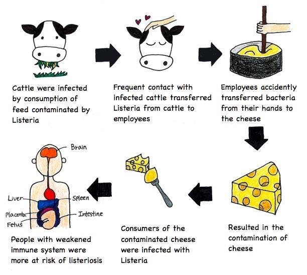
Figure 3: Description of how the listeriosis outbreak happened. Source: Jenna Lee (2017).

Measures taken to end the outbreak

Two measures were proposed to minimize consumption of L. monocytogenes contaminated dairy foods and to lower the event of foodborne illnesses to the public. 1) Strict regulation and ongoing inspections should decrease the cross-contamination during the dairy processing procedures. Stringent monitoring and cleanliness would prevent the growth and dissemination of L. monocytogenes in the cheese products during post-pasteurization in soft-ripened cheeses. For example, dairy plants should limit the use of unpasteurized milk or raw milk to make soft ripened cheeses because pasteurization (high heat in short time) can kill L. monocytogenes. 2) Information about listeriosis was provided to advise the vulnerable populations such as the pregnant women, the elderly and the immunocompromised. L. monocytogenes is in many natural environments; it presents in the soil, water, vegetation, animal feedstuffs and food processing equipment. Hence, good practicing manners could easily eliminate the foodborne illnesses in human.

Aftermath

After the outbreak, British Columbia provincial authorities recommended the dairy processing factory involved in the outbreak to test for L. monocytogenes before selling their products. Also, they made a new requirement for periodic testing of soft ripened cheese products made outside BC for L. monocytogenes. All dairy processing factories were required to submit food safety plans, product lists for their operations, and how the environment of their factory looked like. BC further enhanced inspection, recommending diary and public health inspectors to regularly test for the presence of L. monocytogenes in the facilities, report the source of water the facilities were using, and check whether the ripening solutions were controlled under the food safety plans.

References

Galanis E, Shyng S. 2009. Listeriosis outbreaks in Canada in 2008 [Internet]. Delta (BC): BC Food Protection Association; [cited 2017 Nov 15]. Available from: http://bcfpa.net/Attachments/Presentations/Listeriosis%20outbreaks%20in%20Canada%20in%202008%20(E%20Galanis%20&%20S%20Shyng)%2019%20Jan%202009.pdf.

Kovacevic J, McIntyre LF, Henderson SB, Kosatsky T. 2012. Occurrence and distribution of listeria species in facilities producing ready-to-eat foods under provincial inspection authority in British Columbia, Canada. Journal of food protection. 75(2): 216-224.

McIntyre L, Wilcott L, Naus M. 2015. Listeriosis outbreaks in British Columbia, Canada, caused by soft ripened cheese contaminated from environmental sources. BioMed Research International. 2015:12.

Rodrigues CS, Cordeiro de Sá CGV, Barros de Melo C. 2017. An overview of listeria monocytogenes contamination in ready to eat meat, dairy and fishery foods. Ciência Rural, 47(2).

Sauders BD, D’Amico DJ. 2016. Listeria monocytogenes cross-contamination of cheese: risk throughout the food supply chain. Epidemiology and Infection. 144(13):2693-2697.

Taylor M, Bitzikos O, Galanis E. 2008. Listeriosis awareness among pregnant women and their health care providers in British Columbia. BCMJ. 50(7): 398-399.

Bordetella pertussis

by Wing Chi Cheng and Ramy Elmasry

Introduction

Pertussis, also known as whooping cough (uncontrollable violent coughing), is an infection of the respiratory system originating from the bacterium Bordetella pertussis. It was first isolated and grown in 1906 by Bordet and Gengou. Bordet was later awarded the Nobel prize in Physiology or Medicine in 1920, in part, for his substantial research of B. pertussis as the causative agent of whooping cough. This bacterial pathogen colonizes and multiplies in the upper and lower respiratory tracts. It is highly contagious as it is transferred by coughing or sneezing through airborne droplets.

Disease

Bordetella pertussis primarily targets the respiratory tract as the main site of infection. The infection can be transmitted through coughing or sneezing aerosols. Upon inhalation of contaminated aerosols, the bacteria adhere to the epithelium, which is the protective tissue that surrounds the respiratory airways such as the nasopharynx and trachea. After the attachment process, B. pertussis will start to multiply and produce virulence factors that help the microorganism thrive and cause disease. The collective effect of these factors allows the bacteria to cause damage to the host and elude its immune system.

For instance, they are the main players responsible for the breakdown of cilia found on the respiratory tract (Figure 1). Cilia are membrane protrusions on the surface of respiratory epithelial cells that play a key role in the first line of defense. They mechanically sweep and mediate the clearance of mucus which trap foreign microorganisms and dust particles to be excreted out of the body. Thus, it is the buildup of mucus caused by the deterioration of the cilia that results in the violent coughing. This paroxysmal cough is often followed by a strong gasp for air, which is characterized by the classic “whooping” sound due to narrowed and mucus-filled respiratory airways.  Healthier individuals tend to be better at dealing with B. pertussis and display mild symptoms (sneezing, runny nose, low fever) due to their strong immunity.

Figure 1: Drawing showing the effect of Bordetella pertussis on the ciliated epithelial cells in the respiratory tract. A, normal state of epithelium with regular ciliary function; B, degradation of cilia and breakdown of cells following toxin secretions by B. pertussis. (Source: Wing Chi Cheng)

Epidemiology

Over the course of history, Bordetella pertussis has proven to be a very effective pathogen, causing high numbers of morbidity and mortality. Being an endemic disease, pertussis affects regularly developing and developed countries. Sporadic cases also often tend to happen across the globe. This species of Bordetella is human-specific as it is not found in animals or in the environment. Immunocompromised individuals or those with underdeveloped immune systems are more susceptible to having pertussis. This means that newborns and infants under the age of 5 are much more at risk, especially because the last dose of the vaccine against B. pertussis is administered when children are 4-6 years old.

In 2008, there was an outbreak of 16 million cases of whooping cough worldwide, whereby 195,000 were reported dead. Most of the victims were from countries with little access to vaccines. Researches have shown that pertussis differs from other respiratory diseases as it has a higher tendency to cause disease during autumn and winter (seasonally). Even developed countries such as Canada can be affected by this disease. This was the case in 2015 for instance, in Manitoba and New Brunswick where there were 51 and 56 observed cases respectively. These outbreaks are generally due to the presence of unvaccinated people within the community.

Virulence factors

As Bordetella pertussis travels through the respiratory airways, it begins to attach to the protective epithelial cells that line it. It then secretes a series of toxins that damage the host tissues. Toxins are molecules that are synthesized by the bacterium in order to help in its colonization of the host and allow it to evade the immune system.

One of the most important toxins that is specific to this microorganism is the pertussis toxin (PT). Once this toxin is secreted by the bacteria, it binds to the membrane of the host cell and, through special receptors, becomes engulfed by it in a process called endocytosis (Figure 2a). PT then enters the inside of the epithelial cell while enclosed in a bag-like structure called an endosome. A portion of the toxin is then secreted from the endosome and released into the host cell cytosol (Figure 2b) where it begins altering intracellular functions and inflicting damage.

Figure 2: Insertion of the pertussis toxin into host cells. A, attachment to the epithelial cell surface and endocytosis; B, enclosing in endosome and secretion of cytosolic toxin domain. (Source: Ramy Elmasry)

Early in the infection, the pertussis toxin causes a major delay in the recruitment of white blood cells to the site of infection by blocking chemokine production, a signal molecule that is normally sent from the invaded cells to announce danger (Figure 3a). This delay allows the bacteria to multiply and achieve further colonization. It continues by making the host cells hypersensitive to histamine, a compound that promotes leakiness in the vascular membrane (Figure 3b). This leakiness causes the contents of the blood vessels to be absorbed by the tissue below the epithelial cells and causes swelling. This swelling inflicts pressure on the epithelial layer and promotes mucus production, causing the respiratory airways to narrow, making it more difficult to breathe in extreme cases.

Figure 3: Effects of the pertussis toxin on the host system. A, further multiplication and colonization of B. pertussis thanks to a delayed white blood cell response following chemokine inhibition; B, narrowing of respiratory airways caused by mucus buildup and epithelium pressure triggered by the release of blood vessel contents following capillary leakiness after hypersensitivity to histamine. (Source: Ramy Elmasry)

Treatment

B. pertussis is generally susceptible to erythromycin, azithromycin and clarithromycin antibiotics. These help fight against the bacteria by altering or inhibiting protein synthesis, which prevents formation of crucial toxins and virulence factors. These treatments have been proven to be successful in inhibiting B. pertussis and shortening the infection only if administered in the early stages of the disease (first 1-2 weeks).

However, the best way to treat Bordetella pertussis is to prevent it altogether. There are two major types of vaccines that exist that are administered to infants and children at specific ages that have proven to be very successful in prevention: a whole-cell vaccine (DTP) and an acellular vaccine (DTaP). They are named as such because they are prepared as compound vaccines, protecting against Diphtheria (D), Tetanus (T) as well as Pertussis (P) diseases. The whole-cell vaccine, as the name indicates, contains killed whole bacterial cells while the acellular one contains only non-functional virulence factors and inactivated toxins, called toxoids, such as PT. Currently, the favored vaccine that is the most recommended in North America is the acellular format (DTaP) coupled with a booster vaccine (Tdap).

References

Carbonetti NH. 2010. Pertussis toxin and adenylate cyclase toxin: key virulence factors of Bordetella pertussis and cell biology tools. Future microbiology. [accessed 2017 Nov 16]; 5: 455-469. https://www.ncbi.nlm.nih.gov/pmc/articles/PMC2851156/. doi:10.2217/fmb.09.133.

Epstein D. 1932. The action of histamine on the respiratory tractThe Journal of Physiology. [accessed 2017 Nov 18]; 76. http://onlinelibrary.wiley.com/doi/10.1113/jphysiol.1932.sp002931/abstract. doi: 10.1113/jphysiol.1932.sp002931.

Kilgore, P. E., Salim, A. M., Zervos, M. J., & Schmitt, H. 2016.Pertussis: Microbiology, Disease, Treatment, and Prevention. Clinical Microbiology Reviews. [accessed 2017 November 15];  29(3): 449–486. https://www.ncbi.nlm.nih.gov/pmc/articles/PMC4861987/. doi:  10.1128/CMR.00083-15.

Locht C, Coutte L, Mielcarek N. 2011. The ins and outs of pertussis toxin. FEBS Journal. [accessed 2017 Nov 17]; 278: 4668–4682. http://onlinelibrary.wiley.com/doi/10.1111/j.1742-4658.2011.08237.x/full. doi:10.1111/j.1742-4658.2011.08237.x

Mattoo S, Cherry JD. 2005. Molecular Pathogenesis, Epidemiology, and Clinical Manifestations of Respiratory Infections Due to Bordetella pertussis and Other Bordetella Subspecies. Clinical Microbiology Reviews. [accessed 2017 Nov 15]; 18(2): 326-382. http://cmr.asm.org/content/18/2/326.full?linkType=FULL&resid=18/2/326&journalCode=cmr#sec-20. doi: 10.1128/CMR.18.2.326-382.2005

Melvin JA, Scheller EV, Miller JF, Cotter PA. 2014. Bordetella pertussis pathogenesis: current and future challenges. Nature reviews Microbiology. [accessed 2017 Nov 13]; 12(4): 274-288. https://www.ncbi.nlm.nih.gov/pmc/articles/PMC4205565/. doi:10.1038/nrmicro3235.

Zlamy, M. 2016. Rediscovering Pertussis. Frontiers in Pediatrics. [accessed 2017 November 15]; 4:52. https://www.ncbi.nlm.nih.gov/pmc/articles/PMC4896922/. doi:  10.3389/fped.2016.00052.

Porphyromonas gingivalis

by Thomas Donoso and Dennis Park

Introduction

Porphyromonas gingivalis is the opportunistic pathogen largely responsible for periodontitis, an inflammatory oral disease that results in gum damage and tooth loss. The bacterium resides in the oral cavity of humans with hundreds of other bacteria which make up the normal microbiota. This article will explore the pathogenicity of Porphyromonas gingivalis and its several virulence factors.

Disease

Porphyromonas gingivalis is an opportunistic pathogen, meaning it only infects when an imbalance in the normal host microbial flora occurs. Normally, P. gingivalis resides stably with billions of other bacteria in the oral cavity. However, when the ratio or number of the normal microbes is disrupted, it quickly takes advantage of the situation to invade the underlying oral tissue. Inside the oral tissue, P. gingivalis is able to prevent being killed by lysosomes – sacs filled with corrosive and damaging molecules. This gives it enough time to replicate and spread from cell to cell. As the bacteria replicates and spreads, it progressively degrades the nearby tissues using its protein degrading enzymes, one of which is called gingipains, and induces the host’s inflammatory response. This results in the formation of “pockets” around the teeth that is characteristic of periodontitis. As shown in figure 1, a healthy oral cavity has gingiva (gum) that is tightly wrapped around the teeth. However, with periodontitis, the gums are retracted due to proteases and inflammation, therefore resulting in holes and gaps around the teeth. This weakened junction between teeth and gum is what causes the eventual tooth loss during periodontitis.

Fig 1. Periodontitis decays gum tissue resulting in periodontal pockets. Healthy teeth are wrapped tightly around by gum tissue. With periodontitis, inflammation and bacterial proteases cause the tissue surrounding the tooth to recede and decay, resulting in “pockets” or gaps. Source: Thomas Donoso and Dennis Park.

Epidemiology

The prevalence of periodontitis is a global phenomenon, with different rates among regions, habits, sex, and race. In 1999, 35% of adults over the age of 30 in the United States were diagnosed with periodontitis. This percentage is correlated to an increase in age. Youth do carry P. gingivalis, however, as studies show that this bacteria was found in cavities with a similar prevalence in all ages. Ultimately, the disease has less effect on younger populations (specifically, less than 30 years old). On top of age, cases of periodontitis have a higher occurrence in those of African, Caribbean, and Mexican descent. In terms of habit, smokers and those who have poorly controlled diabetes are also more susceptible to periodontitis. Lastly, sex is also a contributing factor, as males are more prone to periodontitis than females.

Virulence systems

Porphyromonas gingivalis invades oral tissues using structures called the fimbriae, short hair-like extensions on the surface of the bacterium. The fimbriae attaches to molecules called integrins, specialized receptors on the host tissue, and causes the internal cytoskeleton to be rearranged in the host cell (figure 2A and 2B). The cytoskeleton can be thought of as bones: it supports the cell structure and is involved in cell motility.

Once inside the host cell, the bacterium is enclosed in a phagosome – a sac like compartment (figure 2C). Inside the phagosome, the bacterium cannot survive for long due to two reasons. Firstly, it has limited nutrients to grow, and secondly, the phagosome will soon fuse with a lysosome (another sac filled with corrosive and damaging molecules) and thus, the bacteria will die.

Therefore, P. gingivalis induces the host to undergo autophagy, a highly regulated process that is used to recycle cellular components (figure 2D). During autophagy, the host creates a membrane around cellular components that is to be digested and recycled. The enclosure is later fused with a lysosome, which degrades the components.  For reasons not well understood, P. gingivalis can induce the host to undergo autophagy by increasing expression of molecules that regulate this process, such as Beclin-1. The structure that arises from the membrane enclosure around the P. gingivalis (due to autophagy) is called the autophagosome.

Ultimately, inside the autophagosome, the bacteria uses amino acids from the host and replicates using the nutrients (figure 2E). Unfortunately for the bacteria, the autophagosome too will eventually fuse with the lysosome. However, P. gingivalis has cleverly designed methods to delay maturation and fusion of the autophagosome with the lysosome. 

The process mentioned above involves various virulence factors, tools used to help induce disease, but one particular virulence factor of interest are the protein degrading enzymes called gingipains produced by P. gingivalis. It can either be membrane bound or secreted.

It is believed that transfer into the autophagosome (figure 2C to 2E) is in part due to the gingipains breaking down the inner membrane of the autophagosome, allowing entry for the bacterium. Also, it is loosely evidenced that gingipains are involved in delaying the fusion between autophagosome and lysosome. For example, experiments by Yamatake et al. 2007 show P. gingivalis mutants that lack gingipains are less observed in autophagosomes and are killed much faster by lysosomes.

Fig 2. Porphyromonas gingivalis uses its fimbriae to attach to integrin molecules on the host surface and enter the cell. Upon fimbriae and integrin binding, the cytoskeleton of the host cell rearranges, allowing the pathogen to enter. Once inside, the bacterium induces autophagy in the host to create a nutrient rich niche to replicate in. Source: Thomas Donoso and Dennis Park.

Lastly, the most striking fact about gingipains is the wide variety of effects it can have on the host. It is believed that majority of the virulence from P. gingivalis comes from gingipains. Below are some of the effects it can have on a susceptible host and why it contributes to the virulence of P. gingivalis.

Host molecule Effect
T-Cells* Cleavage of T-cell receptors prevent proper presentation of antigen (the target of the specific immune system)
Pro-inflammatory cytokines** Pro-inflammatory cytokines are cleaved to weaken the inflammatory response
Anti-inflammatory cytokines Anti-inflammatory cytokines are cleaved to weaken immune regulation by the host
Complement*** Complement molecules are cleaved to prevent recruitment of phagocytes (digestive molecules) and opsonization (marking for digestion)
Blood coagulation Fibrinogen, a protein involved in blood clotting, is degraded to prevent coagulation

Table 1. Gingipains upset the proper regulation of the host immune response and bodily functions. Gingipains have a wide range of effects within the host. It can prevent proper T-cell function, inflammatory responses and complement activation. Furthermore, it can prevent coagulation to increase migration and replication for the bacterium. * T-Cells are cells of the immune system that help fight off pathogens. It does so either by activating proper responses to deal with the pathogen or by killing cells infected with the pathogen. ** Cytokines are small proteins that act as messengers between cells and tissues. *** Complements are soluble proteins in the blood that can form pores on bacterial membranes or coat bacteria for other immune cells to kill.

Treatment

Before infection, the best method to avoid disease is prevention. Good oral hygiene has been shown to decrease the risk of peritonitis drastically. However, when it is contracted, antibiotics and antiseptics are often prescribed to treat  P. gingivalis infection. Positively charged chlorhexidine is an example of an antiseptic used for dental infections, as it kills a broad variety of different bacteria by binding and destroying the negatively charged cell wall. Chlorhexidine is easy to use as it can be administered through a gel or mouthwash, but it may cause irritation.

The alternative, antibiotics, need to be administered through a systemic administration route. This means that the antibiotic needs to be administered through digestion or injection to allow it to reach the deep tissues of the gums. An example of antibiotics used in the treatment are macrolides, such as erythromycin, and ampicillin, which inhibit the synthesis of proteins.  

References

Albandar, J. M., Brunelle, J. A., & Kingman, A. (1999). Destructive periodontal disease in adults 30 years of age and older in the United States, 1988-1994. Journal of periodontology, 70(1), 13-29.


Bélanger, M., Rodrigues, P. H., Dunn, Jr, W. A., & Progulske-Fox, A. (2006). Autophagy: a highway for Porphyromonas gingivalis in endothelial cells. Autophagy, 2(3), 165-170.


Bostanci, N., & Belibasakis, G. N. (2012). Porphyromonas gingivalis: an invasive and evasive opportunistic oral pathogen. FEMS microbiology letters, 333(1), 1-9.


Cho, Y. J., Song, H. Y., Ben Amara, H., Choi, B. K., Eunju, R., Cho, Y. A., … & Koo, K. T. (2016). In vivo inhibition of Porphyromonas gingivalis growth and prevention of periodontitis with quorum-sensing inhibitors. Journal of periodontology, 87(9), 1075-1082.


Demmer, R. T., & Papapanou, P. N. (2010). Epidemiologic patterns of chronic and aggressive periodontitis. Periodontology 2000, 53(1), 28-44.


Dorn, B. R., Dunn, W. A., & Progulske‐Fox, A. (2002). Bacterial interactions with the autophagic pathway. Cellular microbiology, 4(1), 1-10.


Gerits, E., Verstraeten, N., & Michiels, J. (2017). New approaches to combat Porphyromonas gingivalis biofilms. Journal of oral microbiology, 9(1), 1300366.


Greene, J. C. (1963). Oral hygiene and periodontal disease. American Journal of Public Health and the Nations Health, 53(6), 913-922.


Griffen, A. L., Becker, M. R., Lyons, S. R., Moeschberger, M. L., & Leys, E. J. (1998). Prevalence of Porphyromonas gingivalis and periodontal health status. Journal of clinical microbiology, 36(11), 3239-3242.


How, K. Y., Song, K. P., & Chan, K. G. (2016). Porphyromonas gingivalis: an overview of periodontopathic pathogen below the gum line. Frontiers in microbiology, 7.


Maezono, H., Noiri, Y., Asahi, Y., Yamaguchi, M., Yamamoto, R., Izutani, N., … & Ebisu, S. (2011). Antibiofilm effects of azithromycin and erythromycin on Porphyromonas gingivalis. Antimicrobial agents and chemotherapy, 55(12), 5887-5892.


McClellan, D. L., Griffen, A. L., & Leys, E. J. (1996). Age and prevalence of Porphyromonas gingivalis in children. Journal of clinical microbiology, 34(8), 2017-2019.


Mysak, J., Podzimek, S., Sommerova, P., Lyuya-Mi, Y., Bartova, J., Janatova, T., … & Duskova, J. (2014). Porphyromonas gingivalis: major periodontopathic pathogen overview. Journal of immunology research, 2014.


Park, M. H., Jeong, S. Y., Na, H. S., & Chung, J. (2017). Porphyromonas gingivalis induces autophagy in THP‐1‐derived macrophages. Molecular oral microbiology, 32(1), 48-59.


Yamatake, K., Maeda, M., Kadowaki, T., Takii, R., Tsukuba, T., Ueno, T., … & Yamamoto, K. (2007). Role for gingipains in Porphyromonas gingivalis traffic to phagolysosomes and survival in human aortic endothelial cells. Infection and immunity, 75(5), 2090-2100.


Yilmaz, Ö., Verbeke, P., Lamont, R. J., & Ojcius, D. M. (2006). Intercellular spreading of Porphyromonas gingivalis infection in primary gingival epithelial cells. Infection and immunity, 74(1), 703-710.


Burkholderia pseudomallei

by Jade Lee

Introduction

Burkholderia pseudomallei also known as Pseudomonas pseudomallei is a gram-negative bacterium that causes Melioidosis or Whitemore’s disease. It can grow on a variety of media and forms wrinkled, pinkish colonies (see figure 1). B. pseudomallei exits as a saprophyte in wet soils and rice paddies but is capable of infecting humans and many animal species (including sheep, goats, horses, swine, cats, dogs, cattle). A saprophyte is an organism which gets its energy from dead and decaying organic matter.

Figure 1: Colonies of B. pseudomallei on Ashdown’s agar showing the typical pink crinkled colonies after 72 hours of incubation at 37°C. Source: BioMed Central Veterinary Research, 2014.

Disease

Disease occurs when the bacteria enters the body via cuts and sores in the skin or via inhalation of dust or droplets and very rarely by ingestion of contaminated water. Direct human-to-human and animal-human transmission is rare but may occur after contact with blood or bodily fluids. Indeed, infected animals and humans can shed B. pseudomallei in urine, feces, wound exudates, nasal secretions and milk, these secretions will depend on the site of infection. B. pseudomallei is capable of remaining latent for up to 29 years (longest documented period of latency) because it is a facultative intracellular pathogen and can escape the host’s immune system by invading and multiplying in phagocytic and nonphagocytic cells. Phagocytic cells (neutrophils, monocytes and macrophages) are cells of the immune systems whose role is to take up and digest invading pathogens. The nonphagocytic cells that the bacteria can invade are epithelial cells; epithelial tissues are thin tissues that cover all the exposed surfaces of the body. The bacterial invasion of these cells can result in their lysis. Melioidosis causes localized disease, such as pneumonia and abscesses. The symptoms are non specific: fever (high grade), headache, vomiting, nausea abdominal pain, cough, and lung abscess. B. pseudomallei can also spread to secondary sites, including organs such as liver, spleen or brain, or to the blood, resulting in septicaemia (blood poisoning). The infection can also be asymptomatic.

Epidemiology

Melioidosis is primarily a disease of tropical climates, specifically found in Southeast Asia and northern Australia. B. pseudomallei was recovered in 20.4 % of soil samples in northeastern Thailand and accounts for 20% of community-acquired (infection contracted outside of a health care setting) blood poisoning or septicemias in this region where 50% of those affected die. It usually acts as an opportunistic pathogen, meaning that it can infect a host when the host’s resistance is compromised. Indeed, people that are usually at risk for Melioidosis are those with conditions such as diabetes, kidney disease, lung disease, cancer, heavy alcohol consumption and those on immunosuppressive therapy including steroids. However, healthy people can acquire the disease if they work in muddy soil without adequate hand and foot protection. Children are usually less likely to get the disease than adults but they can still get it during the wet season, especially those with a weakened immune system.

Virulence systems

Initial infection occurs at the epithelial cell layer of either the wounded skin or the mucosal surface. The attachment to these cells is mediated by a thin polysaccharide layer around the bacteria or capsule, and via a type IV pilus. Type IV pili are strong, flexible filaments that are like grappling hooks that can attach to the surface of other cells, and while they retract, they pull the bacterium towards the point of attachment. The capsule of B. pseudomallei can also help the bacterium evade the host’s immune system by resisting phagocytosis. Once the epithelium cells or phagocytic vacuoles are invaded, B. pseudomallei is able to escape from endocytic vacuoles or phagosomes (membrane-bound cellular organelles that engulfed the bacteria) via the type III secretion called bsa. This is a syringe like molecule that injects toxins through the wall of the membrane of the host cell. These toxins enable the bacteria to escape the phagosome and inhibit autophagy, a process that allows host cells to deal with intracellular infection by packaging, degrading and recycling cellular contents. B. pseudomallei is then able to spread from cell to cell by recruiting to its surface host actin and other proteins present in the host cells, which initiates the assembly of an actin tail. The continous assembly of the actin tail supplies force to propel the organism through the cytoplasm of the infected cell and into neighbouring cells (see figure 2). In phagocytic cells B. pseudomallei is also able to neutralize reactive oxygen species and proteases (enzymes that degrade proteins) that normally destroy the pathogens. Finally, the pathogen can also produce haemolysins (which destroy red blood cells), lipases (enzymes that destroy lipids) and siderophores (complexes that have the ability to sequester iron from host cells). These secreted factors have cytotoxic effects on eukaryotic cells and cause further damage to the host’s tissues.

Figure 2: Invasion of host cells by B. pseudomallei: upon entry in the host cell, the bacteria lyses the phagocytic vacuole with its Type III secretion system, once in the cytoplasm the bacteria assembles an actin tail at one of its poles, this assembly propels the bacteria to other adjacent cells. Source: Jade Lee (2017).

Treatment

Antibiotics are less effective against B. pseudomallei because of its intracellular capacity. The pathogen is resistant to penicillin, ampicillin first- and second-generation cephalosporins, gentamicin, tobramycin, and streptomycin. The intensive treatment phase involves intravenous administration of ceftazidime for 10-14 days or longer. This antibiotic has a bactericidal action that inhibits enzymes responsible for cell-wall synthesis in Gram- bacteria. A vaccine is under development but economic constraints may make vaccination an unrealistic option for many regions where Melioidosis is an issue.

References

Department of Agriculture, Center for Food Security and Public Health. 2003. Melioidosis. Ames (IA): Department of Agriculture, Center for Food Security and Public Health; [accessed 2017 Nov 11]. http://www.nj.gov/agriculture/divisions/ah/diseases/melioidosis.html

Adler NRL, Govan B, Cullinane M, Harper M, Adler B, Boyce JD. 2009. The molecular and cellular basis of pathogenesis in melioidosis: how does Burkholderia pseudomallei cause disease? FEMS Microbiology Reviews. [2017 Nov 11]; 33(6): 1079–1099. doi: 10.1111/j.1574-6976.2009.00189.x.

Cheng AC, Currie BJ. 2005. Melioidosis: Epidemiology, Pathophysiology, and Management. Clinical Microbiology Reviews. [2017 Nov 11]; 18(2): 383-416. doi: 10.1128/CMR.18.2.383-416.2005.

Burtnick MN, Brett PJ, Nair V, Warawa JM, Woods DE, Gherardini FC. 2008. Burkholderia pseudomallei Type III Secretion System Mutants Exhibit Delayed Vacuolar Escape Phenotypes in RAW 264.7 Murine Macrophages. Infection and Immunity. [2017 Nov 11]; 76(7): 2991-3000. doi: 10.1128/IAI.00263-08.


Klebsiella pneumoniae

by Wenlie Xie and Huixuan Jia

Introduction

Klebsiella pneumoniae was first described by a German microbiologist and pathologist Carl Friedländer in 1882. K. pneumoniae is found in the normal flora of skin, mouth and intestines, where it does not cause disease. Under opportunistic conditions such as a weakened host immune system or an altered microbiota, K. pneumoniae enters the hosts and causes several disease including pneumoniae, bloodstream infection and wound infection (Figure 1).

Figure 1: A photomicrographic view of a Hiss capsule-stained culture specimen revealing the presence of numerous Klebsiella pneumoniae bacteria. Source: https://phil.cdc.gov/Details.aspx?pid=14342.

Disease

Klebsiella infections are mainly acquired nosocomially, meaning that they are mostly encountered in hospitals or healthcare setting. Hospitalized patients with compromised immune systems are more susceptible: after two weeks of hospital admission, K. pneumoniae colonization in the patient’s intestine can increase up to two- to four-fold. This is mainly due to antibiotic medications that the patients are receiving, especially broad-spectrum or multiple antibiotics. Since the normal commensal micro-organic environment is damaged, this creates a favorable niche for K. pneumoniae colonies to grow. According to the Center of Disease Control and Prevention (CDC) of the USA, healthy people need not to worry about Klebsiella infections.

Under healthcare settings, K. pneumoniae is transmitted between individuals from contaminated invasive medical equipments, such as ventilators (breathing machines) or intravenous (vein) catheters. When K. pneumoniae reaches tissues or organs, it causes diseases including septicemia (blood poisoning), pneumonia, urinary tract infection (UTI), and soft tissue infection. Symptoms of infections vary from different infection sites. For example, hospital-acquired K. pneumoniae induces clinical symptoms such as fever and chills, shortness of breath, decreased blood pressure and faster heart rate, nausea and vomiting, etc. If the bacteria is introduced into the bloodstream, patients can develop symptoms such as sharp headache, nausea, dizziness and impaired memory.

Epidemiology

Canada has only reported sporadic cases and limited outbreaks in hospitals from Ottawa and Montreal. In Europe, K. pneumoniae is reported as the second most common cause of bloodstream infections in adult population. Middle-aged and older people with debilitating diseases, such as diabetes mellitus are more susceptible to the infection. The ability of K. pneumoniae to spread rapidly also can lead to hospital outbreaks in neonatal units. The mortality rate is typically around 100% for patients with alcoholism as alcoholic patients suffer from weakened immune systems, which can increase their susceptibilities to tissue-damaging pathogens.

One noticeable outbreak was the 2011 Montreal Jewish General Hospital outbreak caused by a highly drug-resistant strain called KPC-producing K. pneumoniae. In this outbreak, 27 patients were identified to be infected and among them, 1 patient died from the infection. The outbreak shows that new antibiotic-resistant strains of K. pneumoniae are appearing. Antibiotic resistance is a huge problem because it leads to higher medical costs, prolonged hospital stays, and increased mortality rate. Feces are the most significant source of patient infection, followed by contact with contaminated medical equipment.

Virulence system

The most important virulence factor contributing the pathogenesis of K. pneumoniae is their ability to form a thick layer of biofilm, which consists of a large number of bacteria embedded in an extracellular matrix. Majority of the K. pneumoniae strains are biofilm-producing. The biofilm greatly enhances the bacteria’s ability to attach to abiotic environment such as medical equipments, and living organisms such as tissues. Under the healthcare setting, in order to invade human, K. pneumoniae uses pili (hair-like appendages for attachment) and capsular polysaccharides to attach on the surface of intrusive medical equipments, e.g. the catheter of a patient. This initial attachment allows the growth and colonization of the bacteria in an extracellular matrix on the surface of the catheter. At this stage, the exponentially-growed bacteria is sessile and might not be pathogenic. When the biofilm is matured and the sessile K. pneumoniae bacteria differentiates into planktonic form, K. pneumoniae sheds and free to attach to tissues or enter the bloodstream, causing infections. The biofilm-producing strains of K. pneumoniae often contributes to recurrent infections, since antibiotics only clear the infections within the body but not on the biofilm source.

Upon entering the body, K. pneumoniae uses lipopolysaccharides (LPS) and capsule against the body’s front-line immune defense, such as the complement system. Complement system is one of the host’s lines of defence, consisting of over 30 proteins. LPSs are long polysaccharide filaments on the bacterial outer membrane, which could trigger the activation of the complement system. A proposed mechanism is that LPS is masking under the capsule polysaccharides, of which the surface structure does not activate host complement system (Figure 2a). The longest O-polysaccharide chain from the LPS reaches the exterior milieu and preferentially fixes C3b, a component from complement system. This causes the deposition of C3b onto LPS molecules at distant sites from the bacterial cell membrane (Figure 2b). This inhibits the attachment of C3b to the surface thus the assembly of membrane attack complex is prevented (Figure 2c).

Figure 2: How K. pneumoniae uses LPS and capsule to avoid the host’s complement system. See text for details. Source: Wenlie Xie and Huixuan Jia.

Treatment

K. pneumoniae infection can be treated with antibiotics if the strain is not drug-resistant. The best choices of antibiotics are third-generation and fourth-generation cephalosporins, quinolones and carbapenems. To examine the effectiveness of antibiotics, microbiological laboratory tests can be done.  However, the use of antibiotics alone is usually not enough. Surgery removing is often needed after the patient is started on antibiotics. For patients with more severe infections, the treatment should be more prudent. The method involves 48–72 hours of multiple therapies initially,  followed by a switch to a specific monotherapy.


References

Branswell, H. (2011). The Canadian Press. https://globalnews.ca/news/156114/tricky-new-superbug-making-inroads-in-canada-montreal-hospital-battled-outbreak-2/

Centers for Disease Control and Prevention. (2012). Klebsiella pneumoniae in Healthcare Settings. https://www.cdc.gov/hai/organisms/klebsiella/klebsiella.html

Martin, R.M., et al. (2016). Molecular epidemiology of colonizing and infecting isolates of Klebsiella pneumoniae mSphere 1, e00261-16.

Prince, S.E., et al. (1997). Klebsiella pneumoniae. Heart Lung 26, 413-7.

Podschun, R. & Ullmann, U. (1998). Klebsiella spp. as Nosocomial Pathogens: Epidemiology, Taxonomy, Typing Methods, and Pathogenicity Factors. Clin Microbiol Rev 11, 589–603.  

Seifi, K., Kazemian, H., Heidari, H., Rezagholizadeh, F., Saee, Y., Shirvani, F., & Houri, H. (2016). Evaluation of Biofilm Formation Among Klebsiella pneumoniae Isolates and Molecular Characterization by ERIC-PCR. Jundishapur Journal of Microbiology, 9(1), e30682. http://doi.org/10.5812/jjm.30682

Silvia, L., Poirel, L. & Bonomo, R. (2013). Clinical epidemiology of the global expansion of Klebsiella pneumoniae carbapenemases. Lancet Infect Dis 13, 785–796

Tian, L. & Tan, R. (2016). Epidemiology of Klebsiella pneumoniae bloodstream infections in a teaching hospital: factors related to the carbapenem resistance and patient mortality. Antimicrob Resist Infect Control 5, 48

Pasteurella multocida

by Adriana Kalaska and Jessica Garofalo

Introduction     

Pasteurella multocida is the most common cause of a disease known as Pasteurellosis. This disease is zoonotic as it infects humans through an animal vector. P. multocida is found in the normal oral and nasal bacterial communities of many species. The bacteria is named after Louis Pasteur who first isolated the stain in 1880 from birds suffering from cholera.  

Disease

Pasteurellosis is transmitted to humans by cat or dog bites and licks via a wound in the skin. Symptoms of skin infection usually appear after 24-48 hours. However, people with weak immune systems cannot clear this initial infection effectively. This allows P. multocida to spread to the blood and sometimes to the brain, causing swelling, fever, blood poisoning, and even death. In animals, colonies of P. multocida can also become invasive and spread to the lungs under stressful conditions when the host immune system is weakened. This opportunistic pathogen is able to overwhelm the host defenses. Phagocytes, cells that normally ingest and destroy the bacteria, cannot kill P. multocida efficiently due to the bacteria’s protective capsule, which is what leads to the progression of respiratory diseases. If an animal has a heavy bacterial burden, airborne transmission of the pathogen to others in the herd is possible (see Figure 1). In rare cases, P. multocida infection can also cause severe inflammation, that can lead to cellular damage.

Figure 1:  Different transmission pathways of P. multocida. Left: The evolution of commensal P. multocida into upper respiratory tract infections in farm animals when faced with stressors such as dramatic temperature changes and lack of or improper nutrition. Right: Transmission of P. multocida found in the oral and nasal cavities in common household pets to humans. Created by Garofalo & Kalaska, images sourced from: https://openclipart.org.

Epidemiology

Roughly 300 000 annual visits to emergency rooms in the U.S. are due to animal bites or scratch wounds. Respectively, 50% and 75% of dog and cat bites result in soft tissue infections caused by pasteurella species in humans, as these are the most common carrier animals. The very young (underdeveloped defenses), the elderly (tired defenses), and individuals with underlying conditions (diabetes, chronic diseases) are more susceptible to proliferative infection. Additionally, anyone who frequently handles animals without handwashing is at greater risk of contraction. Overall, death in humans due to P. multocida is extremely rare.

Pasteurellosis also affects domesticated animals such as cattle, buffalo, sheep, goats, horses, pigs, poultry, rabbits, and rats. Some manifestations of this bacteria in these species are: fowl cholera, hemorrhagic septicemia, pneumonia, respiratory atrophic rhinitis, and purulent rhinitis. These infections cause high mortality rates reaching up to 50% in animals with clinical disease. In particular, it causes 30% of total cattle deaths worldwide. Consequently, pasteurellosis causes substantial economic deficits. It is estimated to result in losses of $1 billion in the North American beef industry alone.

Virulence Factors

The main factor that allows P. multocida to infect and spread within hosts is the bacterial capsule that is made up of complex carbohydrate combinations. The capsule masks specific surface molecules of the bacteria, called antigens, that are recognized by the host’s immune system. Furthermore, variability of this structure makes it hard for the immune system to target the bacterial cells, as any memory produced by special immune cells to one form of the capsule will not be specific for new versions. Immune system memory is mediated by  T cells and B cells, which respond to specific antigen. T cells activate B cells which then produce antibodies. Antibodies bind and surround bacteria and bring them to other immune cells to be degraded, which is an important process known as opsonization.

The capsule of most bacterial species protects them from the natural defenses of the skin such as antimicrobial peptides, which are small molecules that form pores or bipass the bacterial membrane to kill the pathogen. Without a capsule, P. multocida is also sensitive to proteins of the blood, called the complement. These tag the cell and attract other complement proteins or antibodies to attach, creating a pore in the membrane. They may also attract macrophages which perform phagocytosis: the engulfing and destruction of bacterial cells using toxic chemicals that are found in a separate compartment called the phagosome.

Thus, this capsule allows the bacteria to spread and invade tissues without being detected by the host (See Figure 2). Additionally, it is believed that the bacterial capsule is also capable of mimicking the host. This means that the components making up the layer surrounding the membrane of the bacteria resemble components of the host cells. P. multocida uses this as another method to evade phagocytes, as the host immune cells are programmed not to recognize and target proteins that resemble themselves.

Figure 2: The function of the capsule. A mutant bacteria without the capsule is easily detected and destroyed by the macrophage phagosome. Encapsulated bacteria mimicking the host are protected from detection and proliferate. Created by Garofalo & Kalaska.

The capsule of P. multocida exists as 5 different types called A, B, C, D, and E. The type of capsule determines the species of animal that the bacteria infects as well as the specific disease that it causes. Furthermore, the thickness of all capsules depends on the level of some nutrients, such as iron, that the bacteria has available to it. Finally, some research has revealed that capsules may play a role in the adhesion of P. multocida to the host cells, facilitating attachment and allowing the bacteria to remain inside of tissues and reproduce.

Treatment

In humans, animal bite infections are treated with broad range antibiotics. This is done to target multiple bacterial species that have the ability to survive in the low oxygen environment of a deep wound. Pasteurella species are susceptible to penicillin, but the preferred treatment is a mixture of amoxicillin/clavulanate or ampicillin/sulbactam to attack the combination of bacteria in bite wounds. These antibiotics target cell wall synthesis which is necessary for bacterial growth and division.

In domesticated animals, treatment with antibiotics is expensive and shown to be inefficient. Research into vaccines has produced a semi-efficient live attenuated vaccine, containing a mutated strain of P. multocida that lacks multiple genes required for infection. This allows the host to produce memory cells against this pathogen and upon future infections they will produce a rapid targeted response to clear it. Unfortunately, these vaccines have resulted in some systemic infections leading to death of vaccinated animals.

References

Ahmad, T. A., et al. (2014). Development of immunization trials against Pasteurella
multocida. Vaccine, 32, 909–917.

Giordano, A., et al. (2015). Clinical Features and Outcomes of Pasteurella multocida Infection. Medicine 94, 1-7.

Harper, M., and Boyce, J.B. (2017). The Myriad Properties of Pasteurella
multocida Lipopolysaccharide. Toxins 9, 254.

Marcantonio, Y. C., et al. (2017). Disseminated Pasteurella multocida infection:
Cellulitis, osteomyelitis, and myositis. IDCases 10, 68–70.


Anaplasma phagocytophilum

by Geraldine Millan and Diana Suarez

Introduction:

Anaplasma phagocytophilum is a gram negative bacteria that can cause disease in a wide variety of mammals including cattle, domestic and wild animals. This bacteria has also shown to infect humans by tick bite resulting in the infectious disease: human granulocytic anaplasmosis (HGA). In the United States, this disease had an incidence of 6.1 cases per million persons in 2010.  Although the fatality cases are rare (less than 1 %) it can cause death if is not treated with antibiotics in an early stage of the infection. A. phagocytophilum has a small size of 0.2-1.0 μm and it infects neutrophils. Once the bacteria is inside white blood cells it replicates until it reaches a high population density to spread to other host cells.

Disease:

A. phagocytophilum causes a zoonotic disease called human granulocytic anaplasmosis. HGA is an infectious disease transmitted by an infected thick which comes in contact with humans and share the bacteria through its saliva. HGA was first identified in a human subject during the 1990’s when a patient from Wisconsin, United States, died two weeks after a tick bite resulting in severe sickness. Human granulocytic anaplasmosis causes serious illness and even death in healthy children and adults because it is difficult to diagnose due to its similar signs and symptoms with viral infections such as the flu. Therefore, treatment is often neglected which leads to severe complications. The symptoms are detected within a week or two after the bite of an infected tick. It causes fever, headaches, fatigue, cough, chills, shaking and many other symptoms with the following being the most serious and life-threatening; leukopenia (a decrease in the number of white blood cells in the blood) and thrombocytopenia (low level of platelets in the blood). These physiological modifications cause individuals to be at a greater risk of infections and decrease their effectiveness of forming blood clots. A. phagocytophilum infects neutrophils which are cells that play an important role in the immune system response.  When neutrophils are infected their overall efficiency of fighting bacterial infections decreases which make the host more susceptible for getting disease.

Epidemiology:

Human beings are accidental hosts of A. phagocytophilum because the bacteria usually infects wild animals. Ticks will feed on wild animal’s blood and get infected at their turn. Once the ticks are infected, they can transmit the bacteria to other mammals through their saliva which makes them the main vector for human granulocytic anaplasmosis. The black-legged tick (Ixodes scapularis) and the Western black-legged tick (Ixodes pacificus) are the two species involved in the transmission of this disease.

Some cases have been reported concerning the transmission of the disease between humans by blood transfusion. According to the center for disease and prevention in the United States, the number of cases with tick-borne rickettsial disease has increased from 348 cases in 2000 to 1761 cases in 2010. In Canada, this disease has not been reported in humans, but animals have been known to be affected.

The signs and symptoms of HGA vary from person to person which makes it harder to detect and diagnose it at an early stage of the infection. The disease has been documented to be deadly in less than 1% of the infected patients. The risk of fatal cases is more significant in individuals with compromised immunity such as HIV positive patients, patients who have had their spleen removed, or patients undergoing immunosuppressive therapy (eg. cancer chemotherapy or organ transplant patients).

Virulence factors:

For this bacteria to be able to invade neutrophils, as it can be seen in figure 1,  it has developed a unique adaptation and pathogenic mechanism. A. phagocytophilum lacks lipopolysaccharide and peptidoglycan which are bacterial components easily identified by the host immunity that can induce an immune response. The absence of these components are an important factor for the avoidance of the host immune system by the bacteria . It also obtains cholesterol derived from host cells to maintain its membrane integrity and most importantly to look like a host cell. A. phagocytophilum’s entry mechanism into neutrophils is  successful due to the presence of a type IV secretion system (T4SS) that releases molecules into the host cells improving the effectiveness of colonization and infection. The virulence factors for adherence secreted by the T4SS are essential for the infection process because they enable the bacteria to adhere to the white blood cell’s surfaces and ultimately colonize them. The pathogen is phagocytosed, eaten,  by neutrophils and actively transported inside of the white blood cell. At the beginning of the bacterium’s life cycle, it populates an early endosome, a membrane-bound compartment inside of eukaryotic cells, where it has access to nutrients allowing for growth and division. It grows into small groups called morulae by using the process of binary fission which is the division of cells by asexual reproduction. This process can be seen in figure 2. It acquires nutrients by hijacking vesicles coming from organelles of nutrient-rich sources: the trans-Golgi and endoplasmic reticulum.

Figure 1: Morulae detected in a neutrophil on a peripheral blood smear, associated with A. phagocytophilum infection. Source: https://www.cdc.gov/anaplasmosis/symptoms/index.html

Figure 2: Steps required for A. phagocytophilum to establish disease in humans. Source: Diana Suarez.

Normally during  phagocytosis, a bacteria is engulfed in an internal compartment called a phagosome and it is eventually  killed by the production of reactive oxygen species (ROS). A. phagocytophilum is protected against this deadly mechanism because it uses two strategies. The first strategy of the bacteria is to be an O2− scavenger which means that it can detoxify these reactive oxygen species. However the specificities of this strategy are yet to be understood. The second one is that the bacteria resides in a protective endosome that does not allow fusion with the lysosome an organelle containing enzymes that digest and disintegrate cells.

Other mechanisms that allow A. phagocytophilum to thrive inside neutrophils are the inhibition of  apoptosis for a period of 48 to 96 hours and the induction of autophagy.  Apoptosis is the process of programmed cell death. It is a response created by the innate immune system against bacterial infection. The inhibition of apoptosis allows the bacteria to replicate inside the cell for a longer period of time. Autophagy allows host cells to degrade and recycle cellular components. In this case, autophagy in neutrophils will create more space for the bacteria to grow as the host membrane is remodeled. In addition, there will be more nutrients available for replication and growth of the bacteria. “A. phagocytophilum subverts two important innate immune mechanisms of neutrophils, apoptosis and autophagy, by inhibiting and inducing, respectively, to keep the host cell alive and create a safe haven” (Rikihisa, 2011).

Treatment:

The treatment for human granulocytic anaplasmosis consists in the use of antibiotics. There is no vaccine against  A. phagocytophilum in humans and, therefore, the only method to prevent HGA is to avoid getting tick bites. Doxycycline is the most commonly prescribed antibiotic because it is effective in treating the disease. However, antibiotic treatment has to be taken as soon as possible when the disease-related symptoms are perceived. Pregnancy can also complicate the antibiotic treatment. The dosage for adults is usually 100 mg  every 12 hours and for children under 45 kg is 2.2 mg per kg of body weight administered twice a day. The treatment usually lasts 7 to 14 days, and symptoms such as fever will disappear within 24-72 hours after starting taking the antibiotics. Tetracycline antibiotic is also used to fight the bacterial infection. Tetracycline binds to the bacterial 30s ribosomal subunit and blocks synthesis of proteins preventing the spread of the bacteria.
References

Rikihisa Y. (2011). Mechanisms of Obligatory Intracellular Infection with Anaplasma phagocytophilum. Clinical Microbiology Reviews, 24(3):469-489. doi:10.1128/CMR.00064-10

Carlyon, J. A., Latif, D. A., Pypaert, M., Lacy, P., & Fikrig, E. (2004). Anaplasma phagocytophilum Utilizes Multiple Host Evasion Mechanisms To Thwart NADPH Oxidase-Mediated Killing during Neutrophil Infection. Infection and Immunity, 72(8), 4772-4783. doi:10.1128/iai.72.8.4772-4783.2004

Rikihisa, Y. (2010). Anaplasma phagocytophilum and Ehrlichia chaffeensis: subversive manipulators of host cells. Nature Reviews Microbiology 8, 328–339. doi:10.1038/ nrmicro2318

Truchan, H. K. (2014). Anaplasma phagocytophilum nutritional virulence mechanisms target the host cell secretory pathway. Virginia Commonwealth University Scholars Compass.

Center for Disease Control and Prevention. (2016). Anaplasmosis Symptoms, Diagnosis, and Treatment. National Center for Emerging and Zoonotic Infectious Diseases, Division of Vector-Borne Disease.

Minnesota Department of Health. (2017). Human Anaplasmosis Information for Health Professionals.

MOH Key Laboratory of Systems Biology of Pathogen.(2003-2017).Virulence Factors of Pathogenic Bacteria: Anaplasma. Institute of Pathogen Biology, CAMS & PUMC.

Huang, B., Hubber, A., Mcdonough, J., Roy, C., Scidmore, M., Carlyon, J. (2010). The Anaplasma phagocytophilum-occupied vacuole selectively recruits Rab-GTPases that are predominantly associated with recycling endosome. Cellular Microbiology. doi: 10.1111/j.1462-5822.2010.01468.x

Illustration references

Figure 1: Morulae detected in a neutrophil on a peripheral blood smear, associated with A. phagocytophilum infection. Source: https://www.cdc.gov/anaplasmosis/symptoms/index.html

Figure 2: Steps required for A. phagocytophilum to establish disease in humans by Diana Suarez.


Haemophilus influenzae

by Julien Leung and Noah Brosseau

Introduction

Haemophilus influenzae is a human-restricted bacterium that was first isolated by Robert Pfeiffer in 1892 following a string of respiratory infections in young children. It resides exclusively in the upper respiratory tract of most humans as part of the native bacterial population and lives in harmony with the host. In this environment, the bacteria metabolize host-derived sugars to fuel basic functions. Furthermore, this bacterium is pleomorphic, meaning that it can alter its shape in response to changes in the environment (Figure 1).

Figure 1: Drawing representation Haemophilus influenzae colony. They are Gram-negative non-motile rods. The elongated Haemophilus influenza is an example of pleomorphism. Source: Julien Leung and Noah Brosseau.

Disease

Transmission of H. influenzae occurs through the inhalation of bacteria-contaminated cough droplets or by direct contact with respiratory secretions. In most cases, colonization by H. influenzae does not cause disease. However, in individuals who are immunocompromised, such as infants under two years old with immune system that are not fully developed, or immunosuppressed, for example patients infected by another pathogen,  due to underlying conditions, colonization may result in pneumonia, a lung infection that causes inflammation and difficulty in breathing. Moreover, spreading of the bacteria to different areas of the body results in the development of a variety of invasive diseases such as bacteremia, meningitis, epiglottitis, otitis media, and sinusitis (Figure 2).

Figure 2: Drawing representation of Otitis media, Sinusitis and Pneumonia symptoms which are potential diseases caused by Haemophilus influenzae. Whether it is internal or external, the most common symptom is inflammation, which is usually recognized by swelling, redness or accumulation of fluids. Source: Julien Leung and Noah Brosseau.

In contrast to many pathogens, H. influenzae does not produce toxins. Instead, disease in humans is caused by the inability of the immune response to target the bacteria with 100% accuracy. As a consequence, the immune system inadvertently damages surrounding tissues, which facilitates bacterial invasion and colonization. Infection is initiated by the attachment of the bacteria to respiratory surfaces, a process that is facilitated in patients with underlying chronic respiratory conditions. They are more prone to infection. Once adherence to the host is established, H. influenzae may invade cells of the respiratory tract and macrophages, cells of the immune system that eat and break down foreign infectious invaders.

Epidemiology

In most individuals, first exposure to H. influenzae occurs during early infancy. In the first year of life, approximately 20% of infants are colonized. By the age of 5, over 50% of children will harbour this bacterium and most healthy adults (near 75%) will have upper respiratory colonization. In children, H. influenzae colonization is a dynamic process, with turnover of different strains occurring every few weeks or months. Hence, children may be carriers of multiple strains at any given moment, while adults are generally colonized by a single strain. The nasopharynx, where this bacterium resides naturally in healthy individuals, serves as a potential reservoir of infection.

Young children in daycare centres are most at risk and incidence of infection and colonization are high. In these cases, systemic infection can potentially lead to colonization of the central nervous system, eventually causing meningitis, a major threat in young children. The possible symptoms to recognize if a children has meningitis includes sudden high fever, stiff neck, no appetite, headache and vomiting. If it is not treated in time, meningitis can lead to severe complications such as hearing loss, learning disabilities, kidney failure and in the worst case scenario, death.

The mean annual infection rate from 2009 to 2014 was 0.6 per 100,000 people. However, this number increased to 3 cases per 100,000 in infants under a month and 5 cases per 100,000 in elderly individuals over 60 years old. Moreover, in children under 4 years of age, the rate of infection reached 1.7 per 100,000 on average.

Virulence system

During infection, H. influenzae deploys a variety of strategies that help it evade the immune system, adhere to host tissues, and invade cells. This bacterium uses long noodle-like structures called pili to initiate host tissue attachment (Figure 3). Acting like ‘bacterial Velcro’, these appendages bind to proteins on the surface of host cells and mediate loose attachment. As the bacterium approaches the host cell, it uses a set of surface proteins called adhesins, which act like bacterial glue by tightly binding to receptors on the host cell surface (Figure 3). This is an essential step in bacterial infection and is required for tissue colonization. Some strains of H. influenzae are also shielded by a capsule, a jelly-like substance composed of sugars (Figure 3). This capsule acts as a sort of camouflage, allowing the bacteria to evade host antibodies, which ‘tag’ the bacteria in order to facilitate recognition by other components of the immune system. Avoiding antibody binding ultimately enables bacteria to escape engulfment and digestion by macrophages, which preferentially target antibody-bound bacteria. Furthermore, some bacteria release IgA1 protease, a protein that cuts the IgA1 antibody in two, rendering it inactive (Figure 3). The inactivated antibody is then unable to complete its function of binding and trapping the bacteria in mucus secretions. This ultimately facilitates colonization of mucus tissues in the upper and lower respiratory tracts. Finally, H. influenzae bacteria possess a lipooligosaccharide (LOS) membrane layer made of a short-chain of sugars. This protective membrane is present in all H. influenzae bacteria and compensates for capsule absence in some strains. The LOS can imitate its surroundings, resembling the camouflaging actions of a chameleon, allowing the bacterium to avoid being detected by the immune system. In addition, variation of the LOS membrane occurs between different bacteria and during the life of the individual bacterium. This change in membrane appearance increases the difficulty for the immune system to pinpoint the bacterium’s location.

Figure 3: Drawing representation of potential virulence factors exhibited by Haemophilus influenzae. These virulence factors vary depending on the strain of the microorganism. Drawing size and quantity is not a direct representation of reality.  Source: Julien Leung and Noah Brosseau.

Treatment

In most cases, oral β-lactam antibiotics that inhibit synthesis of the bacterial cell wall are appropriate first-line therapy for H. influenzae infection. However, in individuals infected with β-lactam-resistant H. influenzae strains, this therapy must be coupled with compounds that inhibit the bacteria’s resistance mechanism. Administering β-lactam antibiotic in conjunction with tetracyclines, quinolones, and macrolides sufficiently stop growth of the bacteria. Because treatment of H. influenzae respiratory tract infection with antibiotics is often partially successful, the use of vaccines to avoid initial colonization has become standards for children in industrialized countries and is usually administered at 2 months, 6 months, and 12-15 months. While this may fight off some strains of the bacterium, vaccines does not cover all the strains.

References

Centers for Disease Control and Prevention. 2016. Haemophilus influenzae disease (including Hib). [online] https://www.cdc.gov/hi-disease/clinicians.html [accessed on Nov 18 2017]

Centers for Disease Control and Prevention. 2017. Epidemiology of Invasive Haemophilus influenzae Disease. [online] https://wwwnc.cdc.gov/eid/article/23/3/16-1552_article [accessed on Nov 18 2017]

Hallström T, Riesbeck K. 2010. Haemophilus influenza and the complement system. Trends Microbiology. 18(6):258-65. DOI: 10.1016/j.tim.2010.03.007

Jordens JZ, Slack MP. 1995. Haemophilus influenzae: then and now. European Journal of Clinical Microbiology and Infectious Diseases. 14(11): 935-48. [online] https://www.ncbi.nlm.nih.gov/pubmed/8654443 [accessed on Nov 18 2017]

King P. 2012. Haemophilus influenzae and the lung. Clinical and Translational Medicine. 1:10. DOI: 10.1186/2001-1326-1-10

Kostyanev TS, Sechanova LP. 2012. Virulence Factors and Mechanisms of Antibiotic Resistance of Haemophilus Influenzae. 54(1): 19-23. Medical University, Department of Medical Microbiology. DOI: 10.2478/v10153-011-0073-y

Murphy TF. 2003. Respiratory infections caused by non-typeable Haemophilus influenzae. Current Opinion in Infectious Diseases. 16(2): 129-34. DOI:10.1097/01.aco.0000065079.06965.e0

Vibrio parahaemolyticus

by Kourosh Ghaeli

Introduction

The death of 20 people in the 1950’s lead to the discovery of the bacterium Vibrio parahaemolyticus (Figure 1). This incident took place in Japan, when the consumption of sardines infected with the foodborne pathogen became fatal. The bacterium prefers to take refuge in warm saltwater, and is naturally found in marine products such as mollusks and oysters.

Figure 1: Scanning electron microscopic image of Vibrio parahaemolyticus (pink). Source: Public Health Image Library (PHIL).

Disease

V. parahaemolyticus is found in raw crustaceans and fish. Shellfish such as oysters that are filter feeders also contain a high amount of this bacterium. The pathogenic bacteria are transmitted to humans by ingestion of uncooked seafood. Contaminated food with said bacteria causes symptoms of gastrointestinal distress such as diarrhea, abdominal cramps, vomiting, nausea, as well as low-grade fever. The bacteria attack the intestinal cell lining  through the use of various pathogenic tools. The neutrophils, which are considered to be the “big eater” cells of the immune system, will then travel to the site of infection and defend our body against the pathogen. It is important to note that in healthy individuals the immune system can overcome the bacteria within 3-4 days. Furthermore, V. parahaemolyticus will also cause infections upon exposure to open wounds by infecting skin cells at the site of infection. This is mostly seen in fishermen, who are routinely exposed to the bacterium. By entering the blood stream of patients this bacterium can lead to the activation of the immune system and subsequently the induction of inflammation throughout the individual’s body. The widespread inflammation then causes widening of blood vessels and increases blood flow throughout the body, which can lead to multiple-organ failure and eventually death. This is mainly seen in immune-compromised individuals, whose immune systems are not able to effectively kill the bacteria.

Epidemiology

In 1950,the first outbreak of V. parahaemolyticcus occurred in Japan, where 272 patients were infected. In countries where eating raw seafood is common, most food-borne infections are associated with V. parahaemolyticcus. The first outbreak to occur in North America was in 1971 in the United States whereby ingestion of contaminated crabmeat was the cause. Since this outbreak, 42 additional outbreaks associated with the bacterium have been reported in the United States and, according to CDC Food Net, V. parahaemolyticus infects approximately 5000 people annually throughout the country. Furthermore, the national mortality rate has been reported to be 4% on average. In general, the majority of V. parahaemolyticcus outbreaks occur between April and October because warmer temperatures promote rapid multiplication of the bacteria, thus initiating its spread. In addition to North America, East and South Asia, outbreaks caused by V. parahaemolyticus have been documented in Europe, including an episode in France in 1997.

Virulence systems

As mentioned earlier, during an intestinal infection by V. parahaemolyticus, neutrophils will travel to the site of infection to pick up and eat the bacteria. This process is known as phagocytosis. However, V. parahaemolyticus also has many other diverse pathogenic tools such as a type 3-secretion system (T3SS), which protects the bacteria against neutrophils. The system does this by forming a pore in the outer layer of the neutrophil by a needle-like structure (Figure 2).  Once the pore is formed, it can then inject molecules, called effector proteins, that enable the bacteria to modify the immune cell so that it can no longer attack it.  In particular, the T3SS directly delivers VopQ inside neutrophils which causes these “big eater” cells to digest themselves and, as a result, the bacteria are protected against attack.  Furthermore, VopS and VPA0450 are additional effector proteins that may be inserted inside intestinal cells by the same T3SS system.  These will cause detachment of the skeleton structure that holds the shape of these cells together, which in turn causes them to become damaged and, ultimately, die.

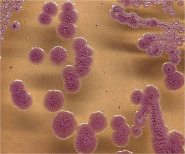
Figure 2: Transfer of effector proteins from bacterium to inside of host cells by type-3 secretion system. VopQ is red, VPA0450 is pink, and VopS is yellow. Adapted from “Vibrio parahaemolyticus cell biology and pathogenicity determinants” by Boberg et al,2011 .

Treatment/Prevention

The bacteria are vulnerable to heat. Cooking the seafood prior to consumption could prevent further infections by the bacteria.In the majority of the cases the symptoms last up to 3 days. It is suggested that infected patients should rest and drink sufficient amount of liquids in order to replace the water lost by diarrhea. Even though V. parahaemolyticus is susceptible to antibiotic treatments, there is a lack of evidence to show that antibiotics can shorten the period of infection. However, in the case of severe infections, which may include high fever, oral antibiotics such as ampicillin or tetracycline should be used. Vaccines have been developed in order to prevent later vibrio infections. These vaccines were tested on mice, where they were found to be 100% effective. They are currently under investigation for use in humans.

References

Broberg, C. A., Calder, T. J., & Orth, K. (November 01, 2011). <b>Vibrio parahaemolyticus</b> cell biology and pathogenicity determinants. Microbes and Infection, 13, 992-1001.

Burdette, D. L., Yarbrough, M. L., Orvedahl, A., Gilpin, C. J., & Orth, K. (January 01, 2008). Vibrio parahaemolyticus orchestrates a multifaceted host cell infection by induction of autophagy, cell rounding, and then cell lysis. Proceedings of the National Academy of Sciences of the United States of America, 105, 34, 12497-502.

Daniels, N. A., MacKinnon, L., Bishop, R., Altekruse, S., Ray, B., Hammond, R. M., Thompson, S., … Slutsker, L. (January 01, 2000). Vibrio parahaemolyticus infections in the United States, 1973-1998. The Journal of Infectious Diseases, 181, 5, 1661-6.

Qadri, F., Alam, M. S., Nishibuchi, M., Rahman, T., Alam, N. H., Chisti, J., Kondo, S., … Nair, G. B. (January 01, 2003). Adaptive and inflammatory immune responses in patients infected with strains of Vibrio parahaemolyticus. The Journal of Infectious Diseases, 187, 7, 1085-96.

Vengadesh eLetchumanan, Vengadesh eLetchumanan, Kok Gan eChan, & Learn-Han eLee. (December 01, 2014). Vibrio parahaemolyticus: A Review on the Pathogenesis, Prevalence and Advance Molecular Identification Techniques. Frontiers in Microbiology, 5.

Vibrio parahaemolyticus infection – including symptoms, treatment and prevention. (n.d.). Retrieved November, 2017, from http://www.sahealth.sa.gov.au/wps/wcm/connect/public content/sa health internet/health topics/health conditions prevention and treatment/infectious diseases/vibrio parahaemolyticus infection/vibrio parahaemolyticus infection – including symptoms treatment and prevention.

Wu, Y., Wen, J., Ma, Y., Ma, X., & Chen, Y. (December 01, 2014). Epidemiology of foodborne disease outbreaks caused by Vibrio parahaemolyticus, China, 2003-2008. Food Control, 46, 1, 197-202.

Zha, Z., Li, C., Li, W., Ye, Z., & Pan, J. (December 06, 2016). LptD is a promising vaccine antigen and potential immunotherapeutic target for protection against Vibrio species infection. Scientific Reports, 6, 1.)


Campylobacter jejuni

by Sereena Moore

Introduction

Campylobacter jejuni is a gram-negative bacteria that causes gastroenteritis in humans. C. jejuni was first isolated in 1972. Since then, it has been known to be one of the main causes of gastroenteritis in the world. It is part of the gut microbiota of many animals including chickens, cattle, goats, dogs, ducks and pigs. However, it causes infection in humans by colonizing the intestinal tract.

Disease

The disease that results from infection by C. jejuni is called Campylobacteriosis. The most common ways that C. jejuni is transmitted is by consuming raw meat, poultry, contaminated water, unpasteurized milk or contacting contaminated animals (Figure 1). Symptoms of the infection are inflammation, watery/bloody diarrhea, fever, weight loss and abdominal cramps.

Figure 1: The most common modes of transmission of Campylobacter jejuni to humans. Source: Sereena Moore.

C. jejuni infection is also linked to other diseases. Some gastrointestinal disorders that may occur after infection are irritable bowel syndrome, pancreatitis, gastrointestinal hemorrhage, functional dyspepsia, and cholecystitis. Furthermore, infection is also connected to diseases in other parts of the body such as Guillain-Barre syndrome, Miller Fisher syndrome, meningitis, bacteremia, sepsis, arthritis, and endocarditis. These happen most often in young children, elderly or those who are immunocompromised. Guillain-Barre syndrome (GBS) is the most common, where 40% of people with GBS were previously infected by C. jejuni. GBS is a neurological condition where antibodies attack the myelin sheath, which is an insulting layer that covers peripheral nerves. Symptoms include limb weakness, problems with respiratory and cranial muscles and even paralysis. GBS and C. jejuni infections are hypothesized to be linked because structures on C. jejuni and proteins from the myelin sheath are structurally similar. Thus, antibodies in the host that were produced against C. jejuni begin to attack the myelin sheath and cause demyelination of the nerve.

Epidemiology:

Campylobacteriosis affects people all over the world. The first infection was officially recorded in the 1980s. Currently, C. jejuni is the cause of gastroenteritis in about 400-500 million people in the world each year. It is the most common disease that causes diarrhea, and studies show that it happens more often than infections from other pathogens, such as Salmonella, Shigella, and E. coli. Infections have recently increased in developed countries in North America, Europe and Australia. About 2.5 million people are infected with C. jejuni each year in the United States. In developing countries, infections from C. jejuni are more common in young children. The infection is also more common to those who are immunodeficient, especially those who have HIV. Outbreaks do not happen often, however, they can occur in places where many people have ingested raw milk or contaminated water.

Virulence:

Currently, the virulence factors used by C. jejuni are not completely understood. However, some mechanisms are known. First, C. jejuni has flagella, which are structures that aid in movement through the gastrointestinal tract, colonization and invasion of the gastrointestinal epithelial layer. The flagella are made up of amino acids that are modified by O-linked glycosylation, which is an addition of a carbohydrate onto the oxygen molecule of an amino acid. This modification is necessary during the assembly of the flagella and has shown to be crucial in the flagella’s functions of movement, colonization and invasion of host cells. Moreover, C. jejuni has a spiral shape that it also uses, along with the flagella, to pass through and invade the mucosa layer of the intestine (Figure 2). Once in the intestine, C. jejuni must bind to the epithelial layer to colonize the hosts intestine, using structures called adhesins. Finally, C. jejuni secretes a toxin called cytolethal distending toxin (CDT). CDT can either stop the hosts cell cycle during replication, or cause programmed cell death. It also helps trigger a response from the host immune system resulting in the release of interleukin-8, which is a cytokine released to induce inflammation and attract immune cells to the area.

Figure 2: Scanning electron micrograph of the spiral shape and flagella of Campylobacter jejuni. It uses its spiral shape, and flagella to pass through the mucosa and epithelial cells of the humans’ intestine, for chemotaxis and adhesion. Source: Esson et al. 2016. Genomic variations leading to alterations in cell morphology of Campylobacter spp. Nature Scientific Reports. 6. https://www.nature.com/articles/srep38303#f1

Treatment:

Normally, treatment for C. jejuni infection is not required, as the infection normally goes away by itself. However, there are a few different treatment options. Fluid replacement may be used because of the large amount of water lost due to the diarrhea. Furthermore, antibiotics may be needed in some cases, such as for those who have severe symptoms, or who are immunocompromised. C. jejuni is known to be resistant to different types of antibiotics including quinolones, cloxacillin, vancomycin and B-lactams. Resistance is hypothesized to be occurring due to the usage of these antibiotics in veterinary medicine, especially in poultry.  However, macrolides, such as erythromycin and azithromycin, have shown to be successful in treating serious cases of Campylobacteriosis. Macrolides interrupt protein synthesis in the bacteria, which causes them to stop growing.

Preventative measures are currently being taken to control the transmission of C. jejuni. These include preventing the spread of C. jejuni between chickens, the use of antimicrobial peptides in chickens to kill bacteria, vaccines and safer food handling practices at the slaughterhouse and processing.

References:

Acheson D, Allos BM. (2001). Campylobacter jejuni Infections: Update on Emerging Issues and Trends. Clinical Infection Diseases. 32(8):1201-1206. DOI: 10.1086/319760.

Altekruse SF, Stern NJ, Fields PI, Swerdlow DL. (1999). Campylobacter jejuni – An Emerging Foodborne Pathogen. Emerging Infecious Diseases. 5(1): 28-35. DOI: 10.3201/eid0501.99010

Dasti JI, Tareen AM, Lugert R, Zautner AE, Gross U. (2010). Campylobacter jejuni: A brief overview on pathogenicity-associated factors and disease-mediating mechanisms. International Journal of Medicl Microbiology. 300(4): 205-211. DOI: 10.1016/j.ijmm.2009.07.002.

Deun KV, Haesebrouck F, Heyndrickx M, Favoreel H, Dewulf J, Ceelen L, Dumez L, Messens W, Leleu S, Immerseel FV, Ducatelle R, Pasmans F. (2007). Virulence properties of Campylobacter jejuni isolates of poultry and human origin. Journal of Medical Microbiology. 56: 1284-1289. DOI: 10.1099/jmm.0.47342-0

Esson D, Mather AE, Scanlan E, Gupta S, de Vries SPW, Bailey D, Harris SR, McKinley TJ, Meric G, Berry SK, Mastroeni P, Sheppard SK, Christie G, Thomson NR, Parkhill J, Maskell DJ, Grant AJ. (2016). Genomic variations leading to alterations in cell morphology of Campylobacter spp. Nature Scientific Reports. 6. DOI: 10.1038/srep38303

Fouts DE, Mongodin EF, Mandrell RE, Miller WG, Rasko DA, Ravel J, Brinkac LM, DeBoy RT, Parker CT, Daugherty SC, Dodson RJ, Durkin AS et al. (2005). Major Structural Differences and Novel Potential Virulence Mechanisms from the Genomes of Multiple Campylobacter Species. PloS Biology. 3(1): e15. DOI: 10.1371/journal.pbio.0030015

Kaakoush NO, Castano-Rodriguez N, Mitchell HM, Man SM. 2015. Global Epidemiology of Campylobacter Infection. Clinical Microbiology Reviews. 28(3): 687-720. DOI: 10.1128%2FCMR.00006-15.

Ternhag A, Asikainen T, Giesecke J, Ekdahl K. 2007. A Meta-Analysis on the Effects of Antibiotic Treatment on Duration of Symptoms Caused by Infection with Campylobacter Species. Clinical Infectious Diseases. 44(5): 696-700. DOI: 10.1086/509924

Young KT, Davis LM, DiRita VJ. 2007. Campylobacter jejuni: molecular biology and pathogenesis. Nature Reviews Microbiology. 5: 665-679. DOI: 10.1038/nrmicro1718.