Category Archives: Uncategorized

Yersinia enterocolitica

by Ruolin Yan

Introduction

Yersinia enterocolitica is a food-borne pathogen which  causes a disease called yersiniosis in animals and humans, infants and young children are particularly affected. To be more specific, the most frequent form of yersiniosis is gastroenteritis or inflammation of the gastrointestinal tract. It is widespread in nature, and is present in aquatic systems, soil and gastrointestinal tracts of a variety of higher vertebrates, particularly pigs.

Disease

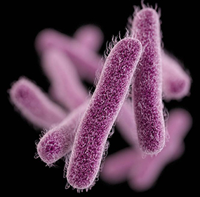
After ingestion of contaminated food, most often pork,  Y. enterocolitica first travels to the small intestine using a structure called flagella (Figure 1) and invades the intestinal epithelial cells using M cells as an entry point. M cells are special epithelial cells located in a region called Peyer’s patches that sample the gastrointestinal tract for potential disease-causing bacteria or pathogens. After invasion, the bacterium replicates and reproduces inside Peyer’s patches, which are lymphatic tissue found in the small intestine. The bacteria can also spread from there to mesenteric lymph nodes, which is also part of the lymphatic system. This causes mesenteric lymphadenitis, which is characterized by inflammation of the lymph nodes. Overall, Y. enterocolitica causes a range of disorders ranging from mild gastroenteritis to more severe lymph node inflammation. Symptoms include fever, diarrhea, vomiting and abdominal pain.

Figure 1: Y. enterocolitica travelling to the small intestine using flagella (a structure that allows the bacteria to move). Source: Ruolin Yan.

Epidemiology

Yersiniosis can be transmitted from animals to humans, this is called zoonosis, which is animal infectious disease that can be transmitted to humans. Pork and pork products are an important reservoir for transmission. However, dog, cat, sheep and wild rodent are also possible reservoirs of pathogenic Y. enterocolitica. The most common transmission route is speculated to be pork products contaminated with pig feces.

Most cases of yersiniosis are sporadic, that is, they do not share a common source.  Y. enterocolitica is a major cause of gastrointestinal disease in many developed countries. In United States, incidence of Yersiniosis appears to be lower compared with many European countries. However, in many developing countries such as Bangladesh, Iraq, Iran and Nigeria, gastrointestinal disease including Yersiniosis is also observed to be highly prevalent, which is related to food safety problem.

Virulence factors

After ingestion of contaminated food, Y. enterocolitica is challenged by high acidity of the stomach, this problem is resolved by an enzyme produced by the bacteria which raises the pH. Then, the bacterium travels to the small intestine, attaches to the epithelial layer, and invades the intestinal barrier via M cells. Adherence and invasion is mediated by two genes, inv (invasion) and ail (attachment).

Once established inside Peyer’s patches, bacteria are normally killed by neutrophils there, which are cells that are specialized in digesting and clearing bacteria. This is accomplished by a process called phagocytosis. During phagocytosis, neutrophils engulf bacteria. At this point, bacteria are inside a membrane compartment called phagosome. Subsequently, they will be killed and digested by enzymes, etc.  However, in the case of Y. enterocolitica, they resist phagocytosis. This is due to several proteins that are exported by them and injected directly into neutrophil, such as Yops and LcrV proteins. This is achieved by type 3 secretion system, which is a secretion system that forms a channel spanning from the interior of the bacteria to interior of the neutrophil (Figure 2).

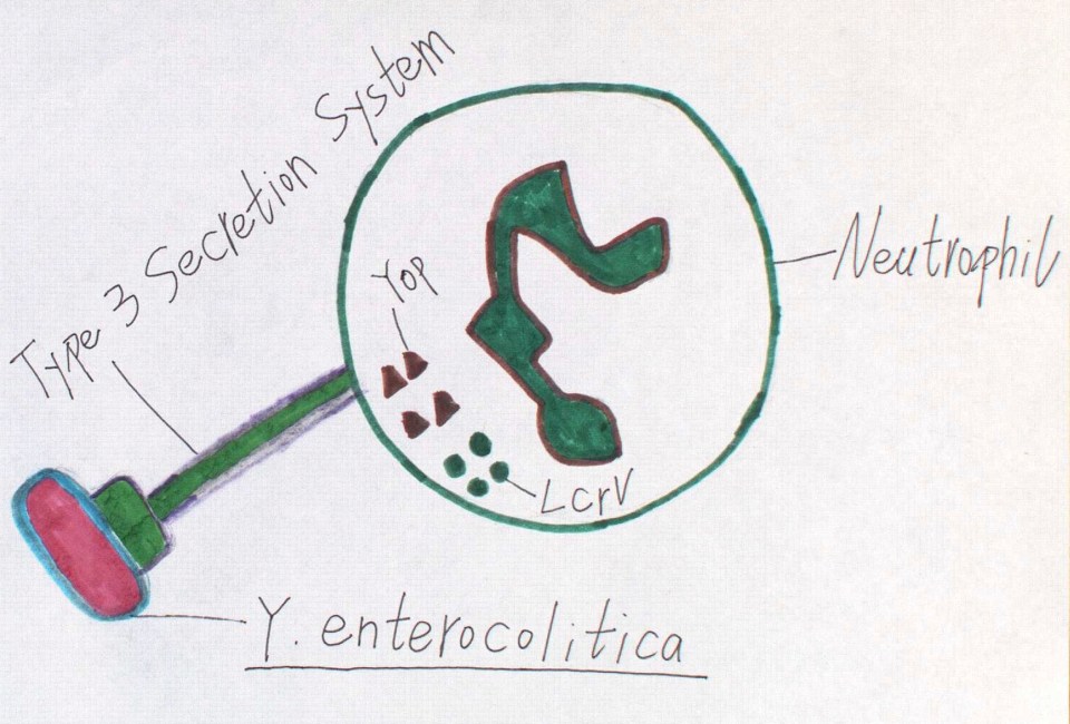
Figure 2: Y. enterocolitica resisting phagocytosis by neutrophil through injection of Yop and LcrV proteins via a type 3 secretion system. Source: Ruolin Yan

Even though Y. enterocolitica multiplication usually happens outside the cell, evidence  suggests that it can survive and multiply in macrophages (another type of cell capable of performing phagocytosis) found in Peyer’s patches, which is likely to be the most important at early stage of infection.  Normally, once inside the phagosome through phagocytosis, pumping of toxic compounds and release of free radicals kills the bacteria. This requires decrease in pH inside the phagosome  in order to activate the proteins. Nevertheless, in the case of Y. enterocolitica, decrease in pH is inhibited, probably through lowering the activity of V-ATPase, an enzyme involved in pumping protons in and decreasing the pH.

Treatment

Yersiniosis is usually diagnosed by detecting bacteria in  stools. Generally, most intestinal illnesses do not require treatment, because patients generally recover without treatment. However, in cases of severe or complicated infections, use of antibiotics should be considered. There is a range of antibiotics to which Y. enterocolitica is sensitive to, one of them is tetracycline. Tetracycline is bacterial protein synthesis inhibitor, which acts by binding to bacterial ribosome, which is a structure necessary for protein synthesis. As a result, Y. enterocolitica are not able to synthesize proteins, including those that are necessary to fight phagocytosis. To protect yourself from Yersiniosis, it is very important not to eat uncooked or undercooked pork. It is also important to avoid consuming unpasteurized milk and milk products.

References

Białas, N., Kasperkiewicz, K., Radziejewska-Lebrecht, J., & Skurnik, M. (2012). Bacterial cell surface structures in Yersinia enterocolitica. Archivum Immunologiae et Therapiae Experimentalis, 60(3), 199-209.

Chopra, I., & Roberts, M. (2001). Tetracycline antibiotics: mode of action, applications, molecular biology, and epidemiology of bacterial resistance. Microbiology and molecular biology reviews, 65(2), 232-260. doi: 10.1128/MMBR.65.2.232-260.2001

Center for Disease Control and Prevention (CDC). (2016). Yersinia enterocolitica (Yersiniosis).  Retrieved from https://www.cdc.gov/yersinia/faq.html‫

Dhar, M. S., & Virdi, J. S. (2014). Strategies used by Yersinia enterocolitica to evade killing by the host: thinking beyond Yops. Microbes and infection, 16(2), 87-95. https://doi.org/10.1016/j.micinf.2013.11.002

Falcao, J. P., Brocchi, M., Proença‐Módena, J. L., Acrani, G. O., Correa, E. F., & Falcao, D. P. (2004). Virulence characteristics and epidemiology of Yersinia enterocolitica and Yersiniae other than Y. pseudotuberculosis and Y. pestis isolated from water and sewage. Journal of applied microbiology, 96(6), 1230-1236. doi: 10.1111/j.1365-2672.2004.02268.x

Fredriksson-Ahomaa, M., Stolle, A., & Korkeala, H. (2006). Molecular epidemiology of Yersinia enterocolitica infections. FEMS Immunology & Medical Microbiology, 47(3), 315-329. https://doi.org/10.1111/j.1574-695X.2006.00095.x

Gebert, A., Fassbender, S., Werner, K., & Weissferdt, A. (1999). The development of M cells in Peyer’s patches is restricted to specialized dome-associated crypts. The American Journal of Pathology, 154(5), 1573-1582.

Rahman, A., Bonny, T. S., Stonsaovapak, S., & Ananchaipattana, C. (2011). Yersinia enterocolitica: Epidemiological studies and outbreaks. Journal of pathogens, 2011. http://dx.doi.org/10.4061/2011/239391


Watanabe, K., Watanabe, N., Jin, M., Matsuhashi, T., Koizumi, S., Onochi, K., … Mashima, H. (2014). Mesenteric lymph node abscess due to Yersinia enterocolitica: case report and review of the literature. Clinical journal of gastroenterology, 7(1), 41-47.

Pujol, C., & Bliska, J. B. (2005). Turning Yersinia pathogenesis outside in: subversion of macrophage function by intracellular yersiniae. Clinical Immunology, 114(3), 216-226. https://doi.org/10.1016/j.clim.2004.07.013


The 2017 Campylobacter Outbreak Across the United States

by Rebecca Novac and Corina Orsini

Introduction

In September of 2017, federal health officials in the US began investigating a multistate outbreak of Campylobacter infections. After sequencing the genome of the bacteria found in several infected individuals and their animals, it was determined that the same antibiotic resistant Campylobacter was present in all of them. While there were no recorded deaths associated with this outbreak, there were 67 cases across 15 states (Figure 1), leading to 17 hospitalizations. This is a recent outbreak and therefore not much is known about the disease causing species. However, we propose that Campylobacter jejuni is the most probable cause of this outbreak.

Figure 1: Number of people infected with campylobacteriosis in each state of the United States during the 2017 Campylobacter outbreak. Source: Center for Disease Control and Prevention, Case Count Maps (2017).

Description of the disease

Campylobacteriosis is a zoonotic disease most often caused by Campylobacter jejuni (Figure 2). It is transmitted through the fecal-oral route and even a very low dose can lead to bloody diarrhea, abdominal pain, fever, nausea and vomiting. C. jejuni is able to easily cause disease due to its ability to move away from the host immune system and towards nutrients with its polar flagella. Companion animals often get the disease from eating raw or undercooked meat. They then continue to shed the bacteria in their feces for 2-7 weeks, whether they show symptoms of the intestinal disease or not, which is how they pass it on to human hosts. Campylobacter species are very resistant to antibiotics and animals can become persistent shedders despite antibiotic therapy. While most people can recover without specific treatment other than heavy fluid consumption while the diarrhea lasts, severe cases of the disease demand treatment with a broad spectrum antibiotic such as erythromycin. This is most common in those with weak immune systems such as the elderly, AIDS patients, chemotherapy patients, etc. The antibiotic resistant properties of this specific strain may complicate treatment and prevent high risk patients from recovering from the illness. While there are no recorded deaths associated with this outbreak as of now, The pathogen has the potential to be deadly in the future if there are no new antibiotics discovered. The class of antibiotics currently being used are commonly found in poultry feed which has been proposed as a main contributor to the development of resistance.

Figure 2: A scanning electron microscopy picture of Campylobacter jejuni (in blue) and its flagella (in brown). Source: University of Leicester.

Source of the outbreak

In this outbreak, 93% of the infected individuals were directly linked to puppies from an Ohio-based Petland store and the others reported contact with puppies from other sources. Therefore, the Campylobacter bacteria was present in one of the puppies sold at the Petland store, which then spread the bacteria to the other puppies. Since, this is a zoonotic disease, the infected puppies transmitted the bacteria to all humans in contact with them, including employees at Petland and people who purchased, or visited a puppy at this pet store.

Cause of the outbreak

This outbreak of Campylobacteriosis occured because one, or more puppies already infected with the bacteria, most likely from eating raw meat and living in a crowded environment, were brought from commercial breeders to the Petland store. Although most infected puppies showed signs of infection, some did not, which increased the risk of transmitting the bacteria to others. Once in the store, the infected puppies continued to shed the bacteria in their feces. All other animals who came in contact with their stool contracted the disease, and they too began to shed the bacteria in their feces, continuing the cycle. Meanwhile, the humans in contact with these puppies and their stool contaminated their hands with Campylobacter. If these individuals did not properly wash their hands and proceeded to touch their face, or the food that they were eating, then the bacteria was transferred to the humans (Figure 3). Once in the human, the bacteria is also shed in their stool; however, further transmission from human to human is very unlikely because we do not usually interact with human feces.

Figure 3: The transmission of the Campylobacter bacteria from canines to humans. Source: Rachel Novac.

How the outbreak was ended

As soon as the employees at Petland were aware of the Campylobacter outbreak, they began setting up sanitary stations throughout the store and stuck to a strict sanitation protocol made by veterinarians. In addition, the infected animals were isolated from those who were not yet showing any signs of the disease until the infection cleared. Moreover, public health recommendations were made to pet owners, which include being aware of the risk factors associated with feeding pets raw food, prompt cleaning and sanitation after the animal has defecated and washing your hands well after handling puppies.

Aftermath

The CDC did not recommend any changes to Petland processes after reviewing kennel documentation, but the company is still not getting off the hook. Petland is now facing a class-action lawsuit from the Animal Legal Defense Fund who is accusing the business of guaranteeing the health of puppies that it knew were prone to illness and defects. Many cities have put laws in place to ban the selling of puppy mill puppies and Petland is the only major pet shop chain left that is selling dogs from commercial breeding operations. This outbreak will likely put pressure on the company to make the transition to selling puppies from a more reliable source.

References

Campanolo ER, Pilipp LM, Anshaw NL. 2016. Pet- associated Campylobacteriosis: A persisting public health concern, Zoonoses Public Health. [accessed 2017 Nov 11]; 0(0): 1-8. http://onlinelibrary.wiley.com.proxy3.library.mcgill.ca/doi/10.1111/zph.12389/epdf. doi: 10.1111/zpj.12389

Centers for Disease Control and Prevention. 2017. Multistate Outbreak of Multidrug-Resistant Campylobacter Infections Linked to Contact with Pet Store Puppies. Atlanta (GA): Centers for Disease Control and Prevention; [updated 2017 Oct 30; accessed 2017 Nov 11]. https://www.cdc.gov/campylobacter/outbreaks/puppies-9-17/index.html

Merck Veterinary Manual. 2016. Overview of Enteric Campylobacteriosis. Kenilworth (NJ): Merck Sharp & Co; [accessed 2017 Nov 11]. http://www.merckvetmanual.com/digestive-system/enteric-campylobacteriosis/overview-of-enteric-campylobacteriosis

Scutti S and LaMotte S.  2017. Pet store puppies spread antibiotic-resistant infection, CDC says. CNN. [updated 2017 Oct 4; accessed 2017 Nov 11]. http://www.cnn.com/2017/09/11/health/puppies-campylobacter-outbreak-cdc/index.html

The Washington Post. 2017. Pet-store puppies linked to bacterial outbreak among people in 7 states, CDC says. Washington (DC): The Washington Post; [updated 2017 Sept 11; accessed 2017 Nov 11]. https://www.washingtonpost.com/news/animalia/wp/2017/09/11/pet-store-puppies-linked-to-bacterial-outbreak-among-people-in-7-states-cdc-says/?utm_term=.31fbdfed35c6

University of Leicester. Campylobacter jejuni. England; [accessed 2017 Dec 14]. https://www2.le.ac.uk/projects/vgec/schoolsandcolleges/Microbial%20Sciences/bacteria-passport/campylobacter-jejuni-1


Coxiella burnetii

by Chantal Coutu and Jason Niness

Introduction

Coxiella burnetii (figure 1), a gram negative intracellular pathogen, is known for causing Q Fever. This zoonotic pathogen was first studied in the late 1930’s after an outbreak of Q Fever affected slaughterhouse workers in Brisbane, Australia; while almost simultaneously being studied in Nine Mile, Montana as an infectious agent in ticks. Typically this pathogen produces symptoms that resemble those of the flu, however an infection by C. burnetii may also lead to pneumonia or hepatitis.

Figure 1: Coxiella burnetii. Source: National Institutes of Health, United States Department of Health and Human Services.

Disease

C. burnetii is spread via the urine, feces, birth products (placenta and amniotic fluid) and the milk of infected animals. It can be transmitted to humans if they come into contact with or inhale contaminated dust from infected animals, such as goats, sheep and cattle. It can also be transmitted from a tick bite or by ingesting unpasteurized milk and dairy products (Figure 2). An infection from C. burnetii can be acute or chronic with symptoms  similar to those of the flu; for example fever, chills, fatigue, stomach pain and muscle pain. However, about half of the people that are exposed to the bacteria do not get sick. Those that develop a severe case typically get inflammation in their lungs (pneumonia) or their liver (hepatitis). C. burnetii may also cause complications during pregnancy such as miscarriage, stillbirth, pre-term delivery or low infant birth weight.  Chronic Q Fever, a more serious infection, can be fatal if not treated with antibiotics. This usually develops in people with pre-existing conditions, such as heart valve disease, blood vessel abnormalities like aneurysms (which are blood filled bulges in the wall of a blood vessel), or if they are immunocompromised. Chronic Q fever has also been linked to endocarditis,  an infection in one or more heart valves, which can be fatal if not diagnosed early.

Figure 2: Possible transmission paths and potential hosts of C. burnetii. Source: Chantal Coutu.

Epidemiology

C. burnetii has shown itself to be more common than previously thought due to its interactions with both domesticated animals and humans. In recent studies, from the United States, C. burnetii was found in 28% of samples taken from various locations including farms and grocery stores. Also, the seroprevalency of Q Fever was found to be 3.1% in U.S. adults, meaning that antibodies against the bacteria were found in their bodies. These two studies indicate that C. burnetii is affecting a larger population of humans than has been reported.

The ability of C. burnetii to infect the host in the form of an aerosol is likely the reason it is so infectious. When it is in the form of a spore, used for survival outside of the host, it is easily picked up by dust particles and carried into the throat and lungs of the host. This mode of infection was recently documented as the likely cause of a 2005 outbreak at an Israeli school, implicating the air conditioning system as the mechanism of transmission. Once within the host, C. burnetii changes to its active form and can invade the host’s tissues.

Virulence systems

Infection by inhalation of C. burnetii targets the alveolar macrophages in the lungs. Infection via the bloodstream or the digestive tract targets the Kupffer cells of the liver. C burnetii enters the cells of the host using a specific receptor called an integrin; either LRI (leukocyte response integrin avβ3) or IAP (integrin-associated protein). After the bacteria has entered the host, a macrophage engulfs the bacteria by a process called phagocytosis. This process forms a sack-like structure around the bacteria  called a phagosome. The phagosomes proceed to fuse with lysosomes, organelles that have a variety of enzymes to help breakdown and digest bacterial invaders, to form phagolysosomes (Figure 3).

Figure 3: C. burnetii infection into a host cell and release out of a cell. Source: Chantal Coutu

Usually, the phagolysosome creates harsh conditions that prevent bacteria from growing and leads to the bacteria’s death. However, C. burnetii is an acidophilic bacterium, meaning its growth is enhanced in acidic conditions. This characteristic is what allows it to overcome the acidic conditions created by the phagolysosome and persist in the host.

Furthermore, the bacteria can create spores which are highly resistant dormant forms of the bacteria to preserve itself and its DNA under extreme conditions or when there is a lack of nutrients. These spores can be released by the host either by cell lysis, the breakdown of the cell, or exocytosis, the release of internal contents enclosed in a cell membrane (Figure 3). These spores can survive in the environment until conditions are adequate to allow for growth again and can be carried by the wind to spread the infection over long distances.

Treatment

Different antibiotics are prescribed depending on the severity of the infection. Patients with acute Q fever can recover without treatment or with an antibiotic such as doxycycline. Patients with chronic Q fever typically require several months of treatment with a combination of antibiotics like doxycycline and hydroxychloroquine or chloroquine or amantadine. Doxycycline and hydroxychloroquine are especially effective as the hydroxychloroquine causes alkalization of the phagolysosome, allowing doxycycline a greater advantage to destroy the bacteria. For patients with medicinal allergies or pregnant women, alternative antibiotics such as moxifloxacin or rifampin may be prescribed. These antibiotics are effective against C. burnetii since, when combined, they reduce the amount of infected cells in the body by sterilizing the bacteria, preventing it from multiplying, and thereby destroying the bacterial cells.

Also, a Q Fever vaccine known as Q-Vax is available in Australia. However, there is limited availability and it causes adverse reactions in those previously infected with C. burnetii. A skin test is required prior to vaccination to test if the patient should receive it or not.

References

Centers for Disease Control and Prevention. (2016). Q Fever. Atlanta, Georgia. https://www.cdc.gov/qfever/index.html

Cooper GM. The Cell: A Molecular Approach. 2nd edition. Sunderland (MA): Sinauer Associates; 2000. Lysosomes. https://www.ncbi.nlm.nih.gov/books/NBK9953/

Maurin, M. and Raoult, D. “Q Fever.” Clinical Microbiology Reviews 12.4 (1999).. https://www.ncbi.nlm.nih.gov/pmc/articles/PMC88923/

“NIAID BSL-3 Priority Pathogens.” Tufts New England Regional Biosafety Laboratory, vetsites.tufts.edu/ne-rbl/resources/niaid-bsl-3-priority-pathogens/.

Tjelle, T. E., Løvdal, T. and Berg, T. (2000), Phagosome dynamics and function. Bioessays, 22: 255–263. doi:10.1002/(SICI)1521-1878(200003)22:3<255::AID-BIES7>3.0.CO;2-R

Toman, Rudolf. Coxiella Burnetii: Recent Advances and New Perspectives in Research of the Q Fever Bacterium. Dordrecht: Springer, 2012. Internet resource.

Corynebacterium pseudotuberculosis

By: Amanda Smith-Stasinopoulos and Nicole Burcar

Introduction

Corynebacterium pseudotuberculosis is a gram positive bacterial pathogen that primarily causes caseous lymphadenitis (CLA) in goats and other small ruminants. CLA produces large glandular swelling, “cheesy glands” (Figure 1); that form externally on the skin around lymph nodes or internally within organs. C. pseudotuberculosis lives inside host cells but can survive in harsh environments for extended periods of time despite not being able to form highly resistant spores or move around. C. pseudotuberculosis was first fully described in 1894 by Preisz for its resemblance to Diphtheria bacillus.

Figure 1: Sheep with cheesy glands caused by C. paratuberculosis (Source: Wikimedia commons)

Disease

In goats and sheep, C. pseudotuberculosis infects superficial and visceral lymph nodes forming an external or internal infection. Lymph nodes are the sites of immune cell activation and act as filters that detect foreign particles. Bacterial pathogens that target the lymph nodes aim to decrease the hosts’ defense against disease establishment. Affected superficial lymph nodes undergo necrosis, uncontrolled cell death, and form abscesses visible on the skin (Figure 1). The pus-like discharge from the abscesses is caused when the bacteria destroys the lymph node tissue, resulting in inflammation. Inflammation attracts neutrophils (a specific immune cell able to engulf foreign particles characteristically found in these types of infections) and carries the bacteria to other locations. The animal can then transmit the disease. Visceral lymph nodes around the lungs, liver and spleen are also commonly affected.

Animals can obtain bacterial particles from a soil environment through lesions in their skin. There have been cases where infected animals cough on superficial wounds of herd mates and spread the disease by releasing bacterial particles. These superficial wounds can be created by activities such as tagging, shearing or castrating, among other activities. Mechanical transmission through vectors such as flies have been identified in cattle and buffalo.

It may take a number of weeks after infection for the disease to start producing symptoms in the animal, known as an incubation period. Affected animals become less productive, in goats this may result in lower milk production and in sheep lower quality wool. The disease has a life-long prevalence and animals infected may show signs like weight loss and abortion, along with recurring abscesses.

C. pseudotuberculosis can cause disease in a number of other species; in horses it causes ulcerative lymphangitis forming severe swelling in the limbs often causing lameness. Pigeon fever is classified by the presence of external abscesses around the chest and can be found in cattle, camels, pigs, buffaloes and humans. It is not common for humans to be infected, normally those that show symptoms of the disease have been exposed to infected animals or have consumed raw goat milk or meat.  Like goats, infected humans have abscesses in the liver and internal lymph nodes.

Epidemiology

C. pseudotuberculosis is found worldwide with varying prevalence in different countries. In Brazil up to 75 % of sheep can be affected, other countries have a lower seroprevalence (number of animals with antibodies against the pathogen in their blood) of the disease making the strains less likely to be spread. In Australia 26% of sheep are affected, and in Canada 21% of sheep brought to slaughterhouses had the disease.

Endemic areas where the disease affects horses in the United States are California and Texas. They have a usual annual disease prevalence of 5-10% but have seen times of large incidence increases and sporadic outbreaks. In 2002 and 2003, thousands of horses were reported as being infected with disease caused by C. pseudotuberculosis in Wyoming, Utah, Kentucky and Colorado, where disease prevalence is historically low. Further, small outbreaks have been reported in areas such as Alberta, Canada in 2013 and British Columbia in 2010 with 350 symptomatic cases.

A long-term study (1989-2001) of dairy cattle in Israel identified 3 clinical forms of C. pseudotuberculosis in 827 animals, the more common form produced ulcer abscesses on the skin and resulted in a 16.3% culling rate (animals removed from the herd) of the infected animals. The other forms of the disease caused mastitis in cattle and infected the visceral lymph nodes.

Virulence factors

The major factor that contributes to the pathogen’s ability to cause damage and disease is an enzyme called phospholipase D. This enzyme is a toxin that is secreted outside the bacterial cell, which classifies it as an exotoxin. Phospholipase D breaks bonds in the membranes of host cells allowing the pathogen to spread to secondary locations. Phagocytes (like neutrophils or macrophages: cells involved in the immune response that engulf foreign particles) engulf these exotoxins and taxi them to regional lymph nodes, where they produce the characteristic lesion of the disease (Figure 2). Phospholipase D is not degraded during transport because it disrupts the phospholipids that make up the compartment inside the macrophage containing the toxin. At one point it will kill the cell since the cell membrane of the macrophage is also made up of these phospholipids.

Figure 2: Corynebacterium pseudotuberculosis pathogenesis. Source: Amanda Smith-Stasinopoulos.

The surface lipids have cytotoxic effects, meaning they can kill host cells, which also adds to the virulence of C. pseudotuberculosis by preventing the macrophages from killing the pathogen and contribute to the formation of the necrotic lesions.

Treatment

The bacteria has different strains that are susceptible to different antibiotics but some antibiotics are able to target all strains. The problem with targeting the disease once it has established in the host, is that the bacteria are encapsulated in the lymph nodes making it difficult to avoid causing harm to the host. Antibiotics are therefore not a viable treatment.

In humans infected lymph nodes are surgically removed. Unfortunately, this is not an economically viable option for goat farmers and infected animals are removed from the herd.

The best option for producers is to prevent the disease by vaccination. A common vaccine uses an inactivated version of phospholipase D. With a proper vaccine protocol the number of abscesses and spread is decreased, however the proper protocol is not followed by many farmers. Many vaccines produced for sheep cannot be used to prevent C. pseudotuberculosis disease in goats. Goats need more frequent booster shots than sheep; every 6 months compared to yearly, after a set of 2 initial doses. The vaccine is more effective when secreted bacterial components are injected with an adjuvant that stimulates an immune response. Some vaccines are made with the whole bacteria but these only show 33% effectiveness and a weak immune response.

References

Baird GJ, Fontaine MC: Corynebacterium pseudotuberculosis and its Role in Ovine Caseous Lymphadenitis. Journal of Comparative Pathology 2007, 137(4):179-210.

Dorella FA, Pacheco LGC, Oliveira SC, Miyoshi A, Azevedo V: Corynebacterium pseudotuberculosis: microbiology, biochemical properties, pathogenesis and molecular studies of virulence. Vet Res 2006, 37(2):201-218.

Guimarães AdS, Dorneles EMS, Andrade GI, Lage AP, Miyoshi A, Azevedo V, Gouveia AMG, Heinemann MB: Molecular characterization of Corynebacterium pseudotuberculosis isolates using ERIC-PCR. Veterinary Microbiology 2011, 153(3):299-306.

Moura-Costa LF, Bahia RC, Carminati R, Vale VLC, Paule BJA, Portela RW, Freire SM, Nascimento I, Schaer R, Barreto LMS et al: Evaluation of the humoral and cellular immune response to different antigens of Corynebacterium pseudotuberculosis in Canindé goats and their potential protection against caseous lymphadenitis. Veterinary Immunology and Immunopathology 2008, 126(1):131-141.

Moussa IM, Ali MS, Hessain AM, Kabli SA, Hemeg HA, Selim SA: Vaccination against Corynebacterium pseudotuberculosis infections controlling caseous lymphadenitis (CLA) and oedematousskin disease. Saudi Journal of Biological Sciences 2016, 23(6):718-723.

Paton, M., Sutherland, S., Rose, I., Hart, R., Mercy, A. and Ellis, T. The spread of Corynebacterium pseudotuberculosis infection to unvaccinated and vaccinated sheep. Australian Veterinary Journal 1995, 72: 266-269. doi:10.1111/j.1751-0813.1995.tb03542.x

Yeruham, I., Elad, D., Friedman, S. and Perl, S. Corynebacterium pseudotuberculosis infection in Israeli dairy cattle. Epidemiology of Infection 2003, 131: 947–955. doi: 10.1017/S095026880300894X

Da Conceição Aquino De Sá, M., Gouveia, GV., Da Costa Krewer, C., Aparecida Veschi, JL., De Mattos-Guaraldi, AL. and Da Mateus Matiuzzi, C. Distribution of PLD and FagA, B, C and D genes in Corynebacterium pseudotuberculosis isolates from sheep and goats with caseus lymphadenitis. Genetics and Molecular Biology 2013, 36(2): 265-268. https://dx.doi.org/10.1590/S1415-47572013005000013

Foley, J.E., Spier, S.J., Mihalyi, J., Drazenovich, N. and Leutenegger, C.M. Molecular epidemiologic features of Corynebacterium pseudotuberculosis isolated from horses. American Journal of Veterinary Research 2004, 65(12): 1734-1737.

Rickettsia rickettsii

by Romeo Proietti

Introduction

Rickettsia rickettsii is the causative agent of Rocky Mountain spotted fever (RMSF). This bacterium is a gram-negative, intracellular bacterium most frequently found in hard-bodied ticks in the environment. If bitten by an infected tick, R. rickettsii can then infect the endothelial cells of mammals, including humans. It is the most lethal of all bacteria in the spotted fever group “rickettsiae.”

Disease

RMSF is called this because it was first identified in the Rocky Mountain region and was found to produce red spots that can sometimes cover the whole body including the palms of hands and soles of feet.  It is caused by R. rickettsii, transmitted to humans mainly through a tick as the vector. Ticks become infected by feeding off the blood of infected animals. The most common types of tick species that the bacteria are found in are the American dog tick, Rocky Mountain wood tick and the brown dog tick. Once a human, or mammal, is bitten by an infected tick, the tick needs to be attached to the host for 6-10 hours for R. rickettsii to be released from the tick’s salivary glands. Once transmitted into the host, it begins to live and multiply in endothelial cells of the body (Figure 1). Endothelial cells line the interior surface of blood vessels and lymphatic vessels.  R. rickettsii can rapidly enter the cells and reduce the immune response of the host so that it can continue to cause infection without being eaten up by the host defensive cells, like macrophages, as quickly compared to having a full immune response. This process of immune system evasion increases its survival in the host and prolongs the time it takes for the digestive cells to take over and eat up the bacteria. The infection of these cells increases vascular permeability of the blood vessels, which can lead to several symptoms such as fever, muscle pain, nausea, vomiting and skin rashes amongst many others.

Figure 1: Microscopic image of Rickettsia rickettsii (red) found in blood vessel endothelial cells (blue/purple). Source: US Department of Health and Human Services/Centers for Disease Control and Prevention (2016).

Epidemiology

Beginning in 2010, RMSF has been placed under the category “Spotted Fever Rickettsiosis” (SFR). The rate of incidence of SFR has been increasing and was over 11 cases per million people in 2014 compared to 2 cases per million people in 2000. Death rate has interestingly decreased to less than 0.5%. Since multiple other species of Rickettsia are part of the SFR group, it is unclear as to how many fatalities are caused by RMSF and R. rickettsii. Since RMSF is first caused by the infection introduced from ticks, anyone can be infected by R. rickettsii.

Throughout the United States, the number of cases begin to spike in the summer months, which coincide with when ticks are most active. In Arizona, R. rickettsii are mainly transmitted by the brown dog tick, infecting domestic and stray dogs which can then be passed on to people. More frequent cases of RMSF are found in men compared to women, as well as in native Americans compared to other groups. This can be attributed to their increased exposure to the ticks in the environment compared to other groups. Those with an increased risk of death by RMSF are children under the age of 10, native Americans, immunocompromised people and individuals who do not receive treatment within the first 5 days of symptoms.

Virulence Factors

During bacterial endothelial infection, R. rickettsii first gets into endothelial cells using surface adhesion proteins, OmpA and OmpB, which help them quickly attach to and pass into endothelial cells, a process called endocytosis (Figure 2a). These surface proteins on the bacteria attract digestive cells called macrophages, which are told to eat things that are foreign to the host. Once engulfed by the macrophage (Figure 2b), the bacteria use a secretion system called T4ASS to inject toxic chemicals into the macrophage to kill it and escape being eaten (Figure 2c). Once the bacteria exit the macrophage and are free in the cytosol of the epithelial cell, they gain nutrients from the host, multiply and steal a type of host protein called actin and use it to their advantage. Actin is a commonly found protein in mammalian host cells that can be combined end to end, starting on one end of the bacteria, with other actin proteins to form a long chain (Figure 2d). This chain of actin acts as a tail and helps the bacteria move within and between endothelial cells. Moving between cells helps them obtain as many nutrients from the host as possible to replicate and to further infect neighboring cells (Figure 2e). Their movement also helps them run away from immune cells, like other macrophages, and antibodies that are looking to kill them.

Figure 2: Host cell interactions with R. rickettsii. a) Endocytosis of Rickettsia rickettsii in an endothelial cell, b) Phagocytosis: eaten up by macrophage, c) Escape and killing of macrophage using T4ASS, d) Production of actin chain/tail on bacteria, e) Movement of bacteria within and between endothelial cells and infection of another epithelial cell. Source: Drawn by Romeo Proietti.

Once the bacteria are ready to leave their host endothelial cell, they release enzymes and proteases that degrade the cell’s membranes and cause large amounts of inflammation and damage to host tissues that can become spread throughout the body.

Prevention and Treatment

The best way to prevent infection of R. rickettsii would be to prevent tick bites. Ticks can be found in wooded areas such as forests, campgrounds and even in some people’s backyards. Wearing clothing that protects skin from encountering brush or grass can help reduce tick bites. Also, spraying clothes with bug repellent can help further reduce the chances of getting bitten by a tick.

If one does get bitten and catch RMSF, doxycycline is the drug used to treat it. Doxycycline acts by preventing the bacteria from reproducing by inhibiting protein synthesis. Without replication, the bacteria cannot continue to infect the host and the immune system will then take over and clear the infection. Treatment of doxycycline for 7-10 should clear the infection and most patients will fully recover.

References

  1. Perlman, S. J., Hunter, M. S., & Zchori-Fein, E. (2006). The emerging diversity of RickettsiaProceedings of the Royal Society B: Biological Sciences273(1598), 2097–2106. http://doi.org/10.1098/rspb.2006.3541
  2. Rocky Mountain Spotted Fever (RMSF). (2017, June 26). Retrieved November 20, 2017, from https://www.cdc.gov/rmsf/index.html
  3. Parola, P., Paddock, C. D., & Raoult, D. (2005). Tick-Borne Rickettsioses around the World: Emerging Diseases Challenging Old Concepts. Clinical Microbiology Reviews18(4), 719–756. http://doi.org/10.1128/CMR.18.4.719-756.2005
  4. Snowden J, Simonsen KA. Tick, Rickettsia Rickettsiae (Rocky Mountain Spotted Fever). Treasure Island (FL): StatPearls Publishing; 2017 Jun-. Available from: https://www-ncbi-nlm-nih-gov.proxy3.library.mcgill.ca/books/NBK430881/
  5. Cunha, B. A. (2017). Rocky Mountain Spotted Fever (RMSF) (M. S. Bronze, Ed.). Retrieved November 20, 2017, from https://emedicine.medscape.com/article/228042-overview#a5
  6. Voth, D. E., Broederdorf, L. J., & Graham, J. G. (2012). Bacterial Type IV Secretion Systems: Versatile Virulence Machines. Future Microbiology7(2), 241–257. http://doi.org/10.2217/fmb.11.150
  7. Walker, D. H. and Ismail, N. (2008). Emerging and re-emerging rickettsioses: endothelial cell infection and early disease events. Nature Reviews Microbiology, 6, 375-386. doi:10.1038/nrmicro1866

Pseudomonas aeruginosa

By Adam Classen and Jennifer Huxham

Introduction

Pseudomonas aeruginosa is a rod shaped gram-negative bacteria (see figure 1) that is often found in wet areas or bodily fluids. The bacterium was first identified by Carle Gessard in 1882. It is an opportunistic pathogen that is one of the leading causes of infections in hospitals. This bacteria can cause a variety of symptoms but it is most commonly associated with pneumonia in immunocompromised or mechanically ventilated individuals. The microbe’s success can be attributed to its extraordinary adaptability and virulence factors.

Figure 1.  Computer generated image of P. aeruginosa with its pili and flagella. Source: Archer USCfDCaP-MIj. 2013. Multidrug-resistant pseudomona aeruginosa. CDC.

Disease

Depending on the site of infection P. aeruginosa can cause a variety of different types of symptoms. Bloodstream infections have symptoms such as; fever/chills, aches, light-headedness, vomiting. Pneumonia is often caused by the microbe and can cause difficulty breathing and coughing with yellow, green or even bloody mucus. P. aeruginosa can also cause urinary tract infection (UTI), leading to painful urination.

Infection of severe burns can also occur. This is attributed to the fact that severe burns cause immunosuppression, making affected individuals more at risk of developing infections by P. aeruginosa.

Cystic fibrosis (CF) is a genetic condition that causes buildup of mucus in the lungs among other things. These individuals are immunocompromised and are at particular risk of lung infection by P. aeruginosa because of their inability to effectively clear mucus from their lungs. Normally, the mucus in the lungs captures microorganisms and is swept out, thus helping to prevent pathogens from infecting the lungs. However, because CF patients are unable to clear lung mucus, the microbes get trapped and remain in the lungs allowing for infection. This infection causes inflammation which can restrict breathing and result in death.

Epidemiology

P. aeruginosa affects mostly immunocompromised individuals and is therefore often caught in hospitals. In the United States of America, it is the number 1 cause of intensive care unit pneumonia, accounts for 10% of all infections contracted in hospitals, and is the 3rd most common way of contracting a UTI in a hospital. It can be spread by people, food and medical equipment. P. aeruginosa is one of the most common forms of burn wound infection. Burn victims that become infected with P.aeruginosa have a 40-50% mortality rate, making this type of infection very dangerous. CF patients have a 40% chance of being infected with P. aeruginosa. Chronic infection of this type is one of the leading causes of death in individuals affected by CF, carrying a 40-60% mortality rate.

Virulence factors

P. aeruginosa is a successful pathogen because of its adaptability and variety of virulence factors. One of which is its ability to form biofilms, which are colonies of bacteria that are clustered together. Biofilms are important for protection against the host immune system. Specifically, these biofilms protect against antibodies (tag foreign particles for destruction), the complement system (forms pores in their membrane and tags the bacteria for destruction), phagocytes (eliminates foreign particles, broken cellular components or bacteria). They also protect against antibiotics and antimicrobial peptides (AMPs). Where AMPs are compounds produced by the host to kill microbes. Biofilms also cause massive inflammation at the site of infection causing damage to the host. This impressive virulence factor allows the bacteria to survive for a prolonged amount of time in the host.Other virulence factors that contribute to its pathogenicity are pili, which are surface proteins that help them attach to the host. As well as flagella, which acts as a tail to help the bacteria propel itself and move around.

P. aeruginosa can cause damage to the host by secreting toxins. Specifically, it uses an exotoxin, which are proteins or lipids that are secreted into the external environment from the bacterial pathogen. This exotoxin inactivates eukaryotic elongation factor 2, a protein that is crucial for protein synthesis. Inhibition of this protein stops the host eukaryotic cells from synthesizing proteins necessary for functioning, resulting in death. It also produces the enzyme ExoU which is secreted extracellularly where it functions, causing lysis (cutting) of host cell plasma membranes. Allowing it to steal iron from the host’s mitochondria, inflicting damage.

P.  aeruginosa has an outer membrane that has low permeability to antibiotics, thus helping protect it. On top of that it has multidrug efflux pumps (see figure 2). These pumps can take antibiotics that do manage to enter the cell and quickly excrete them back out of the cell into the surrounding environment. Stopping them from affecting the bacterial cell.

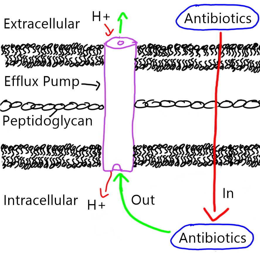
Figure 2.  Efflux pump in the bacterial membrane pumps antibiotics out of the bacteria, passing through the inner, peptidoglycan and outer membranes in an anti-port manner (protons goes in, antibiotics goes out).

Treatment

Wild type P. aeruginosa strains are sensitive to a variety of antibiotics including aminoglycosides and cephalosporins. However, the strains found in hospitals have developed resistance to many types of antibiotics, making them more difficult to treat. In hopes of clearing these infections a combination of 2 drugs is often used. More than one drug is given intravenously to prevent the development of antibiotic resistance and to enhance their effectiveness. However, despite treatment with antibiotics, P. aeruginosa is often able to adapt and survive in the lungs of CF patient for decades. If infection of burn wounds occur surgical removal of infected skin is often needed to help clear infection. As resistance to all commercially available antibiotics is now commonplace with P. aeruginosa.

 References

Bennington-Castro J. Forthcoming 2015. What is pseudomonas aeruginosa? : Everyday Health.

Church D, Elsayed S, Reid O, Winston B, Lindsay R. 2006. Burn wound infections. Clinical microbiology reviews. 19(2):403-434.

Cohen TS, Parker D, Prince A. 2015. Pseudomonas aeruginosa host immune evasion. In: Ramos J-L, Goldberg JB, Filloux A, editors. Pseudomonas: Volume 7: New aspects of pseudomonas biology. Dordrecht: Springer Netherlands. p. 3-23.

EHA consulting group I. Forthcoming 2017. What is pseudomonas aeruginosa? :              Environmental & public health consultants

Estahbanati HK, Kashani PP, Ghanaatpisheh F. 2002. Frequency of pseudomonas aeruginosa serotypes in burn wound infections and their resistance to antibiotics. Burns. 28(4):340-348.

Faucher S. 2017. Mechanisms of Pathogenicity. Mcgill University.

Freidreich M. Pseudomonas aeruginosa infections treatment & management. 2016. MedScape; [accessed 2017]. https://emedicine.medscape.com/article/226748-treatment#d5.

Goldberg JB. 2010. Emergence of pseudomonas aeruginosa in cystic fibrosis lung infections. In: Ramos JL, Filloux A, editors. Pseudomonas: Volume 6: Molecular microbiology, infection and biodiversity. Dordrecht: Springer Netherlands. p. 141-175.

Høiby N, Johansen HK, Moser C, Ciofu O. 2008. Clinical relevance of pseudomonas aeruginosa: A master of adaptation and survival strategies. Pseudomonas. Wiley-VCH Verlag GmbH & Co. KGaA. p. 25-44.

Lister PD, Wolter DJ, Hanson ND. 2009. Antibacterial-resistant pseudomonas aeruginosa: Clinical impact and complex regulation of chromosomally encoded resistance mechanisms. Clinical microbiology reviews. 22(4):582-610.

Pan J, Zha Z, Zhang P, Chen R, Ye C, Ye T. 2017. Serine/threonine protein kinase ppka contributes to the adaptation and virulence in pseudomonas aeruginosa. Microbial Pathogenesis. 113(Supplement C):5-10.

Pirnay J-P, Bilocq F, Pot B, Cornelis P, Zizi M, Van Eldere J, Deschaght P, Vaneechoutte M, Jennes S, Pitt T et al. 2009. Pseudomonas aeruginosa population structure revisited. PLOS ONE. 4(11):e7740.

Saenz-Méndez P, Eriksson M, Eriksson LA. 2017. Ligand selectivity between the adp-ribosylating toxins: An inverse-docking study for multitarget drug discovery. ACS Omega. 2(4):1710-1719.

Shigeki Fujitani, Kathryn Moffett, Victor Yu. 2017. Pseudomonas aeruginosa. Antimicrobe; [accessed]. http://www.antimicrobe.org/b112.asp

2016 Mississippi Federal Prison Outbreak of Botulism

by: Hailey Pelland and Elyse Perrault

Introduction

On June 9, 2016, the Center for Disease Control and Prevention (CDC) was notified of five suspected cases of botulism in inmates at a medium-security federal correctional institution in Yazoo City, Mississippi. A day later, a total of 13 inmates were hospitalized and overall 31 cases were identified, all in men.  Figure 1 shows the reported dates of hooch exposure and symptom onset among the 31 inmates affected.  Generally, it was observed that symptoms appeared 3 days following hooch exposure.  By the end of the outbreak, 24 inmates were hospitalized, including 15 (62.5%) who were admitted to an intensive care unit and nine (37.5%) who required intubation and mechanical ventilation to assist with breathing.  No deaths occurred.

Figure 1: Botulism cases (n = 31) in a federal correctional facility in Mississippi by reported date of hooch exposure and symptom onset between June 1–19, 2016.  Source: McCrickard, L. et al. (2017).

Foodborne Botulism

Foodborne botulism is a paralytic disease caused by ingestion of food contaminated with Clostridium botulinum and the botulinum toxin (BoNT) that this bacterium produces.  As illustrated in Figure 2, one only develops the symptoms of botulism when they ingest the toxin.  On its own, Clostridium botulinum will not cause this disease. The bacterium, which is ubiquitous in the environment, is anaerobic (meaning they grow in the absence of air) and spore-forming.  It is called a spore-former because it develops a “shell” which is capable of protecting the cell under adverse conditions such as extreme heat, dryness and exposure to chemicals. C. botulinum normally grows in soil or in lake sediments and, as mentioned, is itself often harmless to the human body.   However, the botulism disease will occur when its spores have the chance to germinate in foods, leading to bacterial growth and production of the botulinum toxin.  This toxin specifically targets the nervous and muscular systems, causing muscles to go limp all over the body. Symptoms of botulism may include double or blurred vision, drooping eyelids, slurred speech, difficulty swallowing and muscle weakness that radiates down the body.  In more extreme cases, botulism can result in death due to respiratory failure.

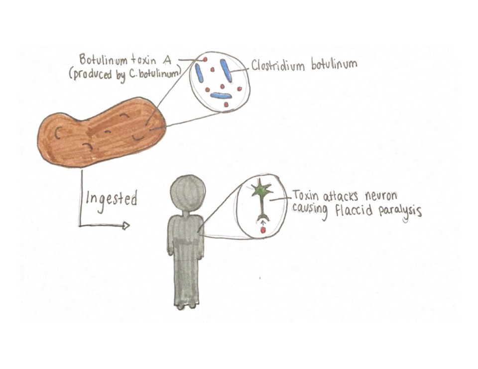
 Figure 2: Illustration of the foodborne botulism pathway showing that BoNT must be produced in food by C. botulinum and, subsequently, ingested by way of the food for symptoms to occur in an individual. Source: Hailey Pelland (2017).

Source of the Outbreak

Staff members of the prison suspected that an alcoholic beverage, illicitly made by inmates and known as “hooch” or “pruno,” was the source of the outbreak.  Among all inmates, 33 reported consuming hooch during June 1–19, 2016 and 31 (94%) had signs or symptoms suggesting botulism.

One patient reported that honey, potatoes, apples and tomato paste from a bulging can were combined, hidden and fermented in a sealed plastic bag (Figure 3) at room temperature for 3–5 days.  Fermentation is the process of converting carbohydrates to alcohol and, in this case, possible sources of Clostridium botulinum included the tomato paste, since a bulging lid is a sign of bacterial contamination, as well as the potatoes or contamination from the environment. Potatoes have been hypothesized as the source of other hooch-associated outbreaks since Cbotulinum lives in the soil and, therefore, its spores can be found on raw potatoes.

Figure 3: A sealed plastic bag containing water, honey, potatoes, apples and tomatoes to be fermented for hooch production.  Source: Elyse Perrault & Hailey Pelland (2017).

Cause of the Outbreak

The exact cause of this outbreak is still uncertain, however the warm anaerobic fermentation process of making hooch creates a predisposition for the production of botulinum toxin if any ingredient happens to be contaminated with C. botulinum or its spores.  This is because the Clostridium bacterium thrive in a warm, wet, low acid, and low oxygen environment, which are favourable conditions for fermentation. It is when C. botulinum is actively growing and replicating that the botulinum toxin is produced.  Therefore, C. botulinum likely produced its toxin in the hooch mixture which was then ingested by the inmates. Botulism is not transferable from person to person, thus, one can only get the disease by actually consuming the toxin themselves.

The fact that prisoners were able to sneak fruit and vegetable scraps from their meals as well as sugar and water in order to produce the hooch is also of significant concern.  The prison in question is a medium security one, where the number of staff is lower than at high-security federal prisons and inmates have more freedom to move between common areas.  Therefore, the inmates here had a greater access to procure the necessary ingredients and hide them in order to make their hooch.

Aftermath

Medical charts of the inmates who were affected in the outbreak were consulted and interviews were conducted to obtain information on hooch exposure, clinical signs and symptoms, medical management and patients’ understanding of botulism.  The responses helped lend insight into possible preventive measures.  In particular, facility staff members must realize the potential for increased hooch consumption during celebratory events since interviews with the inmates revealed that this outbreak coincided with a farewell party for one inmate and the National Basketball Association finals. Furthermore, educating correctional facility staff members and inmates about the risks of consuming hooch and encouraging better communication between staff members and inmates might help decrease this activity from taking place.  Moreover, these measures can help identify and treat persons with botulism quickly and prevent avoidable deaths.


References

Foodborne Illness Outbreak Database. (n.d.). 2016 Outbreak of Botulism Linked to Illicit Alcohol in a Federal Correctional Facility, Mississippi. Retrieved November 15, 2017, from http://outbreakdatabase.com/details/2016-outbreak-of-botulism-linked-to-illicit-alcohol-in-a-federal-correctional-facility-mississippi/?outbreak=botulism&year=2016

McCrickard, L. et al. (2017). Notes from the Field: Botulism Outbreak from Drinking Prison-Made Illicit Alcohol in a Federal Correctional Facility — Mississippi, June 2016. MMWR Morb Mortal Wkly Rep; 65(52): 1491–1492.

Salyers, A.A. & Whitt, D.D. (2002). Bacterial pathogenesis: A molecular approach. Washington, D.C: ASM Press.

Sobel, J., Tucker, N., Sulka, A., McLaughlin, J. & Maslanka, S. (2004). Foodborne Botulism in the United States, 1990–2000. Emerg Infect Dis. 10(9): 1606-1611.

Vugia, D. et al. (2009). Botulism from drinking pruno. Emerg Infect Dis. 15(1): 69-71.


Neorickettsia sennetsu

by Julien Beauchamp

Introduction

Neorickettsia sennetsu, originally classified as Ehrlichia sennetsu, is the etiologic agent of the well-defined human syndrome known as Glandular or Sennetsu Fever. The first recognized infection was diagnosed in Japan in 1954. Cases of Sennetsu Fever seem to be restricted to parts of Western Japan and Southeast Asian countries. N. sennetsu is a member of the family Anaplasmataceae, a group of Gram-negative obligatory intracellular bacteria. This means that they cannot survive outside their eukaryotic host cell. The bacteria invade and multiply in host macrophages and monocytes (specialized white blood cells used to destroying invading microbes).

Disease

N. sennetsu is thought to be transmitted to humans through nematodes encysted in raw or improperly cooked fish. Nematodes are roundworms suited to living inside the digestive tracts of animals. Neorickettsia spp. lack common pili or capsules, used by normal bacteria to bind, and therefore bind to host cells via outer membrane proteins. These are proteins found on the outside surface of cells, used for interacting with the environment. The vector stimulates an inflammatory response when it clings to the intestinal wall. This faciliates the interaction between the bacteria and small veins, arteries, and capillaries. The endothelial cells are an important site of involvement in the disease process. Entry into the host phagocytes is mediated by transglutaminase activity, which is necessary for receptor-mediated endocytosis. Transglutaminase is an enzyme used to bind proteins together. The endocytosis allows the pathogen to successfully establish vacuoles for survival inside the host immune cell. These specialized compartments block the macrophage’s normal activity of fusing it with lysosomes. The appearance of inclusion bodies that appear on the surface of the host cell can lead to the disintegration of the macrophages or monocytes. If the infection progresses, Sennetsu Fever causes enlargement of the liver, spleen and lymph nodes. Other symptoms include fever, chills, headache, sore throat, muscle aches, nausea, vomiting, and insomnia, along with other non-specific flu-like symptoms. Signs of illness begin about 1-2 weeks after consumption.

Figure 1: N. sennetsu presumed life cycle. A, eggs deposited in feces. B, First intermediate host (musculus). C, Cercariae. D/E, Secondary intermediate host. F, Present within parasitic flukes found in the secondary intermediate hosts. H, Ingestion by humans leads to infection of the digestive tract. Source: Julien Beauchamp.

Epidemiology

Sennetsu is only reported in parts of Southeast Asia (Japan, Laos, Thailand, Philippines and Malaysia), where fish is a staple food and eating it raw is common. The disease is described as a type of Human Ehrlichioses. Many forms of veterinary Ehrlichioses have been identified since the early 1900s. In most cases the bacteria were transmitted by ticks, causing disease in horses, sheep, deer, cattle, rodents and dogs. It is known that N. sennetsu shares many antigenic qualities with E. canis, the causative agent of Canine Ehrlichioses. Ehrlichia sennetsu was renamed Neorickettsia sennetsu after being discovered as the first human form of the illness. Sennetsu Fever was first recognized in japan in 1954 on the island of Kyushu and continued to be prevalent in Japan during the rest of the 1950s-60s. Sennetsu Fever was shown to be transmittable to mice and then to humans in Japan. Consumption of grey mullet, a fish indigenous to Japan has been associated with the illness. Also, in Laos, documented cases were linked to eating raw climbing perch.

The disease was described as mononucleosis-like and is often confused for mono. Mononucleosis, or mono, applies to a group of symptoms associated with the Epstein-Barr virus (EBV), which had many similarities with N. sennetsu infection. Fortunately, Glandular Fever is not contagious and will not spread from one person to another. Individuals with reduced immunity, such as those with HIV or who are undergoing cancer treatment may be at a higher risk of contracting Sennetsu Fever.

Virulence factors

Neorickettsia species have complex life cycles involving trematodes and several hosts. Once entering a their human host, the bacteria look to infect the endothelial layers of the GI (gastrointestinal) tract so that they can eventually make their way into the bloodstream and lymphatic system. Despite a small genome of 0.9 Mb, Neorickettsia sennetsu has a wide range outer membrane proteins, dependant on different host cell environments. The internalization, or entering the cell, through specific binding sites is necessary for establishing infection. The binding signals disarm the host cell’s ‘alarm’ against pathogens, allowing entry into the cell. They enter the host phagocyte (macrophage or monocyte) without the help of small filaments used by other species. Therefore, there is no phagocytosis (engulfing by the macrophage); instead they rely on the mentioned transglutaminase activity. This activity allows for receptor-mediated endocytosis. Meaning, the cell can now be inside the cytoplasm of their new home the cells can replicate. The vacuoles they form are called ‘modified parasitophorous endosomes’ because they are negative for lysosomal glycoproteins. These unique containers protect the bacteria from the host cell’s ability to break them down by fusion with lysosomes because of the glycoproteins. The bacteria now looks for a method to acquire nutrients and compete with the host cell. N. sennetsu comes from a group of bacteria with a considerable need of iron. The pathogen helps the host cell regulate the amount of iron-transferrin (Tf) present by synthesizing toxins. Tf regulated the normal amount of iron available. These protein toxins inhibit certain restraining factors normally found in the phagocyte DNA, vastly increasing the amount of available iron. The changes made to the healthy cell’s metabolism and the exit of the bacterial cells through inclusion bodies leads to the death of the host. Both entering the cell and the creation of proteins are important steps in the infection. Additionally, if the white blood cell count decreases the bacteria may also grow in membrane-bound cavities inside bone marrow, lymph nodes, liver, spleen, kidneys, lungs or even cerebrospinal fluid.

Figure 2: Process of a N. sennetsu entering host cell. A, N. sennetsu approaching the cell surface . B, N. sennetsu is partially internalized, being engulfed by a cell ruffle. C, Two morulae are seen still connected by a membrane after division. Source: Julien Beauchamp.

Treatment

N. sennetsu is generally treated with a tetracycline antibiotic, such as doxycycline or minocycline. The antibiotics stop the synthesis of certain proteins, neutralizing the capability of N. sennetsu to inhibit the digestion process of the macrophage/monocyte. Pregnant women may be prescribed another antibiotic since the tetracycline class of antibiotics can be harmful for a fetus. In severe cases hospitalization may be required. Other treatment is largely supportive and symptomatic.

References

Dumler, J. S. (2011). Ehrlichioses and Anaplasmosis. Tropical Infectious Diseases 3, 339-343. doi: 10.1016/B978-0-7020-3935-5.00052-5

Hoilien, C. A., Ristic, M., Huxsoll, D. L. & Rapmund G. (1982). Rickettsia sennetsu in human blood monocyte cultures: similarities to the growth cycle of Ehrlichia canis. Infect. Immun. 35, 314-319.

Kelly, D. J., Lee, M. & Lewis, G. E. Jr. (1985). A light and electron microscopic examination of ehrlichia sennetsu in cultured human endothelial cells. Japan. J. Med. Sci. Biol. 38, 155-168.

Regan, J. J. & Nicholson, W. L. (2012). Etiologic Agents of Infectious Diseases. Principles and Practice of Pediatric Infectious Disease 4, 893-896. doi: 10.1016/B978-1-4377-2702-9.00172-0

Rikihisa, Y. (2003). Mechanisms to Create a Safe Haven by Members of the Family Anaplasmataceae. Annals 990, 548-555. doi: 10.1111/j.1749-6632.2003.tb07425.x

Tachibana N. (1986). Sennetsu fever: the disease, diagnosis, and treatment. Microbiology 1986, 205-208.

Walker, D., et al. (1998). Ultrastructural differentiation of the genogroups in the genus Ehrlichia. Journal of Medical Microbiology 47, 235-251. doi: 10.1099/00222615-47-3-235

Shigella dysenteriae

by Elizabeth Siciliani

Introduction

Shigella dysenteriae is a gram negative, rod-shaped (see figure 1), non-spore forming, facultative anaerobe (capable of both aerobic and anaerobic metabolism, depending on the availability of oxygen), nonmotile bacteria. As depicted in figure 1, this bacterium contains fimbriae, which are 1-2µm long, hair-like structures that allow efficient attachment to the host. The host cell and bacterial cell are both negatively charged in nature, which causes them to repel. Thus, the fimbriae contain adhesins (allow for stickiness) on their tips to contradict this repulsion. It is part of the Shigella genus, which can cause the acute intestinal illness, shigellosis. Shigellosis is a type of diarrhea caused by some bacteria of the Shigella genus, S. dysenteriae included. It is characterized by the phenotype of watery diarrhea at first, followed by the classic dysenteric stool, which is little in volume and grossly bloody. Shigellosis infects humans by invading the epithelial cells and multiplying within them, and eventually destroying them, causing the dysentery. 

Figure 1: Computer-generated image of rod-shaped Shigella bacteria, based on electron micrograph images. The image depicts the fimbriae of Shigella. Source: Public Heath Image Library, Center for Disease Control (CDC).

Disease

Only humans and higher primates can carry Shigella spp.. No other species are affected by this pathogen, thus, it can only be transmitted via the fecal-oral route between humans. Transmission can occur through direct contact or sexually, as well as in contaminated food or water. They are a very infectious species, since as little as 10 microbes can cause disease.

The symptoms of Shigellosis include mucoid, bloody stools with detectable puss, along with abdominal cramps, fever, and tenesmus (the feeling of need to pass a stool despite an empty colon). In the intestinal epithelium, a strong inflammatory reaction will occur, which is responsible for ulcerations and abscess. This is due to both the killing of macrophages (cells of the immune system that are meant to uptake infectious agents and microorganisms) and disruption of the integrity of the intestinal epithelium as intestinal cells are killed.

Certain groups of individuals are at higher risk than others. Young children, travellers, men who have sex with men, and individuals with a compromised immune system.

Epidemiology

Worldwide, Shigella causes an estimated 80-165 million cases of disease per year, and 600,000 deaths.

The bacterium is endemic to temperate and tropical climates, and where hygiene is suboptimal. Compare to other species of Shigella, S. dysenteriae is less of a global health risk. It is most commonly isolated in sub-Saharan Africa and South Asia. Moreover, 56% of S. dyenteriae infections are associated with global travel. The countries that pose the highest risk for contraction of shigellosis are Africa, Central America, South America, and Asia in descending order.

Virulence factors

Shiga Toxin

Shiga toxin has been found to be an important virulence factor of S. dysenteriae. Its roles include inhibiting protein synthesis, inducing bloody diarrhea and causing hemorrhagic colitis and hemolytic uremic syndrome (HUS).

Shiga toxin is an AB5 toxin, which means that it is made up of one A subunit and five B subunits. The A subunit is responsible for conferring the enzymatic activity as it permanently inactivates the ribosome (a protein synthesis complex) of the host cell, and terminates all protein synthesis. The process begins when the B subunits bind to the host cell at a surface receptor, globotriaosylceramide (Gb3) (see figure 2). This initiates an uptake mechanism by the host cell, and eventually the toxin will gain access to the cytoplasm, which is the interior part of the host cell. When the toxin reaches this point, the A subunit can separate from the B5 subunit and elicit its function.

Figure 2: The trafficking of the AB5 shiga toxin. Drawing by: Elizabeth Siciliani. Adapted from: Valério et al. (2010).

Invasion of epithelial cells

There is a process that S. dysenteriae follows to successfully invade and infect the gastrointestinal epithelial cells (see figure 3). The first step is invasion of the M cells, or microfold cells, which are cells in the gastrointestinal epithelium, facing the lumen. They specialize in transporting antigen across to present to the underlying tissue. Antigen can be anything that the M cells “sample” from the intestinal lumen that does not come from our body. Normally, M cells would pass antigen to a macrophage, which is a professional antigen presenting cell (APC). Macrophages reside on the other side of the M cells (see figure 3). The problem arises at the level of the macrophage, when the M cell passes on the bacterium. Rather than the usual response of degradation in the macrophage, which would begin an immune mechanism, the intracellular pathogen specializes in hijacking the degradation pathway and not allowing it to happen. When the macrophage is exposed to shigella, it undergoes apoptosis (programmed cell death) and releases proinflammatory cytokine interleukin 1 (IL-1). Cytokines are small, soluble proteins that mediate inflammatory reactions and immunity.

This process begins with an S. dyenteriae protein, called IpaB protein; which both activates programmed cell death in macrophages, as well as turns pro-IL-1 into the active IL-1 form. By lysing the macrophage, the bacterium has access to the basolateral side of the epithelial cells (see figure 3). This is S. dysenteriae’s site of entry, where it penetrates the epithelial cell by forcing endocytosis. Shigella possesses a Type III secretion system (T3SS), which is a needle-like structure that penetrates the host cell membrane, and is an important virulence factor for cell entry. The T3SS injects a protein called IpaC into host cells, which causes the host cells to produce actin-rich filaments (a component of the cell’s skeleton). By doing this, the host cell is stimulated to produce arm-like structures called pseudopods that take up the bacterium, a process is called phagocytosis, which  would not otherwise happen because epithelial cells are not phagocytic in nature. Once in the cell, the bacterium is in the correct location to multiply and eventually cause necrosis, which is an inflammatory, pathological form of cell death. S. dyenteriae can then spread laterally across the epithelium, spreading infection (see figure 3). They spread by causing an intracellular cascade of events in the host cell, that causes recruitment of the Arp2/3 complex. This complex recruits actin filaments, which make up the skeleton of the cell, defining its shape. The addition of actin is an important mechanism for the bacterium to move from cell to cell sideways, without ever going back out into the intestinal lumen. Staying inside the host cells is a mechanism of hiding from the humoral immune system, which protects from extracellular invaders and toxins. The addition of actin causes the walls of the infected host cell, along with the walls of the neighbouring host cell, to change shape to allow lateral spread of the bacterium (see figure 3). The wall of the cell extends out sideways, in a manner that allows the bacterium to push its way into the neighbouring cell. The strategy used for lateral spread is similar to the strategy used to get in the cell in the first place, where the bacteria manipulates the skeleton of the host cell so that it can initiate and spread infection.


Figure 3: Trafficking of Shigella dysenteriae through host cells. The process is described in the “Invasion of Epithelial Cells section of “Virulence Factors”. Drawing by: Elizabeth Siciliani. Adapted from: Hale TL, Keusch GT (1996).

Treatment

Shigellosis has the potential to resolve itself without treatment, if it did it would occur within 4-7 days. If shigellosis is acquired from international travel, antimicrobial treatment is administered; fluoroquinolone is of common use for shigellosis. However, it is prescribed with caution because of rising rates of multidrug resistance in Shigella spp., which is enhanced with overuse.

There is no existing vaccine unfortunately for Shigella, thus we cannot prevent or preclude disease, other than frequent hand washing, being mindful of cross-contamination in cooking and minimising fecal-oral contact during intercourse.

References

Donnenberg MS. Pathogenic strategies of enteric bacteria. Nature. 2000;406(6797):768-774. doi:10.1038/35021212.

Fontaine A, Arondel J, Sansonetti PJ. Role of Shiga toxin in the pathogenesis of bacillary dysentery, studied by using a Tox- mutant of Shigella dysenteriae 1. Infection and Immunity. 1988;56(12):3099-3109.

Gaston MA, Pellino CA, Weiss AA. Failure of Manganese to Protect from Shiga Toxin. PLoS ONE. 2013;8(7). doi:10.1371/journal.pone.0069823.

Gopal A, Chidambaram IS, Devaraj N, Devaraj H. Shigella dysenteriae infection activates proinflammatory response through β-catenin/NF-κB signaling pathway. Plos One. 2017;12(4). doi:10.1371/journal.pone.0174943.

Hale TL, Keusch GT. Shigella. 4th ed. (Baron S, ed.). Galveston, Texas: Medical Microbiology; 1996.

Keusch G.T., Acheson D.W.K. Shigella Infection. In: Paradise L.J., Bendinelli M., Friedman H. (eds) Enteric Infections and Immunity. Infectious Agents and Pathogenesis. Springer, Boston, MA; 1996.

Madigan MM. Brock Biology of Microorganisms. 14th ed. Harlow: Pearson Education Limited; 2014.

Shigella – Shigellosis. Centers for Disease Control and Prevention. https://www.cdc.gov/shigella/general-information.html. Published October 12, 2017. Accessed November 19, 2017.

Travelers’ Health. Centers for Disease Control and Prevention. https://wwwnc.cdc.gov/travel/yellowbook/2018/infectious-diseases-related-to-travel/shigellosis. Published May 31, 2017. Accessed November 19, 2017.

Valério E, Chaves S, Tenreiro R. Diversity and Impact of Prokaryotic Toxins on Aquatic Environments: A Review. Toxins. 2010;2(10):2359-2410. doi:10.3390/toxins2102359.

Wilson BA. Bacterial pathogenesis a molecular approach. 3rd ed. Washington, DC: ASM Press; 2011.

Neisseria gonorrhoeae

by Malak Sadek and Rana Elyamany

Introduction

Neisseria gonorrhoeae is the causative agent of the sexually transmitted infection gonorrhea which is is the second most prevalent bacterial sexually transmitted infection worldwide. Its formal identification was in 1879 by the German bacteriologist Albert Neisser. Gonorrhea grows mainly in the warm, moist areas of the reproductive tract for both men and women. It can also grow in the mouth, throat, eyes, and anus.

Disease

Neisseria gonorrhoeae infections are acquired in humans by sexual contact. It is able to infect the lower genital tract, urethra in men and cervix in women (see Figure 1 and 2). Infected women may be asymptomatic (show no symptoms), but up to 50% show nonspecific symptoms including odorless mucopurulent, vaginal discharge and vaginal bleeding. Even infections without symptoms can also result in severe consequences. On the other hand, 90% of men with urethral infection have symptomatic mucopurulent penile discharge and dysuria. Gonococci can ascend to the upper genital tract, leading to serious diseases, such as epididymitis in men and cervicitis, endometriosis, and pelvic inflammatory disease in women. When N. gonorrhoeae infects the urogenital tract, it interacts with a variety of cells, including Polymorphonuclear leukocytes (PMNs or neutrophils). PMNs are professional cells of the immune system which often serve as the first line of host defense against the bacterial infection. N. gonorrhoeae can survive and replicate inside PMNs and this interactions likely play a critical role in the pathogenesis of infection (see Figure 3).


Fig1: This illustration depicts a urethral exudate containing Neisseria gonorrhoeae from a patient with gonococcal urethritis. N. gonorrhoeae appears as typical intracellular (pink) diplococcic. Source: Public Health Image Library, Center for Disease Control. Dr. Norman Jacobs (1974).

Figure 1: This illustration depicts a urethral exudate containing Neisseria gonorrhoeae from a patient with gonococcal urethritis. N. gonorrhoeae appears as typical intracellular (pink) diplococcic. Source: Public Health Image Library, Center for Disease Control. Dr. Norman Jacobs (1974).

Fig2: This illustration depicts Neisseria gonorrhoeae in a cervical smear using the Gram-stain technique.. N. gonorrhoeae appears as typical intracellular (pink) diplococcic. Source: Public Health Image Library, Center for Disease Control. Dr. Joe Miller (1975)

Figure 2: This illustration depicts Neisseria gonorrhoeae in a cervical smear using the Gram-stain technique.. N. gonorrhoeae appears as typical intracellular (pink) diplococcic. Source: Public Health Image Library, Center for Disease Control. Dr. Joe Miller (1975)

Figure 3: Microscopic image the presence of intracellular Neisseria gonorrhoeae amongst numerous white blood cells (WBCs) known as polymorphonuclear leukocytes, or PMNs. N. Gonorrhoeae cells are pink diplococcal. Source: Public Health Image Library, Center for Disease Control. Dr. Bill Schwartz (1971).

Figure 3: Microscopic image the presence of intracellular Neisseria gonorrhoeae amongst numerous white blood cells (WBCs) known as polymorphonuclear leukocytes, or PMNs. N. Gonorrhoeae cells are pink diplococcal. Source: Public Health Image Library, Center for Disease Control. Dr. Bill Schwartz (1971).

Epidemiology

Gonorrhea is a very common infectious disease. It is the second most prevalent sexually acquired infection in the United States, with more than 300,000 reported cases per year. The Center for Disease Control and Prevention (CDC) estimates that annually more than 700,000 people in the United States get new gonorrhea infections. The most recent data provided by the (CDC) indicate that reported cases have increased by almost 10% over the last 5 years. In 2013, 106.1 cases of N. gonorrhoeae per 100,000 persons were reported, representing an 8.2% increase in incidence from 2009. Approximately 75 percent of all reported cases of gonorrhea are found in younger persons 15 to 29 years of age. The prevalence of this infection has strong economic effects, as in 2008, the total lifetime direct medical cost of N. gonorrhoeae infections in the United States was estimated to be $162.1 million.

Virulence systems

Neisseria gonorrheae express a set of common mechanisms that allow its adaptation to the immune system and immune evasion. .Gonorrheae first attaches itself to the cells and the tissues using a rod shaped protein structure called Type IV pili that recognizes a molecule of the host and bind to it (the host is the human in whom the bacteria live and cause infection). Therefore Type IV pili is essential for virulence in N. gonorrhoeae because it mediates specific attachment to human mucosal cells, initiating the infectious process. It is known that type IV pili is formed from subunits of a protein called pilin, and a gene called pilE in the genome of N. gonorrhoeae, is responsible for encoding the pilin subunit. Notably, Type IV pili in gonorrhoeae is a surface antigen, an antigen is a substance that causes the immune system to produce large proteins called antibodies to identify and neutralize the bacteria. Because N. gonorrhoeae undergoes antigenic variation, the capacity to generate much more diversity in its surface antigens, it has the ability to resist the killing cells in the host. A mechanism called gene shuffling or gene conversion allows N. gonorrhoeae undergoes antigenic variation events.Through DNA recombination (DNA exchange between genes),pilE gene can exchange a part of itself with pilS gene copy. Subsequently a new variant of pilE protein will be produced which is not the same as the original one (see Figure 4). Producing different types of pili increases the chance of evading the immune system especially the antibodies response. Antibodies should bind to adhesions such as typeIV pili and prevent the attachment of the bacteria to the host cells. Furthermore antibodies increase the attachment of the bacteria to phagocytes (immune system cells that pick up and kill the bacteria by a process called phagoctosis) .Therefore, it is assumed that antibodies reacting with pili type IV could block the infection. However, switching of pili expression states alters bacterial interactions with host cells. Thus varying the antigens that are presented to the host immune system prevents antibodies binding to the bacterial surface and subsequently phagocytosis.

Figure 4: Antigenic variation in Neisseria gonorrhoeae by gene shuffling. The white boxes represent the conserved regions of pilE and pilS. The variable sequences (mc1-mc6) are represented by the yellow boxes for pilS and the pink boxes for pilE. Sma/Cla is DNA sequence that is involved in pilin recombination.

Figure 4: Antigenic variation in Neisseria gonorrhoeae by gene shuffling. The white boxes represent the conserved regions of pilE and pilS. The variable sequences (mc1-mc6) are represented by the yellow boxes for pilS and the pink boxes for pilE. Sma/Cla is DNA sequence that is involved in pilin recombination.

Treatment

N. gonorrhoeae has developed resistance to mainly all antibiotics introduced for treatment of gonorrhea. These drugs include: cefixime (an oral cephalosporin), ceftriaxone (an injectable cephalosporin), azithromycin, and tetracycline. N. gonorrhoeae is able to use number of mechanisms for antibiotics resistance including enzymes to degrade antibiotics. Recently, Centers for Disease Control and Prevention (CDC) recommends only ceftriaxone plus either azithromycin or doxycycline as first-line treatment for gonorrhea.

References:

Hill, S. A., & Davies, J. K. (2009). Pilin gene variation in Neisseria gonorrhoeae: reassessing the old paradigms. FEMS Microbiology Reviews, 33(3), 521–530.

JANET A. M. FYFE, C.S.C., AND JOHN K. DAVIES. 1995. The pilE Gene of Neisseria gonorrhoeae MS11 Is Transcribed from a s70 Promoter during Growth In Vitro. JOURNAL OF BACTERIOLOGY 177, No. 13: 3781–3787.

Lancaster et al (2015) Update on Treatment Options for Gonococcal Infections. Pharmacotherapy: The Journal of Human Pharmacology and Drug Therapy.

Rotman and seifet (2014) The Genetics of Neisseria Species. Annual Reviews 48: 405-431. doi: 10.1146/annurev-genet-120213-092007.

Seifert, A.K.C.a.H.S. 2012. A bacterial siren song: intimate interactions between Neisseria and neutrophils. Nature Reviews Microbiology 10, 178-190. doi: 10.1038/nrmicro2713.

Unemo, M., & Shafer, W. M. (2011). Antibiotic resistance in Neisseria gonorrhoeae: origin, evolution, and lessons learned for the future. Annals of the New York Academy of Sciences, 1230, E19–E28. http://doi.org/10.1111/j.1749-6632.2011.06215.x