Category Archives: Uncategorized

Trueperella pyogenes

By: Miriam Fischer-Henrion and Taïs Veranes

Introduction

Trueperella pyogenes bacteria is part of the normal microbiota of the skin, upper respiratory, gastrointestinal, and urogenital tracts of animal species. The bacterium is usually involved in mixed bacterial infections and is characterized by a strong proteolytic activity. When the defenses of the host are compromised, T. pyogenes acts as an opportunistic pathogen and causes suppurative infections – characterized by pus formation – such as mastitis, endometritis, and abscesses.

Disease

Although there is not much known about the microorganism T. pyogenes, it has been found to transmit to animals through insect bites, abrasions, wounds, and contaminated farming equipment such as milking machines. Even though the mechanisms causing the infection remain poorly understood, T. pyogenes is known to be the cause of various pyogenic diseases involving the production of pus. 

T.pyogenes causes mastitis in cattle. When the bacteria reach or enter the udder tissue, an infection occurs which results in an inflammatory response. The udders become swollen, hard and milk may become watery, or pus may be released. T. pyogenes can infect the udder alone or with other microorganisms present. The bacterium has been found to be transmitted by the insect, Hydrotae irritans, which leads to an outbreak of mastitis in pastured cows every summer. In this case, the udder is swollen, hard and painful with an enlarged teat. The udder secretes thick green/yellow pus. 

In most animal species, the microorganism has been found to cause abscesses in the liver or kidney and occasionally in the lymph nodes in the head and neck region when being activated under the skin. An abscess in the liver or kidney is a pocket of pus, composed of white blood cells and dead cells, that forms in their tissues. The infection also causes swelling and inflammation to the organ which causes pain. In the case of lymph nodes, the abscess swells the neck area making it painful to open the mouth and eat.  

In cattle, endometritis is a frequent disease that comes with a T. pyogenes infection mixed with other bacteria such as E. coli. Endometritis is an infection of the uterus where the functional lining called the endometrium is inflamed. After calving and the epithelium of the endometrium is interrupted, the pathogen can invade and cause damage to the tissue. While this happens, an influx of inflammatory cells enters the tissues and the endometrium becomes inflamed. This infection not only enlarges the uterus but there may also be a discharge of pus from the vulva. Therefore, endometritis has an impact on the fertility of cows.

Epidemiology

T. pyogenes infections are distributed worldwide and follow sporadic patterns. Since the bacteria is a commensal on mucous membranes, the prevalence of the consequent diseases depends on the host species’ stress or trauma experiences. Indeed, T. pyogenes acts as an opportunistic bacterium and exerts its pathogenic functions only when the host’s defenses are weakened. However, the risk factors of the infection’s development are difficult to estimate and not completely understood.

T. pyogenes infections are rare in humans and usually occur in immunosuppressed patients causing endocarditis, pneumonia, sepsis, and ulcers. Since T. pyogenes is not part of the human microbiota, these diseases are zoonotic and therefore transmitted to humans via animal contact. On the other hand, T. pyogenes infections readily occur in both wild and domestic animals. Livestock species – especially cows and pigs – are highly susceptible to these infections which significantly impact animal production industries because of the consequent decrease in reproductive efficiency as well as meat and milk production. The most prevalent type of a T. pyogenes outbreak is summer mastitis in cattle, which has a 50% mortality rate. Affected mammary glands have a low chance of getting cured, which is why the main objectives when treating cattle mastitis are to save the animal and to prevent the spread of the infection to the rest of the udder.

Virulence Systems

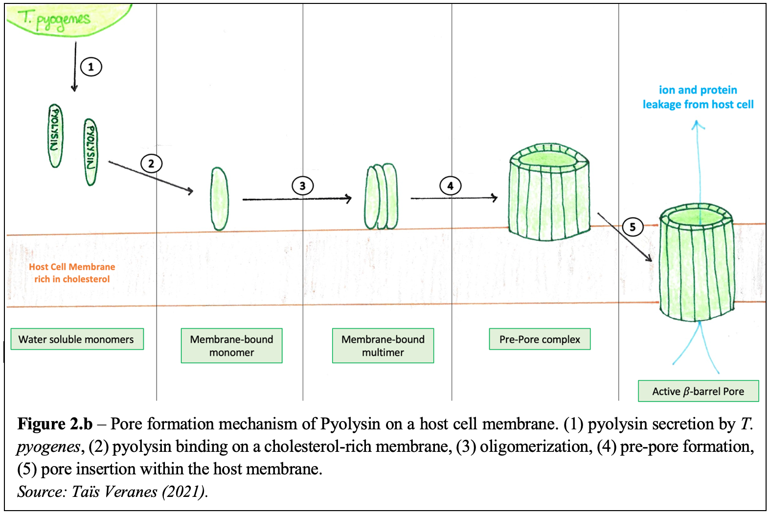
T. pyogenes secretes an exotoxin – called pyolysin – which is considered the primary virulence factor of the bacteria. Pyolysin belongs to the cholesterol-dependent cytolysin (CDC) family which forms large 𝛽-barrel pores in host cell membranes, by a process called cell lysis. Water-soluble pyolysin molecules are first secreted by the bacterium. They then bind to target host membranes containing cholesterol and oligomerize into multimer pre-pore complexes. Pore formation is characterized by the complete insertion of the pre-pores into the cell membrane. To do so, the pyolysin molecules – which are composed of four domains (Figure 2.a.) – must significantly change their conformation to form 𝛽-barrel channels and separate their Domain 2 and Domain 3. As so, 30 to 50nm transmembrane pores are created, allowing various ions and proteins to move out of the host cell consequently leading to cell lysis and death.  The steps following pyolysin’s mechanism of action against host cells are illustrated in Figure 2. b.

Although it is the most potent, pyolysin is not the only virulence factor of T. pyogenes. Indeed, the bacteria express several surface-exposed proteins – such as fimbriae, extracellular matrix-binding proteins, and neuraminidases – involved in the adherence and mucosal colonization of host tissues. Furthermore, T. pyogenes can also form biofilms which increase the bacterium’s resistance against the immune response of the host.

Treatment

T. pyogenes is generally susceptible to beta-lactams antibiotics. However, the microorganism is becoming resistant to some antibiotics such as tetracycline by preventing it from reaching the infection site. In the case of clinical mastitis and endometritis, intramammary antibiotics and non-steroidal drugs are used for mild mastitis; but if the infection spreads throughout the body, then systemic antibiotics are given by intramuscular route are also needed. When treating summer mastitis, the same antibiotics are beneficial to fight against the infection in mild cases; however, for severe cases, anti-inflammatory drugs are added to the treatment plan to relieve the animal of pain and swelling. Severe cases also can lead to amputation of the affected teat to help with drainage of pus and may lead to the animal being culled to avoid the spread of the infection. Treatments for abscesses are the same ones used for clinical mastitis however, in some cases, draining of abscesses of the pus is required. 

References

Deliwala S, Beere T, Samji V, McDonald PJ, Bachuwa G. 2020. When zoonotic organisms cross over – Trueperella pyogenes endocarditis presenting as a septic embolic stroke. Cureus. 12(4):e7740.

Griffin S, Healey GD, Sheldon IM. 2018. Isoprenoids increase bovine endometrial stromal cell tolerance to the cholesterol-dependent cytolysin from Trueperella pyogenes. Biol Reprod. 99(4):749-60.

Guo Y, Liu Y, Zhang Z, Chen M, Zhang D, Tian C, et al. 2020. The antibacterial activity and mechanism of action of luteolin against Trueperella pyogenes. Infect Drug Resist. 13:1697–711

Pokrajac L, Baik C, Harris JR, Sarraf NS, Palmer M. 2012. Partial oligomerization of pyolysin induced by a disulfide-tethered mutant. Biochem Cell Biol. 90(6):709-17.

Preta G, Lotti V, Cronin JG, Sheldon IM. 2015. Protective role of the dynamin inhibitor Dynasore against the cholesterol-dependent cytolysin of Trueperella pyogenes. FASEB J. 29(4):1516-28.

Rzewuska M, Kwiecień E, Chrobak-Chmiel D, Kizerwetter-Swida M, Stefańska I, Gieryńska M. 2019. Pathogenicity and virulence of Trueperella pyogenes: a review. Int J Mol Sci. 20(11):2737.

Ribeiro MG, Risseti RM, Bolaños CAD, Caffaro KA, de Morais ACB, Lara GHB, et al. 2015. Trueperella pyogenes multispecies infections in domestic animals: a retrospective study of 144 cases (2002 to 2012). Vet Q. 35(2):82-7.

Thapa R, Ray S, Keyel PA. 2020. Interaction of macrophages and cholesterol-dependent cytolysins: the impact on immune response and cellular survival. Toxins (Basel). 12(9):531.

Treponema denticola

Introduction

Treponema denticola are a long spiral-shaped, anaerobic bacteria commonly found in the mouth. T. denticola is a gram-negative bacterium, meaning they have a thin peptidoglycan layer and an outer lipid membrane. T. denticola is highly specialized to reside in the gum crevice of the oral cavity and is one of the bacterial species responsible for causing periodontal disease. 

Disease

 Periodontal disease, also known as gum disease, is a severe gum infection that affects the soft tissue surrounding teeth. T. denticola, Porphyromonas gingivalis, and Tannerella forsythia are known as the “red complex”. They are the three most common bacteria present in the biofilm (also known as plaque) associated with chronic periodontitis. Together, they help each other grow and thrive during infection. 

The disease begins with the accumulation of plaque around the teeth. If oral hygiene is poor, plaque can harden under the gum line and become tartar, which can only be removed by a dentist. With time, tartar can cause gingivitis, a reversible, mild version of periodontal disease. Severe periodontal disease occurs once gum tissue breaks down and pockets begin to develop between teeth and gums, filling up with even more plaque. 

The protein-degrading (proteolytic) enzymes secreted by T. denticola break down the host gum proteins. The lysis of gingival cells and subsequent inflammation are the primary causes of periodontitis. Symptoms of periodontitis include pain, redness, swelling, and bleeding of the gums, and a receding gum line. If left untreated, the damage caused by T. denticola can eventually lead to tooth loss and the spread of infection to the jaw bone. 

T. denticola has also been shown to promote oncogenesis, where healthy cells become cancer cells via genetic and/or cellular changes. A specific enzyme produced by T. denticola was identified in the majority of tumor samples and was shown to degrade regulatory proteins involved in the control of inflammation and tumor microenvironment. 

Figure 1. Tooth diagram showing the development of periodontal disease. Source: Mayo Foundation For Medical Education and Research.

Epidemiology 

Periodontal disease is one of the most common infectious diseases around the world. According to the Centers of Disease Control and Prevention, about 47% of all adults over 30 in the U.S. have some level of periodontal disease. The susceptibility also increases with age, with 70% of people over 65 having the disease. Other factors, such as smoking, medications, hormone changes, and underlying health conditions can also increase the risk of developing periodontitis. 

The mouth contains a wide range of natural microbiota. However, periodontal disease often results from the overgrowth of certain bacterial species that are also present in healthy individuals. In fact, studies have shown that it is possible to predict the development of periodontitis based on the abundance of different species, especially those of the Red Complex such as T. denticola

Good oral care including frequent brushing and flossing is the most effective preventative measure against periodontitis. Yearly dentist visits can also help prevent the progression of the disease by removing tartar and plaque that cannot be easily accessed with a toothbrush. 

Virulence factors

T. denticola has a variety of virulence factors that allow it to infect the host. One of the first steps of infection is attachment, and T. denticola cells are covered in a protein called major outer sheath protein, which can bind to many different host proteins. T. denticola has a very strong metabolic association with P. gingivalis. This means that both species can work together to break down proteins, share nutrients, and become overall more efficient. Studies have shown that when cultured together, biofilm formation is enhanced. Biofilm aids attachment to teeth and can help protect bacteria against environmental stress. 

Like other gram-negative bacteria, T. denticola can release small spherical vesicles filled with various molecules such as enzymes and toxins which further help invasion. When released into the environment, they can help break down the tight junctions between host cells and allow the bacterial cells to penetrate deeper tissues. Dentilisin is another very important virulence factor that aids in the progression of disease. It can destroy communication pathways and other structural host cell proteins. 

Once inside, bacteria can use their internalized flagella to swim deeper into the tissue. By being located under the outer sheath, the flagella are concealed from the immune system, avoiding recognition by antibodies. T. denticola can also respond to environmental changes by a process called chemotaxis. Bacteria can move towards different stimuli such as glucose, allowing them to make the most out of their surroundings.

The host’s body will try to fight off the infection to the best of its ability, but T. denticola has ways of suppressing immune responses. This is highly advantageous to T. denticola, as it can remain unnoticed for longer periods of time. Some epithelial cells produce antimicrobial peptides, which are molecules that inhibit different mechanisms and can lead to the destruction of bacterial cells. T. denticola can prevent these molecules from attaching to its outer membrane and therefore entering the cell. It can also pump out these molecules before they cause any damage. Dentilisin produced by T. denticola can also break down different cytokines which are used by the body to alert and activate the immune system. 

 Figure 2. Treponema pallidum. A) The syphilis causing bacterium, T. pallidum, belongs to the same genus as T. denticola. Both have the same spiral-shaped (spirochete) cell structure; B) Cellular components of a typical spirochete bacterium. Adapted from Peeling, et al., (2017). Syphilis. Nature Reviews Disease Primers, 3(1). https://doi.org/10.1038/nrdp.2017.73 

The treatment of T. denticola infections is dependent on how far the infection has spread. For acute infections that have not reached under the gum flaps, excessive plaque can be physically removed from teeth via scaling and a course of antibiotics. For infections that have spread below the gum line and around the root of the tooth, the area may be surgically cleaned and a tissue graft may be performed. This involves removing skin from the roof of the mouth and covering the areas affected by gumline recession caused by T. denticola. For extreme cases when the infection has spread to the bone, a bone graft may be performed. A bone graft procedure is performed by surgically removing the infected bone and then transplanting synthetic material or fragments of your own bone. This essentially “rebuilds” the jaw and allows the bones to regrow over the previously infected areas.

References

Centers for Disease Control and Prevention. (2013). Periodontal disease. Centers for Disease Control and Prevention. Retrieved November 18, 2021, from https://www.cdc.gov/oralhealth/conditions/periodontal-disease.html. 

Dashper, S. G., Seers, C. A., Tan, K. H., & Reynolds, E. C. (2010). Virulence factors of the oral spirochete Treponema denticola. Journal of Dental Research, 90(6), 691–703. https://doi.org/10.1177/0022034510385242 

Foschi, F., Izard, J., Sasaki, H., Sambri, V., Prati, C., Müller, R., & Stashenko, P. (2006). Treponema denticola in disseminating endodontic infections. Journal of dental research, 85(8), 761-765.

Inagaki, S., Kimizuka, R., Kokubu, E., Saito, A., & Ishihara, K. (2016). Treponema denticola invasion into human gingival epithelial cells. Microbial Pathogenesis, 94, 104–111. https://doi.org/10.1016/j.micpath.2016.01.010 

Ishihara, K. (2010). Virulence factors of Treponema denticola. Periodontology 2000, 54(1), 117–135. https://doi.org/10.1111/j.1600-0757.2009.00345.x 

Loesche, W. J., & Grossman, N. S. (2001). Periodontal disease as a specific, albeit chronic, infection: diagnosis and treatment. Clinical microbiology reviews, 14(4), 727-752.

Mayo Foundation for Medical Education and Research. (n.d.). Periodontitis. Mayo Clinic. Retrieved November 18, 2021, from https://www.mayoclinic.org/diseases-conditions/periodontitis/symptoms-causes/syc-20354473. 

Nazir M. A. (2017). Prevalence of periodontal disease, its association with systemic diseases and prevention. International journal of health sciences, 11(2), 72–80.

Nieminen, M., Listyarifah, D., Hagström, J. et al. 2017. Treponema denticola chymotrypsin-like proteinase may contribute to orodigestive carcinogenesis through immunomodulation. Br J Cancer 118, 428–434 (2018). https://doi.org/10.1038/bjc.2017.409

​​​Aeromonas salmonicida

By Ge Gao and Gloria Van

Introduction

In 1894, Emmerich and Weibel documented the first outbreak of Aeromonas salmonicida in trout fish. This bacterial pathogen found in marine and fresh-water environments is well-known for causing furunculosis (Figure 1). Furunculosis is a disease that commonly affects salmonid fish such as salmon, trout, and whitefish. At first, scientists believed A. salmonicida was an exclusive pathogen to salmonids. Now we understand that this bacterium is capable of causing furunculosis and several other diseases among salmonids and non-salmonids species. For over 100 years, this bacteria has globally caused declines in fish hatcheries and extreme financial losses. 

Figure 1: Appearance of A. salmonicida on a blood agar plate. Source: Menanteau-Ledouble et al., 2016

Disease

The severity of A. salmonicida depends on many factors; environmental conditions, its interactions with other organisms, stress levels of fish, and the strength of fish immune systems. Ultimately, A. salmonicida is a clever pathogen that targets fish with wounds or weak immune systems.

They enter fish through their skin, gut, or gills. Once inside their host, they replicate themselves in phagocytic cells such as macrophages. Macrophages are important cells of the immune system that are responsible for digesting and destroying pathogens. They continue replicating and quickly spread across the brain, kidneys, liver, and spleen. Eventually, A. salmonicida leads to furunculosis by suppressing the response of leukocytes. Leukocytes are white blood cells, necessary immune cells that fight off infection and disease. Infected fish showcase a variety of internal and external symptoms such as swollen skin lesions, skin abscesses under the skin called furuncles, intestinal inflammation, and lack of energy/movement (Figure 2).

Figure 2: Summary of infection by A. salmonicida. Source: Gloria Van, 2021

The disease is highly contagious and spreads through the water column and direct contact with infected fishes or its eggs. Cases are most frequently reported in fish hatcheries but can occur in wild populations. Unfortunately, most infected fish will die, some without showing any symptoms at all. Human infections are very rare as the optimal growth temperature of A. salmonicida ranges from 18-24°C.

Epidemiology

A. salmonicida has a worldwide distribution, most commonly causing furunculosis among fish hatcheries. Canada’s Department of Fisheries and Oceans reported 298 cases of furunculosis between 2013-2017.

Controlling this bacterium is challenging. Some countries have succeeded, while others continue to struggle. In 1963, Switzerland and Germany successfully eliminated all traces of furunculosis through strict regulations and increasing the frequency of their inspections. They continue to maintain these standards to avoid any re-introduction of furunculosis. Australia and New Zealand took a different approach by avoiding the importation of any new fish or eggs. On the other hand, furunculosis is still a problem in the United States and many other countries in Europe, such as Sweden and Denmark.

Cases in Salmonids are more likely to be reported and researched due to their popularity and economic importance. As our knowledge of A. salmonicida continues to grow, we understand that it can infect almost every fish species. For instance, regions where the bacterium was previously absent, such as Japan and regions of mainland Asia have since reported A. salmonicida cases. Therefore, it is likely that furunculosis cases are much higher because they are under-reported and under-investigated.

Virulence Factors

The mechanisms of A. salmonicida are complex and consist of multiple virulence factors. The three main factors are the additional layer (A-layer), extracellular products (ECP), and a Type III secretion system (T3SS).

  1. The A-layer is an additional layer on the cell surface encoded by a gene named vapA, and is a complex protein structure composed of lipopolysaccharides (LPS) and other proteins (Figure 3). The A-layer is responsible for macrophage cytotoxicity resistance, helping A. salmonicida attach to fish macrophages and shields it from the fish’s immune system defense mechanisms. Therefore, it helps the bacterium to replicate inside the fish undetected. The A-layer and the bacterial capsule are similar in their functions. The capsule protects the bacteria from being destroyed by immune cells and inflammation. One key difference between the two is that the capsule is made of polysaccharides and/or proteins.
  1. ECP are secreted molecules such as proteins and enzymes. One example is proteases which are enzymes that degrade proteins. Proteases assist the growth of the bacterium by stealing nutrients from the fish. Stealing nutrients leads to skin lesions which are abnormal appearances of skin that appear discoloured and patchy. A. salmonicida produces many different types of proteases, one being AspA. AspA degrades muscle tissue which causes furuncles, skin abscesses under the skin filled with pus. 
  1. T3SS inserts toxic proteins inside fish cells which results in cell lysis. Cell lysis is when the outer membrane breaks down, and intracellular components like DNA, RNA, and proteins are released. This process creates an imbalance in the cell environment which often leads to cell death. T3SS has three major structures:  (1) A secretion apparatus that assists the delivery between protein toxins and their effector, (2) a needle-like channel through the bacterial outer membrane and the host cell membrane, which allows the pathogen to inject their toxic proteins (effectors) into the host cell cytosol, and (3) a translocation tool responsible for the physical movement of the toxins, transferring them from the needle into the fish cell (Figure 4).
Figure 3: Depiction of a virulence factor – the A-layer. Source: Ge Gao, 2021
Figure 4: Schematic overview of the Type III secretion system (T3SS). Source: Frey & Origgi, 2016

Treatment

Prevention is one of the most important treatment strategies as infected fish are highly contagious and hard to control. Scientists have tried many strategies such as selective breeding, vaccinations, and antibiotics. 

Selective breeding is when parents are specifically picked to produce offspring with advantageous traits for survival and reproduction. Since 1954, scientists have worked on breeding fish that are resistant to diseases caused by A. salmonicida. The results were mixed. They observed that when some fish developed slowly, they were more resistant to developing the disease. However, many fish have very different responses to selective breeding so, this strategy is considered somewhat effective but not enough by itself.

Vaccine development for furunculosis began in the 1940s. Many different types of commercialized vaccines were produced. Unfortunately, none were considered effective in controlling furunculosis. Not until a breakthrough occurred in the late 1900s using oil-adjuvant injection vaccines. This type of vaccine contains an ingredient, oil-in-water, which creates an even stronger immune response in fish against A. salmonicida. Oil-adjuvant vaccines are generally accepted and applied in the fish industry. 

In 1952, antibiotics were introduced to treat furunculosis. By the late 1990s, they were the most widely used treatment. Similar to vaccines, there are a wide range of antibiotics available. Antibiotics are effective but there are ongoing concerns about antibiotic resistance. 


All treatments have their advantages and disadvantages. There is no standard treatment that works for all cases of A. salmonicida. The best solution remains to be prevention which is why new approaches for vaccine development continue.

References

Bartkova, S., Leekitcharoenphon, P., Aarestrup, F. M., & Dalsgaard, I. (2017). Epidemiology of Danish Aeromonas salmonicida subsp. Salmonicida in Fish Farms Using Whole Genome Sequencing. Frontiers in Microbiology, 8, 2411. https://doi.org/10.3389/fmicb.2017.02411

Boily, F., Malcolm, G., & Johnson, S. C. (2019). Characterization of Aeromonas salmonicida and furunculosis to inform pathogen transfer risk assessments in British Columbia. 45. Retrieved December 11, 2021, from https://waves-vagues.dfo-mpo.gc.ca/Library/40852611.pdf

Chart, H., Shaw, D. H., Ishiguro, E. E., & Trust, T. J. (1984). Structural and immunochemical homogeneity of Aeromonas salmonicida lipopolysaccharide. Journal of Bacteriology, 158(1), 16–22. https://doi.org/10.1128/jb.158.1.16-22.1984

Cipriano, R. C., Bullock, G. L., & National Fish Health Research Laboratory. (2001). Furunculosis and other diseases caused by aeromonas salmonicida (Ser. Fish disease leaflet, 66). National Fish Health Research Laboratory. Retrieved November 18, 2021, from https://webharvest.gov/peth04/20041029064735/http:/www.lsc.usgs.gov/FHB/leaflets/FHB66.pdf

Desbois, A. P., Cook, K. J., & Buba, E. (2020). Antibiotics modulate biofilm formation in fish pathogenic isolates of atypical aeromonas salmonicida. Journal of Fish Diseases43(11), 1373–1379. https://doi.org/10.1111/jfd.13232

Dallaire-Dufresne, S., Tanaka, K. H., Trudel, M. V., Lafaille, A., & Charette, S. J. (2014). Virulence, genomic features, and plasticity of Aeromonas salmonicida subsp. Salmonicida, the causative agent of fish furunculosis. Veterinary Microbiology, 169(1), 1–7. https://doi.org/10.1016/j.vetmic.2013.06.025

Frey, J., & Origgi, F. C. (2016). Type III Secretion System of Aeromonas salmonicida Undermining the Host’s Immune Response. Frontiers in Marine Science, 3, 130. https://doi.org/10.3389/fmars.2016.00130

Fyfe, L. A., Finley, A., Coleman, G., & Munro, A. L. S. (1986). A study of the pathological effect of isolated Aeromonas salmonidda extracellular protease on Atlantic salmon, Salmo salar L. Journal of Fish Diseases, 9(5), 403–409. https://doi.org/10.1111/j.1365-2761.1986.tb01033.x

Garduño, R. A., Moore, A. R., Olivier, G., Lizama, A. L., Garduño, E., & Kay, W. W. (2000). Host cell invasion and intracellular residence by Aeromonas salmonicida: Role of the S-layer. Canadian Journal of Microbiology, 46(7), 660–668. https://doi.org/10.1139/w00-034

Horne, J. H. (1928). Furunculosis in Trout and the Importance of Carriers in the Spread of the Disease1. Epidemiology & Infection, 28(1), 67–78. https://doi.org/10.1017/S0022172400009396

Janda, J. M., & Abbott, S. L. (2010). The Genus Aeromonas: Taxonomy, Pathogenicity, and Infection. Clinical Microbiology Reviews, 23(1), 35–73. https://doi.org/10.1128/CMR.00039-09

Lee Herman, R. (1968). Fish Furunculosis 1952–1966. Transactions of the American Fisheries Society, 97(3), 221–230. https://doi.org/10.1577/1548-8659(1968)97[221:FF]2.0.CO;2

Menanteau-Ledouble, S., Kumar, G., Saleh, M., & El-Matbouli, M. (2016). Aeromonas salmonicida: Updates on an old acquaintance. Diseases of Aquatic Organisms, 120(1), 49–68. https://doi.org/10.3354/dao03006

Midtlyng, P. J. (1997). 15—Vaccination Against Furunculosis. In E.-M. Bernoth, A. E. Ellis, P. J. Midtlyng, G. Olivier, & P. Smith (Eds.), Furunculosis (pp. 382–404). Academic Press. https://doi.org/10.1016/B978-012093040-1/50020-8

Reith, M. E., Singh, R. K., Curtis, B., Boyd, J. M., Bouevitch, A., Kimball, J., Munholland, J., Murphy, C., Sarty, D., Williams, J., Nash, J. H., Johnson, S. C., & Brown, L. L. (2008). The genome of Aeromonas salmonicida subsp. salmonicida A449: Insights into the evolution of a fish pathogen. BMC Genomics, 9(1), 427. https://doi.org/10.1186/1471-2164-9-427

Sakai, D. K. (1985). Loss of virulence in a protease-deficient mutant of Aeromonas salmonicida. Infection and Immunity, 48(1), 146–152. https://doi.org/10.1128/iai.48.1.146-152.1985

Scott, M. (1968). The pathogenicity of Aeromonas salmonicida (Griffin) in sea and brackish waters. Journal of General Microbiology, 50(2), 321–327. https://doi.org/10.1099/00221287-50-2-321

Valderrama, K., Soto-Dávila, M., Segovia, C., Vásquez, I., Dang, M., & Santander, J. (2019). Aeromonas salmonicida infects Atlantic salmon (Salmo salar) erythrocytes. Journal of Fish Diseases, 42(11), 1601–1608. https://doi.org/10.1111/jfd.13077

Wiklund, T., & Dalsgaard, I. (1998). Occurrence and significance of atypical Aeromonas salmonicida in non-salmonid and salmonid fish species: A review. Diseases of Aquatic Organisms, 32(1), 49–69. https://doi.org/10.3354/dao032049

Woo, P. T. K., Leatherland, J. F., & Bruno, D. W. (2006). Fish diseases and disorders (2nd ed., Ser. Cab books). CABI Pub. Retrieved November 18, 2021, from https://dlib.library.razavi.ir/bitstream/Ebook/89557/2/0851991947.pdf#page=67

How do gut bacterial residents fight whooping cough after taking antibiotics ?

Author: Ziying yuan & Nicolas de Azevedo

Introduction

Bordetella pertussis is a Gram-negative, aerobic, pathogenic bacterium, meaning it possesses a complex cell envelope of the plasma membrane, peptidoglycan cell wall and an outer membrane. It survives and grows in an oxygenated environment, capable of infecting the respiratory system and causing a disease called “whooping cough” in humans. B. pertussis is notorious for severe and uncontrollable coughing that can make it hard to breath or even deadly for babies. It is spread by airborne droplets and infects humans by colonizing the lung cells, then toxin is produced by the bacterium to prevent human cilia from clearing debris from the lungs. The lungs have to cough very hard to expel the debris and bacteria out of the body into the air, which can then infect other people. B. pertussis can affect people of all ages; however, infants, young children, pregnant women and the unvaccinated population remain the most susceptible to pertussis-related morbidity and mortality. Babies at the highest risk of serious disease, are more likely to need hospitalization or even die from whooping cough; about one in every 200 babies under 6 months old who catch whooping cough dies from pneumonia or brain damage.

Vaccine for whooping cough has been widely used since the end of the 20th century. Early administration of antibiotics and corticosteroids treatment immediately after diagnosis can limit the course of the disease, and the therapy is most effective during the incubation period. The side effects of antibiotics varied from case to case.

Findings

In this study, the researchers intervened to change the microbiota in mice using antibiotics treatment, and then measured their resistance against B. pertussis or whooping cough by infecting them. They found out that an intact and balanced microbiota inhibits the colonization of B. pertussis in the lungs, while antibiotics-treated mice showed higher susceptibility to bacterial infection.

In the beginning, the mice were given a slew of four antibiotics for three weeks so that their microbiota could be manipulated as the exposed group. Three days after the antibiotic treatment was done, a sufficient amount of B. pertussis was introduced into the exposed and unexposed groups of mice to mimic the natural bacterial infection in the nose. Then, the exposed group and the unexposed group were compared in terms of their development of symptoms and change in biochemical markers in the body for whooping cough.

Three days after the antibiotics treatment and before B. pertussis being introduced, the lung samples and feces from the antibiotics-exposed mice were compared to those from the non-exposed mice. They found out that the relative abundance of ten main bacteria living in the mice’s feces had drastically changed due to previous antibiotics administration. Noticeably, the antibiotics-exposed mice showed an 87.65% increase in Proteobacteria. Other bacterial populations were affected to different extents, such as the absence of Saccharibacteria. The lungs, however, showed no signs of obvious changes in their bacterial composition, leading to the scientists believing that the oral delivery of antibiotics would not affect the lung microbiota. This inconsistency of population change between the gut and lung tissue could potentially be due to the small sample size used in this experiment. The conclusion drawn from a small sample size is not statistically robust regarding the cause-effect relationship, and the general pattern could be easily overlooked. In other words, the generalizability of the findings in this study to the target population can be a challenge. Moreover, the ten types of gut bacterial residents studied were based on relative abundance so percentage. Without an accurate population count for the microbiota in lungs after antibiotics administration, the conclusion lacks internal validity and reliability. 

 The presence of these bacteria in the feces and lungs is indicative of the gut and the lung’s microbiota (also known as the diverse microorganisms that reside within the aforementioned sections of the body). When the bacteria within the microbiota, also called commensals, were killed, a lack of diversity was found in the fecal matter and thus the gut microbiota. This became a case of dysbiosis, which can be defined as “a compositional and functional alteration in the microbiota that is driven by a set of environmental and host-related factors that perturb the microbial ecosystem to an extent that exceeds its resistance and resilience capabilities”. Multiple studies had shown that the antibiotic treatments could have profound and rapid changes in gut microbiota, some of which could last for years and dysbiosis can be a stable state depending on individuals.  

Figure 1 is an illustration explaining the concept of dysbiosis. The colored bacteria are commensals and the black one is the pathogen. Source: Nicolas de Azevedo, 2021

The next experiment the researchers conducted was to determine if an intact microbiota plays a role in the resistance against whooping cough infection.

Figure 2 is a graph showing the mean of the log₁₀ number of CFU per lung of each mouse across multiple time points post infection of B. pertussis in antibiotic-treated mice (Ab-BP) and non-antibiotic treated mice (BP). CFU means Colony Forming Unit, which is a unit used to describe the concentration of a microorganism or in this case, the concentration of B. pertussis per lung. Source: Adapted from Commensal Microbes Affect Host Humoral Immunity to Bordetella pertussis Infection, by Y. Zhang, 2019, American society for microbiology.

The researchers took samples from the lung tissues to determine the amount of B. pertussis colonizing the Ab-BP mice and BP mice as time went on. The main trend for both series of data points is an increase in B. pertussis from 3hrs to D7 after infection, followed by a gradual clearance of the bacteria from the lungs until D17. However, the Ab-BP demonstrated more serious bacterial colonization than the BP had until D17, notably at 3hrs and D3. This means that previous antibiotic exposure could have a role in lessening resistance to whooping cough in mice, and by extension potentially be caused by the lack of commensal microbes and gut dysbiosis.

The researchers wanted to see if antibiotics could affect the production of antibodies in mice: ten days after B. pertussis infection, the number of antibodies in the mice’s serum (liquid component of blood without clotting factors) was compared between antibiotic treated mice, non-antibiotic treated mice, and control non-infected mice. They observed that in general, a larger quantity of antibodies was collected in the non-antibiotic treated mice, meaning stronger immune response against bacterial infection.

They repeated the same experiment seventeen days after the infection, only to find that the non-antibiotic treated mice have at least more than twice the antibodies compared to their antibiotic treated counterparts. Taken together, these experiments allude to the idea that antibiotics can cause dysbiosis, increase susceptibility to B. pertussis and lower the number of antibodies produced yet the exact mechanisms behind this event are unknown.

Importance of the findings

In this era of increasing demand for antibiotics treatment, the fact that the oral antibiotics can impair the gut health and cause profound and long-term disruption in the composition of gut bacterial population remains a thorny issue because it can lead to increased susceptibility to B. pertussis infection and perturb normal immune response. This research had underscored the importance of the gut microbiome in human immune responses to respiratory infection, which means that by having a better and healthier gut bacterial population, people may have more resistance to whooping cough. Future research should be done to explore novel strategies to recover or optimize the microbiota for robust immune defense. Isolation of specific gut bacteria that are protective against B. pertussis pathogenesis is of great importance to counteract the side effect of oral antibiotics treatment.

Noticeably, cutting down the use of antibiotics is not a simple thing to do in current society. People are becoming more dependent on this fast and simple way of treating a variety of infections. The consequences are a disrupted microbiota and a vulnerable immune system to fight disease. To prevent this, the selection of antibiotics that are less likely to have a long-term effect on the gut microbiota is important. Figuring out the exact cellular and molecular mechanism by which the human microbiota manipulates the immune response to B. pertussis infection is the future focus of the investigation. Hills et al. proposed that the healthy gut flora exert their beneficial effects through the fermentation of dietary fiber to produce short-chain fatty acids (SCFA), so eating a diverse diet rich in whole foods may help build a balanced microbiome.  SCFA promotes immunity and suppress inflammatory process in the intestine and other organs. By consuming dietary fibre which is not digested but fermented by commensal bacteria in the small intestine, SCFA will be released to support the growth of beneficial bacteria in the gut and help fight possible invaders.

Reference

Australian government department of health. (2020, May 27). Whooping cough (pertussis). Retrieved from https://www.health.gov.au/health-topics/whooping-cough-pertussis.

K. L. Sealey, T. Belcher & A. Preston (2016, June 1). Bordetella pertussis epidemiology and evolution in the light of pertussis resurgence. Infection, Genetics and Evolution: journal of molecular epidemiology and evolutionary genetics in infectious disease. 40, pp. 136-143.

N. Shin, T. Wong & J. Bae. (2015, July 22). Proteobacteria: microbial signature of dysbiosis in gut microbiota. Trends in biotechnology, 33(9), pp496-503

S Black (1997, April 16). Epidemiology of pertussis. The Pediatric infectious disease journal, pp. 85-89.

Silvana Bettiol, K. W. (2012). Symptomatic treatment of the cough in whooping cough. Retrieved from https://doi.org/10.1002/14651858.CD003257.pub4.

Y. Zhang, Z. Ran, M. Tian et al (2019, December 19). Commensal Microbes Affect Host Humoral Immunity to Bordetella pertussis Infection. Infection and Immunity, pp. e00421-19. Retrieved from https://journals.asm.org/doi/10.1128/iai.00421-19?permanently=true.

M. Levy, A. Kolodziejczyk, C. Thaiss & E. Elinac. (2017). Dysbiosis and the immune system. Nature reviews. Immunology, 17(4), pp219-232.

L. Dethlefsen & D. A. Relman. (2011). Incomplete recovery and individualized responses of the human distal gut microbiota to repeated antibiotic perturbation. Proceedings of the National Academy of Science of the United States of America, 108 suppl1, pp4554-4561.

The 2017 Canine Leptospirosis Epidemic in Maricopa County, Arizona

By: Marco Marcogliese and Maude Tanguay

Introduction: 

Canine Leptospirosis is categorized as a zoonotic disease which has caused increased concern over the past several decades in a vast majority of countries throughout the world. The disease is mainly caused by spiral, gram-negative bacteria of the genus Leptospira interrogans sensu lato (Sessions & Greene, 2004). Such bacteria can be further categorized into antigenically linked serogroups which are sorted into distinct serovars. Serovars are defined as recognizable variations between bacteria or viruses belonging in the same group (Goldstein et al., 2008). As the disease was initially discovered in 1899, it was believed that the serovars icterohaemorrhagiae and canicola were the bacteria mainly responsible for the majority of clinical cases of canine leptospirosis up until 1960. 

Therefore, bivalent vaccines responsible for targeting the two most popular serovar infections of the disease were administered and had seemed to slowly prevent its dissemination across North America (Rentko et al., 1992). However, over the past two decades, increased incidence of Leptospirosis in dogs linked to novel strains has been increasing, with the most common strains reported in the United States included L. kirchneri serovar gripptyphlosa and L. interrogans serovars pomona and bratislava (Goldstein et al., 2008). To this effect, small outbreaks of canine Leptospirosis have become more prominent in certain states throughout the United States, more specifically in the Maricopa County of Arizona (Jenni et al., 2019). 

Etiology: 

The sudden uptake of reported cases of Canine Leptospirosis has mainly been attributed to exposure to wild reservoir hosts found in rural or suburban areas. The bacteria are transmitted amongst its hosts via both direct contact with other infected species or indirect contact with substances such as water, soil, food, and infected feces (Sessions & Greene, 2004). The main routes of infection involve the penetration of the host’s skin tissue after exposure to contaminated by-products. Once intruding the host’s body, leptospirosis bacteria (i.e. leptospires) invade the bloodstream to infect multiple sites of the host such as the liver, spleen, kidneys and the central nervous system. These bacteria are highly motile and flexible and are composed of a spiral structure with hook-shaped ends (Figure 1), meaning they can spread throughout the body rather quickly and attach to various structures within the body and begin replicating (Goldstein, 2010).  

The amount of damage to such internal organs is dependent on the virulence of the serovar alongside the host’s vulnerability and amount of exposure (Farr, 1995). Severe acute infections of the disease can exhibit bacterial colonization of the renal system as their optimal growth conditions are found within the cells that line the renal tubules. This results in critical renal failure due to tubular damage as the bacterium can destroy such cells which are crucial in lining these important intricate tubules found in the kidney (Sessions & Greene, 2004). The bacterium can then be excreted in the urine and further increase the rates of infection as a result. 

Figure 1: Electron micrograph of L. interrogans serovar icterohaemorrhagiae. Source: Levett, P. N. (2020, December 18). Leptospirosis [Photograph]. Clinical Microbiology Review. https://doi.org/10.1128/CMR.14.2.296-326.2001 

Source of Outbreak: 

Canine leptospirosis has been a known and common zoonotic disease for over 100 years in America (William & Karesh 2017). With cases steadily rising each year in canines, it’s important to understand which factors contribute to the prevalence of outbreaks and to its disproportionate rate of incidence in different areas. Understanding the source of outbreaks is also important for public health since canine leptospirosis can be a zoonotic transmitted disease (Jenni et al., 2019). The environmental factors characterizing different regions are the most noticeable source of outbreaks in multiple studies which have retrospectively studied canine leptospirosis. In a recent meta-analysis, dogs exposed to large environmental water sources were around 68% more likely to be infected (Ricardo et al., 2020). More specifically, stagnant water sources and floods seem to be frequently contaminated with Leptospira serovars. The pathogens which are shed through the urine of infected animals survive for long periods of time when found in water and until they come in contact with a susceptible host’s skin or mucous membranes (Goldstein, 2010). Areas inhabited with wildlife such as suburbs, fields or forests have even higher potential of leptospirosis infection since there are no preventative measures put in place to reduce their rate of infection. Although studies were able to easily identify different variables contributing to outbreaks, the correlation between the risk of being infected with disease with these variables has yet to be proven (White et al., 2017). 

Socioeconomic factors are also a large reason for outbreaks of the disease in dogs. Studies show that dogs living in lower income neighborhoods were not only more exposed to the various environmental factors associated with the disease but also had less access to veterinary care and lower vaccination rates (Ricardo et al., 2020). Historically, areas with lower income often have a lesser understanding and knowledge regarding vaccines which makes them hesitant to vaccinate their pets (Jenni et al., 2019). On the other hand, studies also showed that some middle class areas had higher infection rates. This was suggested to be caused by an increased amount of testing being possible with the higher income of clients in the area (Taylor et al., 2021). 

Cause of Outbreak: 

Being a widely known disease and pathogen, Canine Leptospirosis outbreaks are frequently reported. A notable example of this is the Maricopa County outbreak in Arizona which was reported in 2017. Despite the low amounts of annual rainfalls or the lack of humidity which are unfavorable for the growth and dissemination of the organism, the state of Arizona has been reporting multiple clusters of the infection in the last few years (Ward, 2002).  Knowing that leptospirosis is much less frequent in drier desert environments such as Arizona, public health officials thought crucial to find out the cause of these canine leptospirosis outbreaks.

Veterinarians in Arizona are required to report new cases of leptospirosis yet only 57% of veterinarians surveyed in the state knew when and how to properly report a zoonotic disease. The survey also noticed that small animal veterinarians were the least informed on zoonotic disease reporting. Shockingly, leptospirosis is only reported by 42% of the animal health professionals surveyed (Venkat et al., 2019). Similarly, 12% of the small animal veterinarians of Arizona believed that canine leptospirosis was not a reportable disease (Jenni et al., 2019).

The 2017 outbreak presented many cases where infected individuals presented with one or two clinical signs less commonly associated with leptospirosis such as conjunctivitis and vomiting. Less than 40% of the cases presented with fever, the most common clinical sign of canine leptospirosis (Jenni et al., 2019). This leads to the disease not being tested for as quickly as it should be which increases the chances for an infected animal to contaminate its environment (Langston & Heuter, 2003). Essentially, the diagnosis of this disease in canines is quite challenging as it is affected by the stage of the disease, the vaccination status of the individual, the environment and potential exposure to the pathogen (Reagan & Skyes, 2019). The available tests each have their strengths and weaknesses. Using a combination tends to be the most effective and efficient way to get a quick and accurate result but also more expensive for pet owners (Reagan & Skyes, 2019). 

These studies identified that since the environment in Arizona shouldn’t allow for the pathogen to survive, veterinarians don’t tend to be as vigilant or up to date with the latest protocols regarding the disease as veterinarians in high risk areas. (Jenni et al., 2019). Furthermore, this outbreak was composed mostly of cases from dogs living in urban areas which also indicates that different attitudes and practices among veterinarians in different areas most likely heavily impacted the prevalence of the disease.

Preventative Measures: 

With veterinarians being the first to work with and diagnose the disease, it is important for them to keep up with the latest strategies and protocols regarding diseases such as leptospirosis especially after important outbreaks such as the one in Maricopa County in 2017 (Jenni et al., 2019). 

The first step to ensure the prevention of the disease in animals as well as humans is increasing disinfection and improving hygiene when possibly having had contact with animal urine. These are easy and necessary precautions which should be taken regardless of the area’s risk for leptospirosis (Jenni et al., 2019). Limiting the amount of contact between dogs and any potentially contaminated water source is also an important first step in preventing the disease (Goldstein, 2010).

The most efficient way for veterinarians to prevent canine leptospirosis is by administering vaccines (André-Fontaine, 2006). Since the leptospires are frequently shed by a multitude of wild animals, it is impossible to assume that domesticated animals could achieve herd immunity since so many acquire the disease from water or soil contaminated by infected wildlife. The vaccine allows for dogs to have antibodies against the disease allowing the immune system to be equipped to quickly clear the infection should the animal come into contact with the pathogen. (André-Fontaine, 2006). As seen with the study conducted by Blanchard et al., (Figure 2) introducing annual vaccines which target several of the common serovars associated with Leptospirosis can decrease the ability for the organism to colonize the host and reduce the amount of potential reservoirs in an environment (2021). Leptospirosis vaccines should also be mandatory in places such as kennels and groomers to decrease the chances of spreading the disease to more individuals. (Langston & Heuter, 2003) Infected dogs should be isolated and their urine should be contained and disinfected in order to decrease the chances of infecting another individual. 

Finally, veterinarians should be aware of the latest news regarding the disease, especially with the number of cases rising each year. Improving education and awareness about zoonotic diseases will allow veterinarians to be better prepared at preventing, treating and most importantly managing the incidence rate of canine leptospirosis (Venkat et al., 2019). Creating a standardized vaccination protocol would be beneficial in reducing the impact of the different approaches between veterinarians while also preventing outbreaks in unsuspecting areas such as Arizona. There is also a need for better communication between veterinarians and public health officials to prevent outbreaks and reduce the amount of cases overall. 

Figure 2: Presence of different serovar growth of Leptospirosis bacteria in vaccinated dogs compared to the unvaccinated. Source: Blanchard, S., Cariou, C., Bouvet, J., Valfort, W., Oberli, F., Villard, S., Barret-Hilaire, F., Poulet, H., Cupillard, L., & Saint-Vis, B. (2021, June 18). [Photograph]. Quantitative Real-Time PCR Assays for the Detection of Pathogenic Leptospira Species in Urine and Blood Samples in Canine Vaccine Clinical Studies: A Rapid Alternative to Classical Culture Methods. https://journals-asm-org.proxy3.library.mcgill.ca/doi/10.1128/JCM.03006-20 

Conclusion: 

To conclude, the Maricopa County canine leptospirosis outbreak of 2017 was largely due to veterinarians and health officials not considering the state to be at risk for this disease in dogs due to the dry and desertic climate. However, since wildlife have a large role in spreading the disease, they can cause outbreaks in unsuspecting areas where domesticated animals might be less protected and where prevention against canine leptospirosis isn’t as common. As cases rise, it will be important for veterinarians all over to increase the amount of prevention against canine leptospirosis and to increase awareness about the disease in order to reduce the chances for future outbreaks.

References: 

André-Fontaine G. (2006). Canine leptospirosis–do we have a problem?. Veterinary 

microbiology, 117(1), 19–24. https://doi.org/10.1016/j.vetmic.2006.04.005 

Farr R. W. (1995). Leptospirosis. Clinical infectious diseases : an official publication of the 

Infectious Diseases Society of America, 21(1), 1–8. https://doi.org/10.1093/clinids/21.1.1

Goldstein, R. E. (2010, November 1). Canine Leptospirosis. ScienceDirect. 

https://www.sciencedirect.com/science/article/pii/S0195561610000951?via%3Dihub#bib6

Goldstein, R. E., Lin, R. C., Langston, C. E., Scrivani, P. V., Erb, H. N., & Barr, S. C. (2008, 

May 1). Influence of Infecting Serogroup on Clinical Features of Leptospirosis in Dogs. Wiley Online Library. https://onlinelibrary.wiley.com/doi/abs/10.1111/j.1939-1676.2006.tb02886.x 

Jenni, M. L., Woodward, P., Yaglom, H., Levy, C., Iverson, S. A., Kretschmer, M., Jarrett, N., 

Dooley, E., Narang, J., & Venkat, H. (2019, November 15). Knowledge, attitudes, and practices among veterinarians during an outbreak of canine leptospirosis — Maricopa County, Arizona, 2017. ScienceDirect. https://www.sciencedirect.com/science/article/pii/S0167587719305689 

Langston, C. E., & Heuter, K. J. (2003, July 1). Leptospirosis: A re-emerging zoonotic disease

ScienceDirect. https://www.sciencedirect.com/science/article/pii/S0195561603000263?via%3Dihub 

Reagan, K. L., & Skyes, J. E. (2019, July 1). Diagnosis of Canine Leptospirosis. ScienceDirect. 

https://www.sciencedirect.com/science/article/pii/S0195561619300385?via%3Dihub

Rentko, V. T., Clark, N., Ross, L. A., & Schelling, S. H. (1992, July 1). Canine Leptospirosis: A 

Retrospective Study of 17 Cases. Wiley Online Library. https://onlinelibrary.wiley.com/doi/10.1111/j.1939-1676.1992.tb00345.x 

Ricardo, T., Previtali, A., & Marcelo, S. (2020, August 1). Meta-analysis of risk factors for 

canine leptospirosis. ScienceDirect. https://www.sciencedirect.com/science/article/pii/S0167587720301513?via%3Dihub 

Sessions, J. K., & Greene, C. E. (2004, August). Canine Leptospirosis: Epidemiology, 

Pathogenesis, and Diagnosis. Auburn University College of Veterinary Medicine. http://vetfolio-vetstreet.s3.amazonaws.com/mmah/b1/b3b4ac15fb443ebb9d66e46acc513e/filePV_26_08_606_0.pdf 

Taylor, C., O’Neill, D. G., Catchpole, B., & Brodbelt, D. C. (2021, May 31). Incidence and 

demographic risk factors for leptospirosis in dogs in the UK. British Veterinary Association. https://bvajournals.onlinelibrary.wiley.com/doi/10.1002/vetr.512 

Venkat, H., Yaglom, H. D., & Adams, L. (2019, August 1). Knowledge, attitudes, and practices 

relevant to zoonotic disease reporting and infection prevention practices among veterinarians — Arizona, 2015. ScienceDirect. https://www.sciencedirect.com/science/article/pii/S0167587718305208?via%3Dihub 

Ward, M. P. (2002, December 30). Seasonality of canine leptospirosis in the United States and 

Canada and its association with rainfall. ScienceDirect. https://www.sciencedirect.com/science/article/pii/S0167587702001836?via%3Dihub 

White, A. M., Zambrana-Torrelio, C., Allen, T., Rostal, M. K., Wright, A. K., Ball, E. C., Daszak, P., & Karesh, W. B. (2017, April 1). Hotspots of canine leptospirosis in the United States of America. ScienceDirect. https://www.sciencedirect.com/science/article/pii/S109002331730059X?via%3Dihub

Corynebacterium diphtheriae

by Josh Koh & Seyed Alipour

Introduction

Corynebacterium diphtheriae was identified as the causative agent of diphtheria by Edwin Klebs in 1883. By 1884, Friedrich Löffler isolated C. diphtheriae. Due to Klebs’ and Löffler’s collective work, this bacterium was then known as Klebs-Löffler bacillus. C. diphtheriae is a Gram-positive bacteria surrounded by a thick outermost layer of peptidoglycan (polymer of sugars and amino acids). This bacterium is found throughout the world but is more prevalent in subtropical and developing countries that lack active vaccination programs against its toxin. Most commonly, C. diphtheriae infects humans, but there have been records of rare animal infections. In humans, C. diphtheriae infects and grows outside cells on the skin or the nasopharyngeal cavity, the upper part of the respiratory tract near the throat that lies behind the nose and above the mouth.

Disease

C. diphtheriae is transmitted between humans by contaminated aerosols or by contact with contaminated objects. Even if the infected person is asymptomatic, they can be infectious for up to four weeks. Once on the skin or in the upper respiratory tract, C. diphtheriae attaches to the cells and kills them by releasing its toxin, the Diphtheria Toxin (DT). DT will kill the cells by stopping protein production, allowing C. diphtheriae to use the released resources for its growth and multiplication. Through this mechanism, C. diphtheriae damages the skin and the lining of the upper respiratory tract and throat. 

Symptoms of C. diphtheriae infections usually arise 2-5 days post-infection. DT released by C. diphtheriae causes open sores or shallow ulcers on the skin. The respiratory damage from DT results in sore throat, swollen regions in the neck, and the formation of a thick and grey coating of dead tissue in the affected area – called a “pseudo-membrane” (Figure 1). This pseudo-membrane may cause difficulty breathing or choking. A respiratory infection may also result in mild fever and weakness. In such cases, the disease is referred to as diphtheria. In some rare cases, DT can enter the circulatory system and cause heart, nerve, and kidney damage, as well as organ death. 

Diphtheria diagnosis can be done through the identification of the common symptoms mentioned above and doing a swab test. The swab test includes taking a sample from the back of the throat or the skin lesion and trying to identify if C. diphtheriae and DT are present. 

Figure 1. Symptoms of C. diphtheriae infection.
A: Swollen parts of the Neck. B: Pseudo-membrane in the throat. Source: Government of Canada (2018)

Epidemiology

While skin diphtheria rarely causes severe disease, respiratory diphtheria remained the number one cause of death in children under 14 years of age across Canada until the mid-1920s. Before the introduction of the diphtheria vaccine in 1926, Canada recorded 9057 cases of diphtheria in 1924 – its highest number of annual cases ever. For more information regarding the vaccine, refer to the ‘Treatment’ section. Routine immunization in infants and children began in 1930, leading to a steep decline in diphtheria morbidity and mortality rates in Canada. Consequently, from 1993 to 2018, only 19 cases were reported, and the last recorded death by diphtheria was in 2010. Other industrialized and developed countries began infant and childhood immunization shortly after World War II. Like Canada, immunizations led to a dramatic reduction of diphtheria cases and transmission in those countries.

Recent respiratory outbreaks in developed regions such as Europe and North America are associated with disease-carrying travelers returning from regions where diphtheria is endemic (regularly found among populations of defined locations). Diphtheria is endemic in developing countries with insufficient immunization of children under 15, typically with high population densities and unsanitary living conditions. For instance, countries of the former Soviet Union with low immunization coverage endured over 150,000 cases and 5,000 deaths in the 1990s via respiratory diphtheria. Furthermore, regions where diphtheria is endemic usually have subtropical climates, such as many Asian and African countries (e.g., Haiti, Bangladesh, and Yemen).

Recent studies have suggested that sporadic (irregular and infrequent) cases of respiratory diphtheria occurring in countries with higher immunization coverages mostly affect the population older than 15 years of age, indicating waning vaccine immunity. Even so, the most susceptible populations to diphtheria are children below the age of 15 and the elderly. New outbreaks occur mainly among alcohol and drug abusers. The case mortality rate of respiratory diphtheria without treatments (refer to ‘Treatment’ section) is 30–50%. The introduction of respiratory diphtheria treatments significantly reduced the mortality rate to 5-10%.

Virulence

The first step necessary for C. diphtheriae infection is the colonization of the host (Figure 2). C. diphtheriae uses pili, a sort of hair-like protrusion on its surface, to attach to the host cells. The bacterial DNA encodes for nine different types of pili. The bacteria can switch between these nine different pili to confuse the human immune system so that it can live and grow longer at the infection site. This is called antigenic variation.

The most important virulence factor for C. diphtheriae is the DT. C. diphtheriae will secrete DT once it attaches to the cells lining the throat, the upper respiratory tract, or the skin. As DT is secreted from C. diphtheriae, it is referred to as an exotoxin. DT is composed of two subunits, A and B. The A subunit is the enzymatic subunit that speeds up reactions, and the B subunit is responsible for binding to target cells and releasing the A subunit into the cell. The B subunit attaches to a receptor, a molecule that recognizes specific targets, on the surface of the host cells. This receptor is called the heparin-binding epidermal growth factor precursor (HB-EGF). The binding of DT via the B subunit to the receptor causes the cell to take in the DT-receptor complex. This process is called endocytosis. At this point, DT is in an endosome, a sort of bag, inside the host cell. Proteases, which are enzymes that degrade proteins, then cut DT as the environment in the endosome becomes progressively more acidic. The acidic endosome causes DT to change shape and the B subunit pushes the A subunit into the host cell cytoplasm, which is the solution that fills cells. 

Within the cytoplasm, the A subunit will modify a component of the host cell machinery that is involved in protein synthesis. This component is called EF-2. By modifying this component, DT will make the host cell unable to make proteins needed for its survival. As a result, the DT affected cell will die, providing resources for the growth of C. diphtheriae.

Figure 2. The role of different C. diphtheriae Virulence Factors involved in diphtheria.
DT: Diphtheria Toxin; HB-EGF: heparin-binding epidermal growth factor precursor. Source: Josh and Seyed (2021)

Treatment

Antibiotic treatments in conjunction with the diphtheria antitoxin are very effective in treating skin and respiratory C. diphtheriae infections. C. diphtheriae is susceptible to antibiotics such as penicillin and erythromycin. Penicillin inhibits the enzyme that adds rigidity to the peptidoglycan layers of the bacterium’s cell wall. By inhibiting this enzyme, penicillin will destabilize C. diphtheriae’s cell wall and kill it. Contrarily, erythromycin will stop the synthesis of proteins, namely DT, to neutralize C. diphtheriae’s ability to stop the host cell’s protein production. Furthermore, the diphtheria antitoxin deactivates the unbound DT to prevent damage to the host cells. As respiratory diphtheria may proceed to cause damage to organs – as mentioned in the ‘disease’ section – or death if left untreated, early diagnosis and subsequent treatment mean a better chance of survival. 

While treatments for diphtheria have proven effective, the best method for controlling diphtheria morbidity and mortality is through preventive measures such as immunization. Immunization is established against C. diphtheriae’s toxin via the Diphtheria, Tetanus, and Pertussis (DTaP) vaccine, which contains an inactivated version of the DT called a toxoid. The inactivated DT will not cause an illness but will trigger the human immune system to form antibodies against it for protection against future exposures to DT-producing C. diphtheriae. The vaccine will only confer immunity to DT (the disease-causing agent) and not C. diphtheriae specifically. In 2014, it was estimated that 86% are vaccinated against DT globally. The DTaP vaccine also confers immunity to tetanus and pertussis, two other deadly human diseases. 

References

Clarke, K. E. N., MacNeil, A., Hadler, S., Scott, C., Tiwari, T. S. P., & Cherian, T. (2019). Global Epidemiology of Diphtheria, 2000-2017(1). Emerging infectious diseases, 25(10), 1834-1842. https://doi.org/10.3201/eid2510.190271

Diphtheria: For Health Professionals. (2018, June 21). Government of Canada. Retrieved November 17, 2021, from https://www.canada.ca/en/public-health/services/immunization/vaccine-preventable-diseases/diphtheria/health-professionals.html

Diphtheria. (2021) Museum of Health Care at Kingston. Retrieved Nov 17, 2021 from https://www.museumofhealthcare.ca/explore/exhibits/vaccinations/diphtheria.html

Efstratiou, A., Engler, K. H., Mazurova, I. K., Glushkevich, T., Vuopio-Varkila, J., & Popovic, T. (2000). Current approaches to the laboratory diagnosis of diphtheria. J Infect Dis, 181 Suppl 1, S138-145. https://doi.org/10.1086/315552

Kabanova, A., & Rappuoli, R. (2011). CHAPTER 34 – Diphtheria. In R. L. Guerrant, D. H. Walker, & P. F. Weller (Eds.), Tropical Infectious Diseases: Principles, Pathogens and Practice (Third Edition) (pp. 223-227). W.B. Saunders. https://doi.org/10.1016/B978-0-7020-3935-5.00034-3

Kneen, R., Giao, P. N., Solomon, T., Van, T. T. M., Hoa, N. T. T., Long, T. B., Wain, J., Day, N. P. J., Hien, T. T., Parry, C. M., & White, N. J. (1998). Penicillin vs. Erythromycin in the Treatment of Diphtheria. Clinical Infectious Diseases, 27(4), 845-850. https://doi.org/10.1086/514959

Kyle, R. A., Steensma, D. P., & Shampo, M. A. (2015). Friedrich August Johannes Löffler (Loeffler), German Bacteriologist. Mayo Clinic proceedings, 90(12), e135. https://doi.org/10.1016/j.mayocp.2015.07.032

Murphy, J. (1996). 32 – Corynebacterium Diphtheriae. In S. Baron (Ed.), Medical Microbiology (Fourth Edition). University of Texas Medical Branch at Galveston. https://www.ncbi.nlm.nih.gov/books/NBK7971/

Sangal, V., & Hoskisson, P. A. (2016). Evolution, epidemiology and diversity of Corynebacterium diphtheriae: New perspectives on an old foe. Infection, Genetics and Evolution, 43, 364-370. https://doi.org/https://doi.org/10.1016/j.meegid.2016.06.024

Sharma, N. C., Efstratiou, A., Mokrousov, I., Mutreja, A., Das, B., & Ramamurthy, T. (2019). Diphtheria. Nature Reviews Disease Primers, 5(1). https://doi.org/10.1038/s41572-019-0131-y

Sing, A., Konrad, R., Meinel, D. M., Mauder, N., Schwabe, I., & Sting, R. (2016). Corynebacterium diphtheriae in a free-roaming red fox: case report and historical review on diphtheria in animals. Infection, 44(4), 441-445. https://doi.org/10.1007/s15010-015-0846-y

Symptoms of diphtheria. (2020, May 26). Centers for Disease Control and Prevention. Retrieved November 17, 2021, from https://www.cdc.gov/diphtheria/about/symptoms.html 

Ongoing Syphilis Outbreak in Manitoba

By Sophie Chauvin-Bossé

Introduction


On February 25th, 2019, Dr. Michael Isaac, Manitoba’s provincial health public officer, declares a province wide syphilis outbreak. The outbreak was declared following an alarming increase in syphilis cases, congenital syphilis cases, as well as an increase in women infected. In the span of 6 months (2018-2019), 10 cases of congenital syphilis were reported and treated. Prior to this sudden increase in congenital syphilis cases, none had been reported for many years. The increase of syphilis cases in Manitoba, in both men and women, went from 118 in 2014 to more than 350 in 2018. The case numbers for women, which historically are not at risk for this infection, went from 16 in 2014, to more than 168 in 2018. The number of congenital syphilis cases have continued to increase. 30 cases were reported in just the first 8 months of 2020. The number of cases has been increasing and the outbreak is still ongoing.

Figure 1: Rates of reported infectious cases of syphilis by sex from 2009-2018. A steep increase can be seen in 2018 before the outbreak was officially declared in 2019 by provincial health officials. Source: Image created by author with data provided by the government of Canada (Govt. of Canada, 2020).

Syphilis


Syphilis is a bacterial, sexually transmitted infection, caused by the bacterium Treponema pallidum. The bacterium is able to disseminate quickly into the host upon entry and evades the immune system due to the low presence of surface proteins on the outer membrane. The infectious disease can either be transmitted by sexual contact or by vertical transmission, whereby an infected mother passes on the disease to the fetus during pregnancy. The disease is systemic and is divided into multiple stages: primary, secondary, latent, and tertiary. Depending on the stage of the infection, patients will display different sets of symptoms. The first symptom of the primary stage of syphilis is the appearance of a chancre (sore) where the inoculation took place. The chancre will excrete fluid containing the syphilis bacteria. Other symptoms present in later stages of the syphilis infection are lymphadenopathy (disease of the lymph nodes), rash, fever, genital, and perineal lesions. In the case of congenital syphilis, if the child is not born deceased, symptoms usually appear noticeable shortly after birth. There are a very large number of symptoms possible such as aseptic meningitis, anemia, enlarged liver and spleen and mental retardation. Syphilis is treatable and curable by administering antibiotics, which will kill the bacterium causing the infection. The most widely used antibiotic to treat syphilis is penicillin.

Figure 2: Electron micrograph of the bacterium Treponema pallidum which causes a syphilis infection upon entry into the body. Note the helically coiled shape of the microorganism. Source: Cox, D. (1977). Public Health Image Library (PHIL). Retrieved from https://phil.cdc.gov/details.aspx?pid=1977.

Cause of the outbreak


There are many factors which contribute syphilis outbreaks, such as inadequate sexual education, low socioeconomic status of people, poor housing stability, discrimination, and violence against minority groups. However, the main causes of the current syphilis outbreak in Manitoba are the changes in demographics of the infected and at risk population, a shift in behavior as well as a convergence with the illicit drug epidemic currently occuring.

Previously seen in earlier syphilis outbreaks in Manitoba and other provinces, the vast majority of people infected and at risk of contracting the disease were men who had sex with other men. With the current ongoing outbreak there has been a change in demographics of the people contracting the infection and at risk of the infection. The syphilis outbreak has shifted towards the heterosexual populations, affecting both men and women. The increase in cases in heterosexual men is multifactorial and most notably behavioral which can be brought upon by societal pressures to conform to gender norms. A trend in the heterosexual male population, that has increased their likelihood of contracting the infection, is the increase in number of sexual partners combined with the reduced use of barrier protection during sexual encounters. The syphilis outbreak in Manitoba can be partially attributed by this behavior as it will spread rapidly in the population before detection. Consequently, an increase in cases in heterosexual women can be attributed to their sexual partner’s behavior.

The increase of syphilis cases in both heterosexual men and women can be seen convergent with the ongoing illicit drug and mental illness epidemic occuring in Manitoba. In 2020, it was reported that 20-30% of those diagnosed with syphilis were crystal methamphetamine users. The usage of non sterilized needles for drug use has been a cause of the syphilis outbreak as it is spread through vulnerable populations that have been shown to have reduced access to health care and STI testing.

Now that heterosexual women are a part of the at risk and infected group in this current outbreak, the spread of the infection from pregnant mothers to the fetus is a major contributor to the severity of the syphilis outbreak in Manitoba. The factors causing an increase in congenital syphilis cases is substance use and/or lack of prenatal care. From January 2015 to July 2019, it was reported that 50% of babies born with congenital syphilis had mothers who used crystal methamphetamines. Furthermore, by not accessing proper prenatal care, mothers avoid the routine syphilis screening which is done at least once during the length of the pregnancy.

The ongoing Manitoba syphilis outbreak is still not resolved. The province’s chief medical officer explains that it is mostly likely because of the COVID-19 pandemic and the change in demographics of those infected and at risk. Firstly, in 2020, there has been a decease in people getting tested for syphilis due to the pandemic. With less people getting tested, those infected are more likely to be super spreaders and infect multiple people. Furthermore, now that the heterosexual population is increasingly vulnerable at contracting the infection, case management, contact tracing and awareness campaigns are not as effective as they were set up to target the previously most at-risk population, men who have sex with men.

Response


As a response to the ongoing syphilis outbreak in Manitoba, the provincial government has created a detailed list with all the guidelines for testing and reporting syphilis cases. Since the outbreak in 2019, the guidelines are revised every 6-12 months. For all pregnant women, the current recommendations are to get screen for syphilis in the first trimester. All pregnant women who are or have been diagnosed with syphilis will need to get screened for the infection every month of the pregnancy and again at birth. All pregnant women who are determined to be at higher risk of contracting the infection are subjected to additional screening. Additionally, the provincial health officer of Manitoba has implemented an increase in the activities of safe needle programs to decrease the spread of syphilis by those who use un-sterilized needles to inject drugs. The programs and recommendations by the provincial government are done with the goal of decreasing the number of syphilis cases, slowing the spread of the infection to eventually end the current syphilis and congenital syphilis outbreak.

References

Government of Canada. (2020, November 18). Government of Canada. Canada.ca. Retrieved on November 1 2021 from https://www.canada.ca/en/services/health/publications/diseases-conditions/syphilis-epidemiological-report.html#4

Choudhri,Y., Miller, J., Sandhu, J., Leon, A., Aho, J. (2018, February). Infectious and congenital syphilis in Canada, 2010-2015. Canada communicable disease report = Releve des maladies transmissibles au Canada. doi: 10.14745/ccdr.v44i02a02.

Forese, I. (2020, October 28). Manitoba shatters grim record with sharp rise in newborns with syphilis . CBCnews. Retrieved November 3, 2021, from https://www.cbc.ca/news/canada/manitoba/manitoba-syphilis-grim-record-newborns-sharp-rise-congenital-1.5779397#:~:text=Irvine%2FAssociated%20Press)-,Manitoba%20is%20on%20track%20to%20have%20as%20many%20newborns%20infected,17%2C%202020.

News releases: Province advises of syphilis outbreak in Manitoba. Province of Manitoba. (2019, February 25). Retrieved November 7, 2021, from https://news.gov.mb.ca/news/index.html?item=45074.

Manitoba Health and Seniors Care: Population and Public Health. (2021, October 12). Syphilis Protocol . Government of Manitoba. Retrieved November 10, 2021, from https://www.gov.mb.ca/health/publichealth/cdc/protocol/syphilis.pdf.

Shaw, Y. S., Ross, C., Nowicki, D. L., Marshall, S., Stephen, S., Davies, C., Riddell, J., Bailey, K., Elliott, L. J., Reimer, J. N., Plourde, P. J. (2018, January). Infectious syphilis in women: What’s old is new again? International journal of STD & AIDS. https://doi.org/10.1177/0956462415627397

Tudor, M. E., Al Aboud, A. M., & Gossman, W. (2021, October 18). Syphilis. StatPearls . Retrieved November 15, 2021, from https://www.ncbi.nlm.nih.gov/books/NBK534780/.

Radolf, J. D. (1996, January 1). Treponema. Medical Microbiology. 4th edition. Retrieved November 12, 2021, from https://www.ncbi.nlm.nih.gov/books/NBK7716/.

2020 Plague Outbreak in the Democratic Republic of Congo

By Sheli Malikin and Anikait Panikker

Introduction

In the Democratic Republic of Congo (DRC), the plague has made its appearance multiple times. However, the most recent outbreak had its first case reported on June 12, 2020 in the health zone of Rethy in which a 12 year old girl was infected. Shortly after being reported, the first patient died and was quickly followed by other members of her community.  From the middle of June 2020 to the middle of July of the same year, cases began to rise. Throughout the DRC, 578 cases were reported while 44 of them resulted in death. The plague is an ongoing epidemic that the DRC is attempting to control through various measures.

Description of the Disease

The plague is a zoonotic disease caused by the Gram-negative bacterium Yersinia pestis. Depending on the source and progress of the infection, it can be referenced as either the bubonic, septicemic, or pneumonic plague. Bubonic plague refers to a flea bite that transfers the bacterium and begins to propagate as it enters the lymphatic system where it will begin to replicate and cause infection. If the bubonic plague is left untreated, it will begin to progress, becoming the septicemic plague. The septicemic plague can result from direct flea bite or through touch of infected animal, where the bacterium begins to enter the bloodstream. Furthermore, the plague can continue to develop into pneumonic plague where it harms the lungs and is transmittable through human to human contact. A patient will begin experiencing symptoms such as fever, headache, weakness, and swollen lymph nodes (Buboes) (figure 1). Once left untreated, the symptoms will progress to a severe fever, chills, abdominal pain, bleeding into other organs, and tissues may begin to turn black and die (figure 1). If symptoms continue to be untreated, chest pain, difficulty breathing, cough, and rapid development of pneumonia (figure 1) will be experienced. Without proper administration of antibiotics, mortality rate increases to 100%, whereas with the administration, mortality rate decreases to 50%. There are various factors associated with the vulnerability of this disease. For instance, populations living in rural areas, with poor sanitation, and living in the lower economic class are most vulnerable to the exposure of the plague. Furthermore, malnourishment, iron deficiency, and general infections are key factors enhancing vulnerability.

Figure 1: Visual representation of symptoms for each stage of the plague. Source: Public Health Image Library, Center for Disease Control (2020).

Sources of the Outbreak

Y. pestis is transmitted from diseased rodents to humans by the bite of fleas carrying the plague bacteria (figure 2). Therefore, the movement of rats carrying fleas between provinces in the DRC is the primary source of the outbreak. Though initial cases of the modern plague in Congo were discovered in 2005, it is unclear how the first person to become infected with the disease transmitted it. What is clear is that the unsanitary and poor living conditions many of the population live through are primary drivers for the re-emergence in cases, as it was already an epidemic to begin with. Flea infested rodents come into villages looking for food, allowing the insect to infect domestic animals and livestock before the disease is transmitted onto humans.

Y. pestis invades skin and eventually evades the immune response triggered by the human body. Y. pestis is presumed to be eliminated when neutrophils, a type of white blood cell that travels to the site of infection to destroy microorganisms, phagocytize foreign bacteria. However, Y. pestis evades the first line of defence and is able to live past being phagocytized, because the bacteria can replicate in the neutrophils itself. Furthermore, along with a combination of other mechanisms created by the bacteria, Y. pestis is able to cause critical damage to the human body.

Though human to human transmission is nonexistent when it comes to the bubonic plague, it is important to note that this is not the case in regards to the pneumonic plague. A more severe and the most virulent form of plague, the pneumonic plague is a causation of an untreated bubonic plague spreading to the lungs. Aerosols infected with Y. pestis are able to facilitate inter-human transmission. In addition to mode of infection, pneumonic plague differs from the bubonic plague from the target tissue, being the lungs as opposed to the skin/lymph nodes. Furthermore, Y. pestis growth is enabled due to the generation of a permissive environment generated during the pre-inflammatory phase.

Figure 2: Scanning electron micrograph of Yersinia pestis, which causes bubonic plague, on proventricular spines of a Xenopsylla cheopis flea. Source: National Institutes of Health (2013).

Causes of the Outbreak

Considering the plague thrives on poor sanitation and hygiene practices, the living conditions in rural Congo, where one out of six people live in extreme poverty, inevitably increases the rate of infection. Unfortunately, the largest demographic affected by the disease is characterized by their socio-economic status. Therefore, poorer families who lack resources are especially affected negatively.

Specifically, there are a plethora of families that sleep on the floor due to the lack of beds, thus aggravating unhygienic living conditions. In addition to poor waste disposal that would eventually attract more rodents, there is a lack of secure options to stock food and livestock. This translates into livestock being cohabitants with their owners under the same roof, which would only serve to increase flea populations in the household. These living conditions only exacerbate the infection rates in the DRC and the lack of resources deployed into the country make countermeasures against the disease more difficult.

Response

Though it is unfortunate that because the DRC is a developing nation, the global community does not focus its attention on eradicating a disease which is of utmost importance. However, international groups like the World Health Organization (WHO) and the United Nations Children’s Fund (UNICEF) are providing adequate resources in an effort to alleviate plague cases in the Congo. An initial measure taken to end the outbreak was the deployment of a national rapid response team (RRT) to the affected health zone to conduct an outbreak investigation and implement initial response activities. Moreover, the WHO and other local health partners are cooperating with Congo’s health ministry to support response measures like, contact tracing, case identification, and enforcing quarantine measures. In fact, those who were potentially exposed were given one of the following antibiotics in order to limit further transmission: Doxycycline, Ciprofloxacin, Cotrimoxazole, and Gentamicin.

Even though it is beneficial to see UN agencies partaking in an effort to decrease the amount of plague cases and aid local communities in overcoming this health crisis, the international community needs to play a larger part to ensure complete eradication of this overlooked disease by deploying healthcare personnel, developing contact follow-up procedures, and other adequate resources. 

Aftermath

WHO has suggested preventative methods to limit outbreaks such as the increase in surveillance of animal species responsible for the outbreaks If an outbreak within the animal species is detected, action to control the outbreak will be applied, and environmental management programs will be created in order to gain a better understanding for this disease, as well as better communication between each sector involved in the management control of the plague. Furthermore, UNICEF has started a campaign for the extermination of fleas and rodents, as well as ameliorating houses to be more resistant to the entrance of unwanted insects and rodents. They will also be providing beds for children in order to better their living sanitary conditions. Preventative measures were advised to the communities such as washing their hands, keeping up with good hygiene, avoiding animal carcasses and areas experiencing an outbreak within the animal species, as well as, putting emphasis on precautions taken after a flea bite.

References

Alberts, B., Johnson, A., Lewis, J., Raff, M., Roberts, K., & Walter, P. (2014). Innate Immunity. Nih.gov; Garland Science. https://www.ncbi.nlm.nih.gov/books/NBK26846/#:~:text=Innate%20immune%20responses%20are%20not,activated%20to%20help%20destroy%20invaders.

Centers for Disease Control and Prevention. (2021, November 15). Symptoms. Centers for Disease Control and Prevention. Retrieved November 18, 2021, from https://www.cdc.gov/plague/symptoms/index.html. 

Centers for Disease Control and Prevention. (2019, July 31). Ecology and Transmission. Centers for Disease Control and Prevention. https://www.cdc.gov/plague/transmission/index.html#:~:text=The%20plague%20bacteria%20can%20be,seek%20other%20sources%20of%20blood.

​​Demeure, C. E., Dussurget, O., Mas Fiol, G., Le Guern, A.-S., Savin, C., & Pizarro-Cerdá, J. (2019). Yersinia pestis and plague: an updated view on evolution, virulence determinants, immune subversion, vaccination, and diagnostics. Genes & Immunity, 20(5), 357–370. https://doi.org/10.1038/s41435-019-0065-0

Ditchburn, J.-L., & Hodgkins, R. (2019). Yersinia pestis, a problem of the past and a re-emerging threat. Biosafety and Health, 1(2), 65–70. https://doi.org/10.1016/j.bsheal.2019.09.001

Dunham, W. (2008, January 28). Study shows black death did not kill indiscriminately. Reuters. Retrieved November 18, 2021, from https://www.reuters.com/article/uk-plague-europe-idUKN2846871520080128. 

Huhn, N. (2021, September 2). Plague Outbreak in Ituri Province. Outbreak Observatory. https://www.outbreakobservatory.org/outbreakthursday-1/9/2/2021/plague-outbreak-in-ituri-province

Montana State university. (2021). Plague Outbreak in the Congo – Insects, Disease, and History. https://www.montana.edu/historybug/yersiniaessays/butler.html

Mutreja, A. (2016). Microbial Genomics. Medical and Health Genomics, 101–106. https://doi.org/10.1016/b978-0-12-420196-5.00008-3

Pechous, R. D., Sivaraman, V., Stasulli, N. M., & Goldman, W. E. (2016). Pneumonic plague: the darker side of Yersinia pestis. Trends in microbiology, 24(3), 190-197. DOI https://doi.org/10.1016/j.tim.2015.11.008.

United Nations. (2021, August 24). Bubonic plague putting young lives at risk in DR Congo: UNICEF | | UN news. United Nations. Retrieved November 18, 2021, from https://news.un.org/en/story/2021/08/1098312?fbclid=IwAR0bsKGAvJHSQe6VGhykImphpf353bpZczUOPeC7arhH62a7exMgDZWKkhs. 

World Health Organization. (2020, July 23). Plague – democratic republic of the Congo. World Health Organization. Retrieved November 18, 2021, from https://www.who.int/emergencies/disease-outbreak-news/item/plague-democratic-republic-of-the-congo. 

Venkatesan, P. (2020). Managing infectious diseases in DR Congo: lessons learned from Ebola. The Lancet Microbe, 1(4), 153. https://doi.org/10.1016/s2666-5247(20)30102-6.

Staphylococcus simulans

by Marianne Lessard Mastine & Kimberly Zajac

Introduction

Staphylococcus simulans is an opportunistic animal pathogen often associated with domestic and farm animals such as cows, horses, goats, chickens, dogs, hedgehogs, birds, turkeys, and others. S. simulans is very rarely found on human skin but reports of infections with individuals working in close contact with animals, such as veterinarians and butchers, have been recorded.

Disease

As an opportunistic pathogen, S. simulans generally only cause disease when the infected host’s immune system is compromised. Therefore a weakened host already battling another infection could prompt S. simulans to infect and cause damage to the individual. In addition, S. simulans is coagulase-negative staphylococcus (CoNS) and a Gram-positive bacterium with a thick cell wall made of a peptidoglycan layer. A coagulase-negative staphylococcus is an organism that can often be detected on the surface of human skin as part of its normal microbiome. In animals and humans, a S. simulans infection can cause bacteremia (i.e. bacterial infection in the bloodstream), endocarditis, post-surgical and vertebral osteomyelitis, and prosthetic joint infection. Specifically in animals, S. simulans can cause lameness in broiler chickens, ear infections in dogs, pyoderma (i.e. bacterial skin infection common in dogs), and is predominantly associated with bovine mastitis. In humans, S. simulans infection is associated with nausea, dysuria, and skin and soft tissue infection. Figure 1 illustrates a skin infection on a patient’s large toe. In rare cases, infections caused by S. simulans in humans can result in urinary tract infection (UTI), corneal infection, pleural empyema, and pneumonia.

Figure 1. S. simulans skin infection of the big toe (Source: Shields et al., 2016)

Epidemiology

Reports of S. simulans infections in humans are uncommon and generally appear in individuals who are in frequent contact with animals such as farmers, animal facility workers, veterinarians, butchers, and more. In some cases, animals are not the source of infection and the mode of bacterial contamination by S. simulans is unclear. In general, infections are more common in elders and immunosuppressed individuals whose immune system is weakened. Unfortunately, new evidence suggests that S. simulans is “emerging as an important cause of virulent infections of high mortality in humans” because of an increase in antimicrobial resistance. The new virulent strains can no longer be adequately treated with infections resisting the antibiotics generally used for S. simulans. In animals, infection by S. simulans has been shown to cause mortality in birds, mice, goats, and other animals.

Virulence factors

S. simulans can occur in humans that may have had successively repeated interactions with an animal that is infected. Because of its rareness, there are not many cases that have been studied explaining the pathogenic pathway and how it may infect the host. In 2017, a study examining an S. simulans infection transmitted to a human by broiler chickens tested four independent isolates of the pathogen to identify virulence factors. All four were positive for protease and slime production. Protease is a degradative component secreted by the pathogen to infect, lyse and damage host cells. S. simulans produce large quantities of slime, a factor that greatly enhances the bacteria’s ability to infect its host. Moreover, slime production helps S. simulans protect itself from the immune system of the host by creating an envelope of biofilm that surrounds the bacteria and is difficult to degrade by immune cells. After the bacteria has colonized the host, S. simulans secrete proteases, a destructive substance, that damages host tissues. Slime production (biofilm), particularly for CoNS pathogens, is extremely important. It is with the slime production that the pathogen can colonize smooth surfaces, such as prosthetic devices, catheters, and shunts, and survive in the host. The slime layer produced by S. simulans has an antiphagocytic effect and mediates protection by preventing immune cells from phagocytosing the pathogen and destroying it with degradative enzymes. Furthermore, S. simulans appear to share virulence factors with Staphylococcus aureus, another CoNS pathogen. For example, both bacteria share the following virulence factors: staphylococcal enterotoxins, and tissue necrosis cytotoxin Panton-Valentine leukocidin, and the methicillin-resistance gene, mecA. Although mecA does not contribute to the bacterium’s virulence, an increase in pathogenicity has been noticed in human infections of S. simulans and the methicillin-resistance gene may be at fault. Although antibiotic resistance is not a virulence factor, as the pathogen becomes more resistant to antibiotic treatment, the harder it becomes to treat the infection, and the longer S. simulans may remain in the host and cause tissue damage. Figure 2 illustrates biofilm produced by the S. aureus bacteria, a close cousin to S. simulans, and although not the same pathogen, their biofilm production is very similar and works for the same purpose: protecting the microorganism from the immune system.

Figure 2. S. aureus biofilm (shown as a sticky-like substance surrounding pathogen) used to protect itself from the host’s immune system (Source: Carr, 2005)

Treatment

In general, an S. simulans infection can be effectively treated with ceftriaxone, clindamycin, ciprofloxacin, and sulfamethoxazole-trimethoprim, all of which are antibiotics. Diverse treatments have been tested in different studies, showing S. simulans were resistant to some therapies and susceptible to others. For instance, in addition, the aforementioned antibiotics, the pathogen is susceptible to erythromycin, florfenicol, gentamicin, neomycin, penicillin, streptomycin, tetracycline, vancomycin, and amikacin; again, all antibiotics. For now, most antibiotic treatments are successful at eliminating infection, but the susceptibility of the host to the medication such as allergies and underlying diseases must be taken into consideration.

References

Anderson, J. C., & Wilson, C. D. (1981). Encapsulated, coagulase-negative strain of Staphylococcus simulans. Infection and Immunity, 33(1), 304–308. PubMed. https://doi.org/10.1128/iai.33.1.304-308.1981

Carr, J. H. (2005). Public Health Image Library #7485. https://phil.cdc.gov/Details.aspx?pid=7485

da Silva, E. R., Siqueira, A. P., Martins, J. C. D., Ferreira, W. P. B., & da Silva, N. (2004). Identification and in vitro antimicrobial susceptibility of Staphylococcus species isolated from goat mastitis in the Northeast of Brazil. Small Ruminant Research, 55(1), 45–49. https://doi.org/10.1016/j.smallrumres.2004.01.001

de, M. do C., Bastos, F., Coutinho, B. G., & Coelho, M. L. V. (2010). Lysostaphin: A Staphylococcal Bacteriolysin with Potential Clinical Applications. Pharmaceuticals, 3(4), 1139–1161. Research Library. https://doi.org/10.3390/ph3041139

Drobeniuc, A., Traenkner, J., Rebolledo, P. A., Ghazaryan, V., & Rouphael, N. (2021). Staphylococcus simulans: A rare uropathogen. IDCases, 25, e01202–e01202. PubMed. https://doi.org/10.1016/j.idcr.2021.e01202

Lal, A., Akhtar, J., Ullah, A., & Abraham, G. M. (2018). First Case of Pleural Empyema Caused by Staphylococcus simulans: Review of the Literature. Case Reports in Infectious Diseases, 2018, e7831284. https://doi.org/10.1155/2018/7831284

Males, B. M., Bartholomew, W. R., & Amsterdam, D. (1985). Staphylococcus simulans septicemia in a patient with chronic osteomyelitis and pyarthrosis. Journal of Clinical Microbiology, 21(2), 255–257. PubMed. https://doi.org/10.1128/jcm.21.2.255-257.1985

Penna, B., Varges, R., Medeiros, L., Martins, G. M., Martins, R. R., & Lilenbaum, W. (2009). In vitro antimicrobial susceptibility of staphylococci isolated from canine pyoderma in Rio de Janeiro, Brazil. Brazilian Journal of Microbiology, 40, 490–494. https://doi.org/10.1590/S1517-83822009000300011

Shields, B. E., Tschetter, A. J., & Wanat, K. A. (2016). Staphylococcus simulans: An emerging cutaneous pathogen. JAAD Case Reports, 2(6), 428–429. PubMed. https://doi.org/10.1016/j.jdcr.2016.08.015

Smith, D. A., & Nehring, S. M. (2021). Bacteremia. In StatPearls [Internet]. StatPearls Publishing. https://www.ncbi.nlm.nih.gov/books/NBK441979/

Stępień-Pyśniak, D., Wilczyński, J., Marek, A., Śmiech, A., Kosikowska, U., & Hauschild, T. (2017). Staphylococcus simulans associated with endocarditis in broiler chickens. Avian Pathology, 46(1), 44–51. https://doi.org/10.1080/03079457.2016.1203392

Vibrio coralliilyticus

by Megan Sawatzky and Emily Byrnes

Introduction

Vibrio coralliilyticus is a rod-shaped, Gram-negative bacterium found globally in the marine environment. Gram-negative bacteria have an outer lipid membrane that can help in resisting host and environmental defenses, thus making them more difficult to destroy. V. coralliilyticus is primarily pathogenic to several genera of tropical corals. Corals benefit human communities by supplying food and shelter for fish and other marine animals that are a principal source of food and income and providing coastal protection. Coral reefs act as a buffer between the open ocean and shorelines; without them, shorelines are at risk of erosion and coastal communities are vulnerable to storms.

V. coralliilyticus is a temperature-dependent, opportunistic pathogen. An opportunistic pathogen is one that, while harmless in healthy organisms, may be threatening to those that are weakened by other factors. For example, increasing sea temperatures decrease chlorophyll concentration in the coral’s algal symbionts, which subsequently limits the coral’s food supply and induces stress. This cascade of events makes the coral more susceptible to infection by V. coralliilyticus (Figure 1). V. coralliilyticus directly benefits from the warmer temperatures as well, as there is an upregulation in the pathogen’s motility, resistance, and virulence.

Figure 1. Factors contributing to increased host susceptibility to, and virulence of, V. coralliilyticus. Source: Megan Sawatzky, 2021.

Disease

V. coralliilyticus is associated with disease in a variety of marine organisms, including oyster and mussel larvae, rainbow trout, and most significantly, several genera of corals. The symptoms of disease vary depending on the species/organism that is infected. The disease progression in V. coralliilyticus infections has been principally studied in the coral, Pocillopora damicornis. In P. damicornis, V. coralliilyticus causes temperature-induced bleaching and tissue lysis. Bleaching is the disruption in the obligate symbiotic relationship between the coral and the dinoflagellates, i.e., algae, living in their tissues. This relationship can be damaged from environmental stress, or by a direct attack on the algae by V. coralliilyticus. The algae are the coral’s primary energy source, and when their relationship is damaged, it leaves the coral highly susceptible to other diseases.

When V. coralliilyticus enters the coral host, P. damicornis, it grows in the mucus and tissue. Next, bleaching occurs readily at temperatures between 25-29°C. Coral tissue lysis, promoted by extracellular bacterial proteases (enzymes that break down proteins), is apparent three to five days post-infection and reaches completion after two weeks at temperatures above 27°C. Bleaching and tissue lysis are slowed at temperatures below 25°C, and virulence is significantly impaired at temperatures below 22°C.

In other coral species, V. coralliilyticus is the primary cause of White syndrome (WS). WS, or White Band Disease (WBD), is a term used to describe a class of coral tissue loss diseases characterized by acute and/or rapid tissue lysis leaving the coral as a bare skeleton. In non-coral hosts, V. coralliilyticus infections cause widespread mortalities. V. coralliilyticus is a highly infectious bacterial pathogen that can be transmitted through direct contact between the tissue of an infected and uninfected host. In corals, once contact is made, the bacterium can enter through the mouth or tears in the tissue.

Epidemiology

V. coralliilyticus is commonly located throughout the Indo-Pacific Ocean, as well as the Red and Mediterranean Seas (Figure 2). In the Red Sea, V. coralliilyticus was first shown to infect the coral P. damicornis in 2002. Since then, the bacterium has been the causative agent of numerous outbreaks of bleaching in the species. In the fall of 2006, there was an outbreak in the North-west Mediterranean Sea where V. coralliilyticus caused tissue loss in the coral, Paramuricea clavata. The minimum infectious dose, which is the minimum number of bacteria needed to cause fifty percent tissue lysis in infected corals, was 104 bacterial cells per gram of tissue.

In the Indo-Pacific Ocean, major outbreaks have been reported in Kāne’ohe Bay, Hawai’i, and the West Coast of the United States. In Kāne’ohe Bay, Hawai’i, V. coralliilyticus was a prominent agent in an outbreak of Monitpora WS in the coral, Montipora capitata, from 2010 to 2011. The minimum infectious dose in this outbreak was between 107 and 108 bacterial cells per gram of tissue, however, this was exceeded in many of the infected coral. Outbreaks of V. coralliilyticus have also been observed in non-coral species, such as oysters, causing mass mortalities. In hatcheries on the West coast of the U.S, oyster larvae are frequently killed by this pathogen. Only hatcheries face this threat because V. coralliilyticus is only infectious to the oyster larva in their stages before they stop being free-swimming, which is only the first two to three weeks. In the past couple of years, two hatcheries on the West coast have lost over 80% of their larvae due to V. coralliilyticus.

Other outbreaks have been reported in Nelly Bay, Great Barrier Reef; Majuro Atoll, Republic of the Marshall Islands; and Nikko Bay, Republic of Palau, also all in the Indo-Pacific. Since this is a newly studied pathogen, outbreaks of V. coralliilyticus are still being discovered with different symptoms of infection.

Figure 2. Outbreaks of V. coralliilyticus and corresponding diseases. Source: iStock by Getty Images (map), Emily Byrnes, 2021 (legend).

Virulence

The virulence of a pathogen refers to its ability to cause damage and/or disease in the host; virulence factors are structures, molecules, and/or cell systems that help it do so. Very few virulence factors have been identified in V. coralliilyticus; those that have, demonstrate the temperature-dependent nature of the pathogen. A key temperature-dependent virulence factor of V. coralliilyticus is the flagellum. Flagella are “whip-like” surface structures used by motile bacteria to facilitate chemotaxis, adhesion, and invasion of host surfaces. Chemotaxis is cell/bacterium movement in response to the concentration of certain chemicals in the extracellular environment. V. coralliilyticus, and other Vibrio species, have a single flagellum at one end, known as a polar flagellum.

The first step in any bacterial infection is contact between the pathogen and the host. V. coralliilyticus uses its flagellum to follow a chemical gradient, i.e., move from areas of low to high chemical concentration, and facilitate contact with the coral host. Corals secrete copious amounts of mucus which covers their epidermis (outer “skin” layer) to help protect against surrounding pathogens. Ironically, the mucus of P. damicornis provides the chemical gradient that attracts V. coralliilyticus. With no flagellum, the bacterium is unable to adhere to/invade the coral host and cause disease.

Figure 3. V. coralliilyticus flagellum as a key virulence factor. Source: Megan Sawatzky, 2021.

Treatment

The main treatment plan currently being studied for organisms infected by V. coralliilyticus is bacteriophage, or phage, therapy. In bacteriophage therapy, specific viruses are used to kill a target bacterial pathogen. For V. coralliilyticus infected corals, bacteriophages stop tissue degradation and prevent the dissociation of the coral’s algal symbionts, thus preventing mortality. Many bacteriophages can infect V. coralliilyticus, and bacteriophages belonging to the Myoviridae family, including bacteriophage YC and vB_VcorM-GR28A, are the most effective.

Bacteriophage YC was discovered in Nelly Bay, Great Barrier Reef. It is an effective lytic phage in V. coralliilyticus, meaning it takes over the bacterium and destroys it. The other bacteriophage, vB_VcorM-GR28A, is a lytic phage that has been used for treatment in oyster larvae. Bacteriophage therapy is the main source of treatment against infections caused by V. coralliilyticus at this time.

References

Ben-Haim, Y., & Rosenberg, E. (2002). A novel Vibrio sp. pathogen of the coral Pocillopora damicornis. Marine Biology, 141(1), 47-55. doi:10.1007/s00227-002-0797-6

Ben-Haim, Y., Thompson, F. L., Thompson, C. C., Cnockaert, M. C., Hoste, B., Swings, J., & Rosenberg, E. (2003). Vibrio coralliilyticus sp. nov., a temperature-dependent pathogen of the coral Pocillopora damicornis. International Journal of Systematic and Evolutionary Microbiology, 53(1), 309-315. doi:https://doi.org/10.1099/ijs.0.02402-0

Ben-Haim, Y., Zicherman-Keren, M., & Rosenberg, E. (2003). Temperature-Regulated Bleaching and Lysis of the Coral Pocillopora damicornis by the Novel Pathogen Vibrio coralliilyticus. Applied and Environmental Microbiology, 69(7), 4236-4242. doi:10.1128/AEM.69.7.4236-4242.2003

Cohen, Y., Joseph Pollock, F., Rosenberg, E., & Bourne, D. G. (2013). Phage therapy treatment of the coral pathogen Vibrio coralliilyticus. MicrobiologyOpen, 2(1), 64-74. doi:10.1002/mbo3.52

Gibbin, E., Gavish, A., Krueger, T., Kramarsky-Winter, E., Shapiro, O., Guiet, R., . . . Meibom, A. (2019). Vibrio coralliilyticus infection triggers a behavioural response and perturbs nutritional exchange and tissue integrity in a symbiotic coral. The ISME Journal, 13(4), 989-1003. doi:10.1038/s41396-018-0327-2

Meron, D., Efrony, R., Johnson, W. R., Schaefer, A. L., Morris, P. J., Rosenberg, E., . . . Banin, E.   (2009). Role of Flagella in Virulence of the Coral Pathogen Vibrio coralliilyticus. Applied and Environmental Microbiology, 75(17), 5704-5707. doi:10.1128/AEM.00198-09

Richards, G. P., Watson, M. A., Madison, D., Soffer, N., Needleman, D. S., Soroka, D. S., . . Elkins, C. A. (2021). Bacteriophages against Vibrio coralliilyticus and Vibrio tubiashii: Isolation, Characterization, and Remediation of Larval Oyster Mortalities. Applied and Environmental Microbiology, 87(10), e00008-00021. doi:10.1128/AEM.00008-21

Richards, G. P., Watson, M. A., Needleman, D. S., Church, K. M., Häse, C. C., & Griffiths, M. W. (2015). Mortalities of Eastern and Pacific Oyster Larvae Caused by the Pathogens Vibrio coralliilyticus and Vibrio tubiashii. Applied and Environmental Microbiology, 81(1), 292-297. doi:10.1128/AEM.02930-14

Sussman, M., Willis, B. L., Victor, S., & Bourne, D. G. (2008). Coral pathogens identified for White Syndrome (WS) epizootics in the Indo-Pacific. PLOS ONE, 3(6), e2393-e2393. doi:10.1371/journal.pone.0002393

Ushijima, B., Videau, P., Burger, A. H., Shore-Maggio, A., Runyon, C. M., Sudek, M., . . . Wommack, K. E. (2014). Vibrio coralliilyticus Strain OCN008 Is an Etiological Agent of Acute Montipora White Syndrome. Applied and Environmental Microbiology, 80(7), 2102-2109. doi:10.1128/AEM.03463-13

Vezzulli, L., Previati, M., Pruzzo, C., Marchese, A., Bourne, D. G., Cerrano, C., & Consortium, t. V. (2010). Vibrio infections triggering mass mortality events in a warming Mediterranean Sea. Environmental Microbiology, 12(7), 2007-2019. doi:https://doi.org/10.1111/j.1462-2920.2010.02209.x