
By: Marco Marcogliese and Maude Tanguay
Introduction:
Canine leptospirosis is categorized as a zoonotic disease which has caused increased concern over the past several decades in a vast majority of countries throughout the world. The disease is mainly caused by spiral, gram-negative bacteria of the genus Leptospira interrogans sensu lato . Such bacteria can be further categorized into antigenically linked serogroups which are sorted into distinct serovars. Serovars are defined as recognizable variations between bacteria or viruses belonging in the same group. As the disease was initially discovered in 1899, it was believed that the serovars icterohaemorrhagiae and canicola were the bacteria mainly responsible for the majority of clinical cases of canine leptospirosis up until 1960.
Therefore, vaccines responsible for targeting the two most popular serovar infections of the disease simultaneously were administered and had seemed to slowly prevent its dissemination across North America. However, over the past two decades, increased incidence of leptospirosis in dogs linked to novel strains has been increasing, with the most common strains reported in the United States included L. kirchneri serovar gripptyphlosa and L. interrogans serovars pomona and bratislava. To this effect, small outbreaks of canine leptospirosis have become more prominent in certain states throughout the United States, more specifically in the Maricopa County of Arizona.
Etiology:
The sudden uptake of reported cases of canine leptospirosis has mainly been attributed to exposure to wild reservoir hosts found in rural or suburban areas. The bacteria are transmitted amongst their hosts via both direct contact with other infected species or indirect contact with substances such as water, soil, food, and infected feces. The main routes of infection involve the penetration of the host’s skin tissue after exposure to contaminated by-products. Once intruding the host’s body, leptospirosis bacteria (i.e. leptospires) invade the bloodstream to infect multiple host sites such as the liver, spleen, kidneys and the central nervous system. These bacteria are highly motile and flexible and are composed of a spiral structure with hook-shaped ends (Figure 1). This infers that they can spread throughout the body rather quickly and attach to various structures within the body and begin replicating.
The amount of damage to such internal organs depends on the servovar’s virulence alongside the host’s vulnerability and amount of exposure. Severe acute infections of the disease can exhibit bacterial colonization of the renal system as their optimal growth conditions are found within the cells that line the renal tubules. This results in critical renal failure due to tubular damage. The bacterium can destroy such cells that are crucial in lining these important intricate tubules found in the kidney. Afterwards, the bacterium is excreted in the urine and further increases the rates of infection as a result.
Figure 1: Electron micrograph of L. interrogans serovar icterohaemorrhagiae
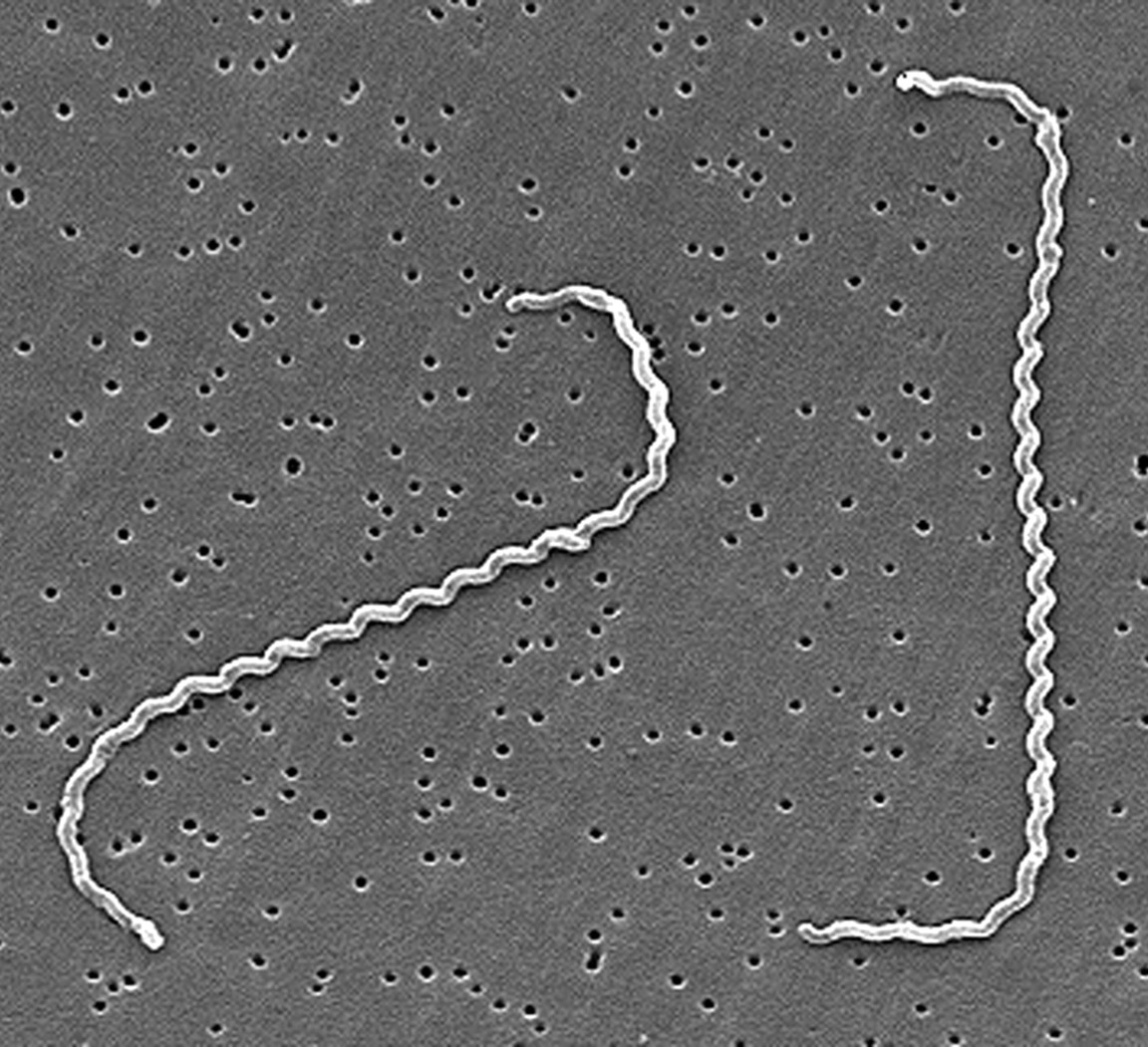
Source: “Centers of Disease Control and Prevention.& R. Weyant (n.d.). Electron micrograph of L. interrogans serovar icterohaemorrhagiae [Photograph]. Public Health Image Library.” https://phil.cdc.gov/details.aspx?pid=1220
Source of Outbreak:
Canine leptospirosis has been a known and common zoonotic disease for over 100 years in America. With cases steadily rising each year in canines, it’s important to understand which factors contribute to the prevalence of outbreaks and its disproportionate rate of incidence in different areas. Understanding the source of outbreaks is also important for public health since canine leptospirosis can be a zoonotic transmitted disease. The environmental factors characterizing different regions are the most noticeable source of outbreaks in multiple studies which have retrospectively studied this disease. In a recent meta-analysis, dogs exposed to large environmental water sources were around 68% more likely to be infected. Stagnant water sources and floods seem to be frequently contaminated with leptospira serovars. The pathogens which are shed through the urine of infected animals survive for long periods of time when found in water, sustaining themselves until they come in contact with a susceptible host’s skin or mucous membranes. Areas inhabited with wildlife such as suburbs, fields or forests have an even higher potential of leptospirosis infection since there are no preventative measures to reduce their rate of transmission. Although studies were able to easily identify different variables contributing to outbreaks, the correlation between the risk of being infected with disease with these variables has yet to be proven.
Cause of Outbreak:
Being a widely known disease and pathogen, canine leptospirosis outbreaks are frequently reported. A notable example of this is the Maricopa County outbreak in Arizona reported in 2017. The state of Arizona has been reporting multiple clusters of the infection in the last few years despite its unfavourable environment. Arizona receives low amounts of annual rainfalls and lacks the humidity encouraging the growth and dissemination of the organisms. Knowing that leptospirosis is less frequent in drier desert environments such as Arizona, public health officials found it crucial to find out the cause of these canine leptospirosis outbreaks.
Veterinarians in Arizona are required to report new cases of leptospirosis. However, a survey showed that only 57% of veterinarians in the state knew when and how to properly report a zoonotic disease. The survey also noticed that veterinarians treating small animals were the least informed on zoonotic disease reporting. Shockingly, leptospirosis is only reported by 42% of the animal health professionals surveyed. Similarly, 12% of the small animal veterinarians of Arizona believed that canine leptospirosis was not a reportable disease.
Socioeconomic factors are also a large reason for outbreaks of the disease in dogs. Studies show that dogs living in lower-income neighborhoods were more exposed to the various environmental factors associated with the disease and had less access to veterinary care and lower vaccination rates. Historically, lower-income areas often have a lesser understanding and knowledge regarding vaccines which makes them hesitant to vaccinate their pets. On the other hand, studies also showed that some middle-class areas had higher infection rates. This was suggested to be caused by an increased amount of testing being possible with the higher income of clients in the area.
The 2017 outbreak presented many cases with novel symptoms less commonly associated with leptospirosis such as conjunctivitis and vomiting. Less than 40% of the cases exhibited signs of fever, the most common clinical sign of canine leptospirosis. This leads to the disease not being tested as quickly as it should be, increasing the chances for an infected animal to contaminate its environment. Essentially, the diagnosis of this disease in canines is quite challenging as it is affected by the stage of the disease, the vaccination status of the individual, the environment and potential exposure to the pathogen. The available tests each have their strengths and weaknesses. Using a combination of potential treatments tends to be the most effective and efficient way to get a quick and accurate result but also more expensive for pet owners.
These studies identified that due to the unfavourable environment in Arizona, veterinarians don’t tend to be as vigilant or up to date with the latest protocols regarding the disease than veterinarians in high risk areas.
Furthermore, this outbreak was composed majoritarily of cases from dogs living in urban areas. This also indicates that different attitudes and practices among veterinarians in different areas most likely heavily impacted the prevalence of the disease.
Preventative Measures:
With veterinarians being the first to work with and diagnose the disease, they need to keep up with the latest strategies and protocols. This is crucial when examining recent outbreaks of diseases such as leptospirosis that occured in Maricopa County in 2017.
The first step to ensure the prevention of the disease in animals as well as humans is increasing disinfection and improving hygiene when there is possible contact with animal urine. These are easy and necessary precautions which should be taken regardless of the area’s risk for leptospirosis. In addition, limiting the amount of contact between dogs and any potentially contaminated water source is also an essential first step in preventing the disease.
The most efficient way for veterinarians to prevent canine leptospirosis is by administering vaccines. Since a multitude of wild animals frequently shed leptospires, it is impossible to assume that domesticated animals could achieve herd immunity. Too many animals acquire the disease from water or soil contaminated by infected wildlife. The vaccine allows for dogs to have antibodies against the disease. These new antibodies allow the immune system to be equipped to quickly clear the infection should the animal come into contact with the pathogen. As seen with the study conducted by Blanchard et al., (Figure 2) introducing annual vaccines which target several of the common serovars associated with leptospirosis can decrease the ability for the organism to colonize the host. This is an effective practice that can reduce the amount of potential reservoirs in an environment as a result. Leptospirosis vaccines should also be mandatory in places such as kennels and groomers to decrease the chances of spreading the disease to more individuals. Infected dogs should be isolated and their urine should be contained and disinfected in order to decrease the chances of infecting another potential host as well.
Finally, veterinarians should be aware of the latest news regarding the disease, especially with the number of cases rising each year. Improving education and awareness about zoonotic diseases will allow veterinarians to prevent, treat and most importantly, manage the incidence rate of canine leptospirosis. Creating a standardized vaccination protocol would be beneficial in reducing the impact of the conflicting approaches between veterinarians. It would also help prevent outbreaks in unsuspecting areas such as Arizona. There is also a need for better communication between veterinarians and public health officials to prevent outbreaks and reduce the number of cases overall.
Figure 2: Comparison of different serovar groups growth associated with bacterial leptospirosis in vaccinated dogs compared to the unvaccinated
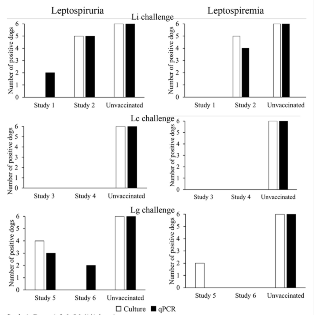
Source: “Blanchard, S., Cariou, C., Bouvet, J., Valfort, W., Oberli, F., Villard, S., Barret-Hilaire, F., Poulet, H., Cupillard, L., & Saint-Vis, B. (2021, June 18). [Photograph]. Quantitative Real-Time PCR Assays for the Detection of Pathogenic Leptospira Species in Urine and Blood Samples in Canine Vaccine Clinical Studies: A Rapid Alternative to Classical Culture Methods.” https://journals-asm-org.proxy3.library.mcgill.ca/doi/10.1128/JCM.03006-20
Conclusion:
To conclude, the Maricopa County canine leptospirosis outbreak of 2017 was largely due to veterinarians and health officials not considering the state to be at risk for this disease in dogs due to the dry and desertic climate. However, since wildlife have a large role in spreading the disease, they can cause outbreaks in unsuspecting areas where domesticated animals might be less protected and where prevention against canine leptospirosis isn’t as common. Therefore, as cases rise, it will be important for veterinarians all over to increase the amount of prevention against canine leptospirosis and to increase awareness about the disease in order to reduce the chances for future outbreaks.
References:
André-Fontaine G. (2006). Canine leptospirosis–do we have a problem?. Veterinary
microbiology, 117(1), 19–24. https://doi.org/10.1016/j.vetmic.2006.04.005
Farr R. W. (1995). Leptospirosis. Clinical infectious diseases : an official publication of the
Infectious Diseases Society of America, 21(1), 1–8. https://doi.org/10.1093/clinids/21.1.1
Goldstein, R. E. (2010, November 1). Canine Leptospirosis. ScienceDirect. https://www.sciencedirect.com/science/article/pii/S0195561610000951?via%3Dihub#bib6
Goldstein, R. E., Lin, R. C., Langston, C. E., Scrivani, P. V., Erb, H. N., & Barr, S. C. (2008,
May 1). Influence of Infecting Serogroup on Clinical Features of Leptospirosis in Dogs. Wiley Online Library. https://onlinelibrary.wiley.com/doi/abs/10.1111/j.1939-1676.2006.tb02886.x
Jenni, M. L., Woodward, P., Yaglom, H., Levy, C., Iverson, S. A., Kretschmer, M., Jarrett, N.,
Dooley, E., Narang, J., & Venkat, H. (2019, November 15). Knowledge, attitudes, and practices among veterinarians during an outbreak of canine leptospirosis — Maricopa County, Arizona, 2017. ScienceDirect. https://www.sciencedirect.com/science/article/pii/S0167587719305689
Langston, C. E., & Heuter, K. J. (2003, July 1). Leptospirosis: A re-emerging zoonotic disease.
ScienceDirect. https://www.sciencedirect.com/science/article/pii/S0195561603000263?via%3Dihub
Reagan, K. L., & Skyes, J. E. (2019, July 1). Diagnosis of Canine Leptospirosis. ScienceDirect.
https://www.sciencedirect.com/science/article/pii/S0195561619300385?via%3Dihub
Rentko, V. T., Clark, N., Ross, L. A., & Schelling, S. H. (1992, July 1). Canine Leptospirosis: A
Retrospective Study of 17 Cases. Wiley Online Library. https://onlinelibrary.wiley.com/doi/10.1111/j.1939-1676.1992.tb00345.x
Ricardo, T., Previtali, A., & Marcelo, S. (2020, August 1). Meta-analysis of risk factors for
canine leptospirosis. ScienceDirect. https://www.sciencedirect.com/science/article/pii/S0167587720301513?via%3Dihub
Sessions, J. K., & Greene, C. E. (2004, August). Canine Leptospirosis: Epidemiology,
Pathogenesis, and Diagnosis. Auburn University College of Veterinary Medicine. http://vetfolio-vetstreet.s3.amazonaws.com/mmah/b1/b3b4ac15fb443ebb9d66e46acc513e/filePV_26_08_606_0.pdf
Taylor, C., O’Neill, D. G., Catchpole, B., & Brodbelt, D. C. (2021, May 31). Incidence and
demographic risk factors for leptospirosis in dogs in the UK. British Veterinary Association. https://bvajournals.onlinelibrary.wiley.com/doi/10.1002/vetr.512
Venkat, H., Yaglom, H. D., & Adams, L. (2019, August 1). Knowledge, attitudes, and practices
relevant to zoonotic disease reporting and infection prevention practices among veterinarians â Arizona, 2015. ScienceDirect. https://www.sciencedirect.com/science/article/pii/S0167587718305208?via%3Dihub
Ward, M. P. (2002, December 30). Seasonality of canine leptospirosis in the United States and
Canada and its association with rainfall. ScienceDirect. https://www.sciencedirect.com/science/article/pii/S0167587702001836?via%3Dihub
White, A. M., Zambrana-Torrelio, C., Allen, T., Rostal, M. K., Wright, A. K., Ball, E. C.,
Daszak, P., & Karesh, W. B. (2017, April 1). Hotspots of canine leptospirosis in the United States of America. ScienceDirect. https://www.sciencedirect.com/science/article/pii/S109002331730059X?via%3Dihub
