
Introduction
Haemophilus parasuis is a Gram-negative, rod-shaped bacterium that causes Glasser’s disease in pigs. While H. parasuis is often found in swines’ upper respiratory tract (URT) as part of their normal microbiota, stressed and immunocompromised pigs are susceptible to infection and subsequent disease, which is characterized by pneumonia, fibrinous polyserositis, polyarthritis and meningitis. In 1910, Glasser was the first to describe the bacterial infection, and by 1943, it was evident that the causal pathogen was H. parasuis. Since then, worldwide outbreaks of Glasser’s disease are on the rise, and infection afflicts almost every large-scale swine production system. H. parasuis is also implicated in increasing the morbidity and mortality of other viral infections, such as Porcine Reproductive and Respiratory Syndrome Virus.
Disease
H. parasuis is most commonly transmitted via direct contact, but is known to also be transferred via aerosol inhalation. Although not well characterized, H. parasuis is thought to infect the pig when its immune system is compromised or when the host experiences stress. The bacteria first attach to and colonize the mucosa of the nasal cavity. Once inside the bloodstream, H. parasuis targets the epithelial cells lining the chest, abdomen, brain cavity, heart sac, and joints (serosal membranes). Once the bacteria adhere to the epithelial cells, it induces cell death, which in turn releases signaling molecules that induce the host’s immune system. This will then cause inflammation and can lead to severe inflammatory injuries, such as swollen joints and purple extremities. Currently, there have been 21 different serovars, variations of H. parasuis with different surface proteins, classified. The different serovars range in virulence with the most virulent being able to invade endothelial cells. However, this invasion is not required for disease to occur.
H. parasuis can cause three clinical forms of Glasser’s disease—a sporadic form in young pigs, a second form characterized by sub-capsular kidney bleeding and sudden death, and a third form as a secondary agent in infections of Circovirus and virus causing swine Reproductive and Respiratory Syndrome. At the beginning of the acute form, pigs often show symptoms of increased body temperature of above 40 °C, lethargy, coughing, anorexia, and incoordination. If H. parasuis crosses the endothelial cells lining the blood-brain barrier and reaches the central nervous system, it can cause meningitis, which is characterized by inflammation of the sacs that surround the brain. This disturbs the nervous system and can cause neurological clinical signals such as tremors and convulsions. If the disease progresses to chronic form, then chronic arthritis and severe fibrosis can occur and stunt growth rate.
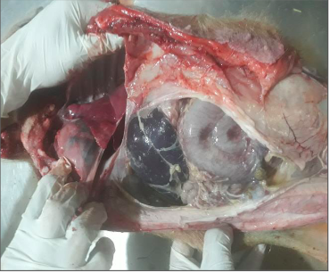
Figure 1: Three months old piglet that died due to Glasser’s Disease. It is exhibiting severe inflammation of the peritoneum (serous membrane lining the cavity of the abdomen and covering the abdominal organs) with accumulation of fluid in the abdominal cavity. (Source: International Journal of Veterinary Sciences and Animal Husbandry, S. Jyoti, R. Nepal, Dr. A. Thapa, Dr. S. Rimal, 2019)
Epidemiology
H. parasuis infection often occurs when an animal is purchased from an infected herd, thus introducing the bacteria to an unaffected herd (Figure 2). An infected herd is susceptible to an infection by a different serovar of H. parasuis because infection by one serovar does not provide immunity towards another serovar of H. parasuis. Therefore, a herd that was previously infected by one serovar can become infected by a different serovar of H. parasuis. The introduction of the new serovar occurs when a pig infected with the different serovar is brought into the herd. For example, a herd that was infected with serovar 1 does not have immunity towards serovar 2. If a pig infected with serovar 2 is introduced to this herd, then the pigs of the herd can become infected with serovar 2. When infection happens, animals of all ages are susceptible of contracting the disease, possibly leading to an outbreak. During an H. parasuis infections outbreak, infection rate of adults can reach 15% and young pigs 50%.
Piglets are often the most afflicted with Glasser’s disease as infected mothers can transfer the bacteria through their breast milk. Piglets colonized by pathogenic variants will become carriers of the disease, but are immune. This is because their immune systems are able to develop while receiving immune protection from proteins in the mother’s milk. These animals will later carry the disease, but will not exhibit any observable symptoms. However, piglets that are not colonized by pathogenic serovar are highly susceptible to infection when they stop suckling. Since their developing immune systems have not been exposed to the pathogenic H. parasuis, the piglets are not able to ward off infection, leading to high rates of Glasser’s disease at the age of 5 to 6 weeks.
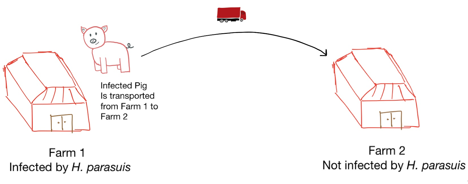
Figure 2: Illustration showing a possible case of transmission of H. parasuis. Source: J. Dujardin, 2019.
Virulence
During infection, H. parasuis firstly invades the lung epithelium using a variety of virulence factors. As the first step in this process, the bacteria adheres to the host epithelium. This is facilitated by fimbriae, which are adhesion proteins on the bacterial surface. Secondly, H. parasuis must avoid the host’s immune system, which has cells trying to engulf and process the bacteria. For example, alveolar macrophages are innate immune cells located in the lungs’ alveoli and they constantly pick up bacteria to try and identify pathogens. When they find an invading microbe, macrophages will initiate an innate immune response. In order to circumvent these cells, H. parasuis has two virulence factors—a capsule and imitation of host cells’ surfaces. The capsule is a protective cover that is part of its outer membrane and protects the bacteria from most host immune responses, including engulfment by macrophages. All host cells are covered in sialic acid; therefore, H. parasuis mimics this by sialylation of their capsule layer. By having this virulence factor, the bacteria evade digestion. H. parasuis does this in an attempt to delay macrophage activity and, ultimately, the immune response.
If the macrophage is successful in engulfing H. parasuis, the cell still has to kill the bacteria via the release of destructive proteins and molecules. However, H. parasuis’s capsule interferes with the macrophage’s ability to mount this response. Therefore, the bacteria are able to live intracellularly in these immune cells. Once inside the macrophage, the bacteria prevent certain inflammatory messages from being produced as well as modify the surface proteins on the macrophage’s surface. This delays the host’s inflammatory response and gives the bacteria time to spread throughout the body. They can then enter the bloodstream and colonize elsewhere, such as the liver, brain and kidneys. In fact, H. parasuis itself is not what causes death when a pig is infected, it is rather the delayed-onset of systemic inflammation, which sometimes can lead to septic shock.
Overall, much is still unknown and not understood regarding H. parasuis. Further identification of virulence factors and their respective mechanisms will help elucidate aspects of its infection. Many aspects of its infection remain to be associated with and explained by identification of more virulence factors.
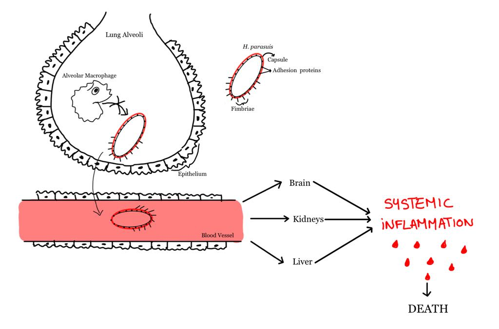
Figure 3: Bacterial infection process in the lung alveoli and consequent spreading throughout the body via the bloodstream. By G. Mezentzeff and V. Guay, 2019.
Prevention and Treatment
Good animal hygiene and nutrition as well as animal management are important factors that can help prevent incidence of infection. Most importantly, transport and the raising of animals need to be closely monitored to prevent the spread of disease to new regions and prevent outbreaks. Antibiotics, specifically prophylactic antimicrobials, can also be used as a method of prevention in piglets. This is useful since H. parasuis is able to colonize piglets as early as less than ten days of age.
Another possible method used to control H. parasuis infection is vaccination, typically through the single dose injection of Parasail HPS injectable vaccine or Ingelvac HP-1 vaccine in the intramuscular space of the animal. Vaccines are administered to both piglets and mothers. The proportion of animals that are infected by H. parasuis is significantly lower in vaccinated than non vaccinated animals. Revaccination should take place right after the piglets stop drinking milk in order to provide the necessary protection against H. parasuis. Idealially, H. parasuis infection is prevented through the combination of vaccination of the sows and piglets and prophylactic antibiotic treatment of newborn piglets.
In the case where an animal is infected with H. parasuis, antibiotics can be used. For example, H. parasuis is successfully treated with synthetic penicillin, which acts to weaken and burst the cell walls of the bacteria. The drugs accomplish this by preventing the bacteria’s peptidoglycan, a cell wall layer, from crosslinking effectively during cell wall synthesis. Enroflox, Excede, and Draxxin, are all injectable over the counter antimicrobial solutions and three of the most effective treatments against H. parasuis. It is recommended to target antibiotic therapy towards the sows, as well as piglets, and to begin treatment immediately after symptoms are observed. The dose administered depends on the severity of the infection: if the disease has spread to the tissues, spinal fluids, or has affected joints, a higher dose is used.
Word count 1466
References
Brockmeier, Susan L., et al. “Virulence and draft genome sequence overview of multiple strains of the swine pathogen Haemophilus parasuis.” PloS one 9.8 (2014): e103787.
Costa-Hurtado, Mar, et al. “Changes in macrophage phenotype after infection of pigs with Haemophilus parasuis strains with different levels of virulence.” Infection and immunity 81.7 (2013): 2327-2333.
Iowa State University Haemophilus parasuis (Glasser’s Disease) Iowa State University. (2019). Retrieved November 13, 2019, from Iastate.edu website: https://vetmed.iastate.edu/vdpam/FSVD/swine/index-diseases/glasser-disease
Nedbalcova, K., et al. “Haemophilus parasuis and Glässer’s disease in pigs: a review.” Veterinarni Medicina 51.5 (2006): 168-179.
Oliveira, Simone, and Carlos Pijoan. “Diagnosis of Haemophilus parasuis in affected herds and use of epidemiological data to control disease.” Journal of Swine Health and Production 10.5 (2002): 221-225.
Oliveira, Simone, P. J. Blackall, and Carlos Pijoan. “Characterization of the diversity of Haemophilus parasuis field isolates by use of serotyping and genotyping.” American journal of veterinary research 64.4 (2003): 435-442.
Oh, Y., Han, K., Seo, H. W., Park, C., & Chae, C. (2013). Program of vaccination and antibiotic treatment to control polyserositis caused by Haemophilus parasuis under field conditions. Canadian journal of veterinary research, 77(3), 183-190.
Pipestone Veterinary Sevices (PVS). (2018). Retrieved November 11, 2019, from https://pipevet.com/glassers-disease-in-swine.
Rapp-Gabrielson, V. J. “Haemophilus parasuis. 6 Disease of Swine th. Ed.: 475-481. Straw, B.8 E. et al. eds.” (1999).
Sack, Meike, and Nina Baltes. “Identification of novel potential virulence-associated factors in Haemophilus parasuis.” Veterinary microbiology 136.3-4 (2009): 382-386.
Solano-Aguilar, G. I., et al. “Protective role of maternal antibodies against Haemophilus parasuis infection.” American journal of veterinary research 60.1 (1999): 81-87.
White, Mark. NADIS – National Animal Disease Information Service. 2012, https://www.nadis.org.uk/disease-a-z/pigs/glaessers-disease/.
