Category Archives: Uncategorized

Leptospira interrogans

By Laetitia Gaurier and Danaelle Page

Introduction

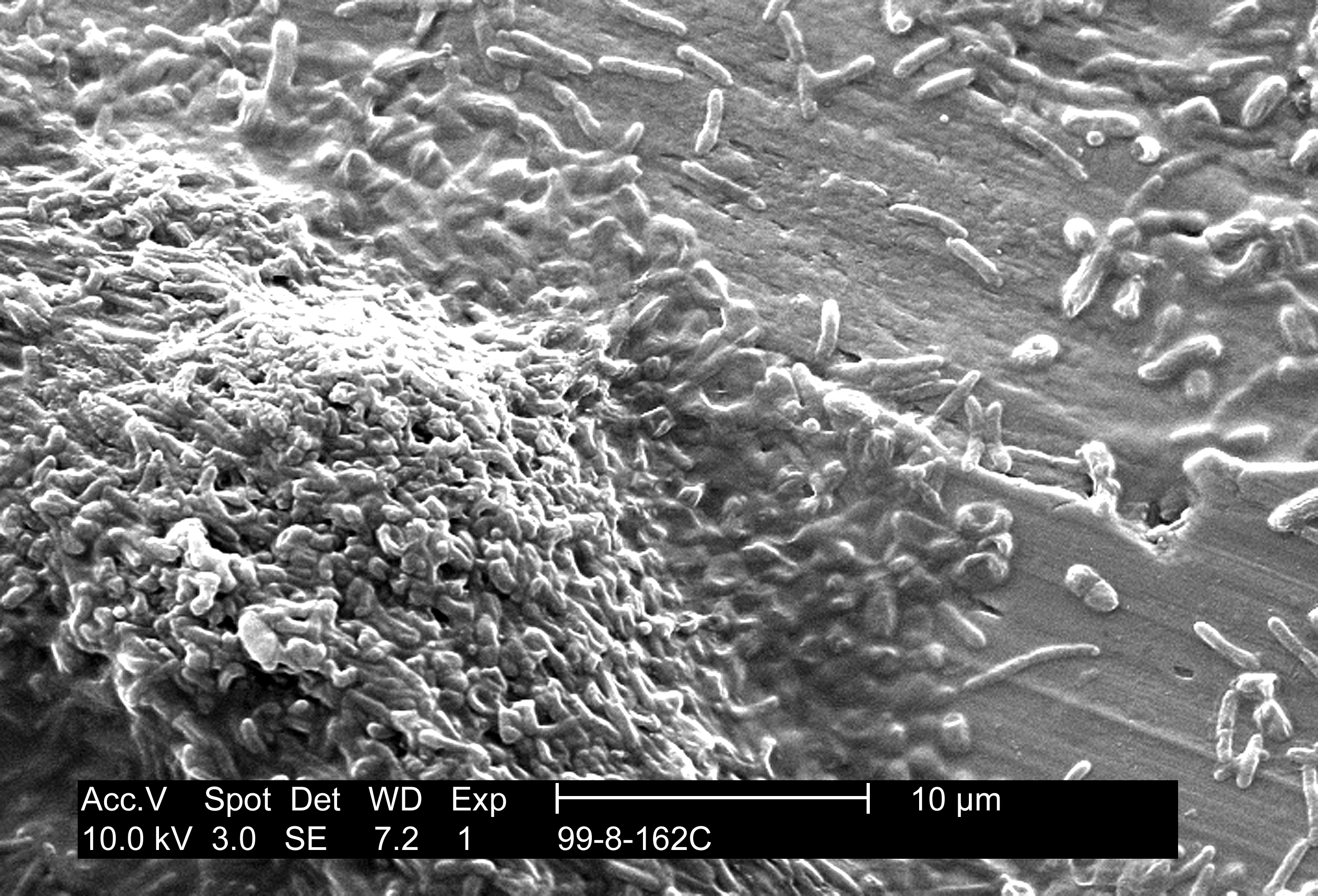
Leptospira interrogans causes leptospirosis. This disease is a zoonosis affecting many animal species and it can be transmitted to humans. These agents of disease, called leptospires, are a type of spirochetes, highly motile cylindrical cell bodies that grow in presence of oxygen and use axial filament rotation for motility (see Figure 1). L. interrogans is found worldwide, but mostly in hot and humid regions (e.g Asia, Latin America and Africa). This species encompasses many serovars, i.e a distinguishable variant of L. interrogans that can be pathogenic (cause disease) or not.

Figure 1. Scanning Electron Micrograph of Leptospira interrogans. Two spirochetes are bound to a 0.2-μm filter. Notice the coiling of the cell. Source: CDC/NCID/HIP/Janice Carr, doi:10.1371/journal.pmed.0030302.g001.

Figure 1. Scanning Electron Micrograph of Leptospira interrogans. Two spirochetes are bound to a 0.2-μm filter. Notice the coiling of the cell. Source: CDC/NCID/HIP/Janice Carr,
doi:10.1371/journal.pmed.0030302.g001.

Disease

L. interrogans can be transmitted to humans by skin or mucosal contact with urine-contaminated soil or fluids from contaminated animals (see Figure 2). Leptospires migrate through the host tissues, invade cells and reach important organs of mammals. They activate macrophages, immune cells that protect the host by eating and digesting anything foreign encountered. Activation happens through binding of bacterial surface receptors that elicit the host immune response. Antibodies specific for lipids exposed on the bacterial membrane (lipopolysaccharides) are secreted against the leptospires and provide protection against reinfection.

Figure 2: Leptospirosis transmission route. Reservoir for L. interrogans transmission include rodents species, wild and domestic animals, as well as contaminated environment. Humans are accidental hosts and do not spread the microorganism in the environment ; they are not reservoirs for transmission. Pathogenic leptospires penetrate wounded skin or mucous membranes, enter the bloodstream and disseminate throughout the body tissue. Adapted from: Ko, A.I., Goarant, C. and Picardeau, M. 2009. Leptospira: the dawn of the molecular genetics era for an emerging zoonotic pathogen. Nature Reviews Microbiology. 7: 736-747. Available from: DOI: 10.1038/nrmicro2208

Figure 2: Leptospirosis transmission route. Reservoir for L. interrogans transmission include rodents species, wild and domestic animals, as well as contaminated environment. Humans are accidental hosts and do not spread the microorganism in the environment ; they are not reservoirs for transmission. Pathogenic leptospires penetrate wounded skin or mucous membranes, enter the bloodstream and disseminate throughout the body tissue.
Adapted from: Ko, A.I., Goarant, C. and Picardeau, M. 2009. Leptospira: the dawn of the molecular genetics era for an emerging zoonotic pathogen. Nature Reviews Microbiology. 7: 736-747. Available from: DOI: 10.1038/nrmicro2208

Symptoms of leptospirosis vary greatly: asymptomatic, fever, affected liver, lungs or kidneys. In extreme cases, it can result in multiple organ failure. This disease presents itself either as Weil’s disease (jaundice form), or as icteric plurivisceral form (jaundice and many organs affected). The latter progresses through three phases:

1) The asymptomatic incubation phase; leptospires gain the bloodstream.

2) The pre-icteric phase or the invasion of the host tissues.

3) The icteric phase where non-specific antibodies appear and symptoms increase and   decrease.

Leptospirosis is usually poorly diagnosed due to the large variety and non-specificity of symptoms. Many methods of diagnosis exist, but the three most common are: polymerase chain reaction (PCR), microscopic agglutination test (MAT) and rapid genus-specific tests. The PCR method detects leptospires’ DNA in the analyzed patient sample. The MAT technique is the reference test; it detects antibodies against leptospires present in the sample. Rapid genus-specific tests are faster than MAT method at detecting unspecific antibodies, but complete diagnosis requires confirmation with the MAT technique.

Epidemiology

Leptospirosis represents a public health threat as a potential epidemic and newly emerging infectious disease. Reservoirs of this zoonosis include rodents, livestock and dogs (see Figure 2). Its incidence is normally higher in tropical and subtropical areas like South America due to climatic conditions; outbreaks from various serovars have been linked to floods, hurricanes or heavy rainfall. The number of cases worldwide is not known precisely, partly due to difficulties in establishing clear diagnosis, but recent estimates indicate more than 500,000 cases annually reaching 10% mortality. Leptospirosis may be an occupational hazard through direct or indirect urine contact. This transmission mode threatens pit workers, outdoors workers like farmers and animal contact workers like veterinarians. This disease also represents a recreational hazard for swimmers. For example, several leptospirosis outbreaks occurred following triathlons.

Virulence factors

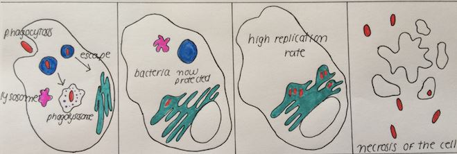
L . interrogans different serovars have their own virulence factors suspected to play a role in leptospirosis causation. Among these virulence factors, we find toxin production. A toxin is a poisonous substance secreted by a pathogenic bacterium to harm host cells. Haemolysins, a toxin category, lyse or burst host red blood cells and are produced by L. interrogans. Bacterium attachment to host epithelial cells (i.e cells covering body surfaces such as skin, mucosa, etc.) also represents a virulence factor. L. interrogans especially attaches to renal epithelial cells, but host immune cells such as macrophages can fight this by engulfing invading bacteria. Surface proteins (lipopolysaccharides) allow L. interrogans to be recognized as pathogenic by macrophage and elicit an immune response. This response consists of antibodies production; those target and attack specifically the infecting leptospires. However, our bacterium defends itself by killing macrophages through elevation of their intracellular calcium levels. Last but not least, host antibodies may harm the host itself since L. interrogans can disappear from bloodstream. Antibodies could start attacking host red blood cells or platelets; this last virulence factor is immune-mediated and aggravates disease symptoms.

Treatment

Various antibiotics usually clear L. interrogans depending on the gravity of the infection (e.g penicillin (or penicillin G), doxycycline, ampicillin and amoxicillin). A few new antibiotics are being tested (e.g cefepime, ertapenem, norfloxacin). All antibiotics used and tested so far seem to control the disease. Also, vaccines made from killed bacteria are used in humans and animals to promote temporary immunity against leptospirosis.

References

Assez, N., Mauriaucourt, P., Cuny, J., Goldstein, P. and Wiel, E. 2013. Ictère fébrile… et si c’était une leptospirose. À propos d’un cas de L. interrogans Icterohaemorrhagia dans le Nord de la France. Annales Françaises d’anesthésie et de Réanimation. 32: 439-443.

Bharti, A.R., Nally, J.E., Ricaldi, J.N., Matthias, M.A. et al. 2003. Leptospirosis : a zoonotic disease of global importance. The Lancet Infectious Diseases. 3: 757-771.

Haake, D.A. and Levett, P.N. 2015. Leptospirosis in Humans. Current Topics in Microbiology and Immunology. 387: 65-97. Available from: DOI: 10.1007/978-3-662-45059-8_5

Lee, S.H., Kim, K.A., Park, Y.G., Seong, I.W., Kim, M.J and Lee, Y.J. 2000. Identification and partial characterization of a novel hemolysin from Leptospira interrogans serovar lai. Gene. 254(1-2): 19-28. Available from: DOI: 10.1016/S0378-1119(00)00293-6

Levett, P.N. 2001. Leptospirosis. Clinical Microbiology Reviews. 14(2): 296-326. Available from: DOI: 10.1128/CMR.14.2.296-326.2001

World Health Organization. 2003. Human Leptospirosis: Guidance For Diagnosis, Surveillance And Control. Malta. Retrieved from: http://apps.who.int/iris/bitstream/10665/42667/1/WHO_CDS_CSR_EPH_2002.23.pdf

World Health Organization. 2010. Report of the first meeting of the Leptospirosis Burden Epidemiology Reference Group. Switzerland, Geneva. Retrieved from: http://apps.who.int/iris/bitstream/10665/44382/1/9789241599894_eng.pdf

Zhang, W., Zhang, N., Wang, W., Wang, F. et al. 2014. Efficacy of cefepime, ertapenem and norfloxacin against leptospirosis and for the clearance of pathogens in a hamster model. Microbial Pathogenesis. 77: 78-83. Available from: http://dx.doi.org/10.1016/j.micpath.2014.11.006

Zhao, J.F., Chen, H.H., Ojcius, D.M., Zhao, X., Sun, D., Ge, Y.M., Zheng, L.L, Lin, X., Li, L.J and Yan, J. 2013. Identification of Leptospira interrogans Phospholipase C as a Novel Virulence Factor Responsible for Intracellular Free Calcium Ion Elevation during Macrophage Death. PLoS One. 8(10): e75652. Available from: DOI: 10.1371/journal.pone.0075652

Brucella abortus

by Christine L. Toma and Justine Hadrava

Introduction

Brucella abortus is an intracellular pathogen causing brucellosis, an infectious disease also referred to as undulant fever, Mediterranean fever or Malta fever. Brucellosis is categorized as a zoonosis, meaning that the infection primarily targets animals, but is also transmissible to humans. The principal hosts of B. abortus are livestock animals, but the clinical effects are more severe in humans. In addition to being of economic importance, B. abortus represents a threat to public health.

Disease

Predominantly in cattle, but also sometimes in sheep, goats and pigs, B. abortus infection is typically localized in the reproductive system of sexually mature animals. Both females and males can be affected, although more severe complications are observed in females. Brucellosis is usually characterized by abortion, premature birth, retained placenta and lowered milk production. Infertility occurs between two weeks to five months following the infection, but animals regain their reproductive capability afterwards. B. abortus transmission to humans is accomplished by direct or indirect contact with infected animals or animal products.

In humans, B. abortus induces severe illnesses involving fever, malaise, anorexia, depression, physical weakness, weight loss, headache and bone and muscle pain. It has the ability to spread to and damage any organ of the body and thus has the potential to cause what is called a systemic infection. In addition, all age groups are affected, contrary to animals. During pregnancy, B. abortus can cause spontaneous abortion, but can also be transmitted to the infant. The complications of the infection can be persistent and last for multiple weeks or months. Since the manifestations of B. abortus infection symptoms are highly variable and non-specific, laboratory tests need to be performed to confirm a diagnosis.

Epidemiology

Transmission between animals results mainly from contact following abortion or by ingestion of pasture contaminated with the bacteria. However, other inoculation source of B. abortus include infected milking devices and artificial insemination with contaminated semen. Susceptibility increases as the animal becomes sexually mature and decreases at least a thousand fold under vaccination. Transmission is also favored in high density herds. Animal brucellosis rarely leads to death, but the abortion rate of infected individuals is between 30% and 80%.

B.abortus is predominantly transmitted to humans from contaminated environments (Figure 1). Therefore, any farmworker, animal attendant, veterinarian, inseminator or worker involved in animal product processing is more susceptible to acquire brucellosis. B. abortus can survive for a long period of time in dung, water, dust, soil, meat and dairy products. Therefore, route of exposure includes inhalation of contaminated dust, dried dung etc., direct contact with an animal or its fluids, or accidental ingestion. For people who are not working with animals or animal parts, eating raw meat or unpasteurized milk or milk products is the major cause of B. abortus infection. The rate of brucellosis varies greatly between locations and can range from 0.01 to more than 200 cases per 100 000 inhabitants. However, in the US, brucellosis occurrence is less than 0.05 per 100 000 inhabitants. In addition, the mortality rate varies from 2% to 5% in untreated individuals and is extremely rare if appropriate treatment is received.

Figure 1: Summary of the major possible transmission routes of B. abortus to humans (figure by Justine Hadrava).

Figure 1: Summary of the major possible transmission routes of B. abortus to humans (figure by Justine Hadrava).

Virulence

Successful colonization of a host by B. abortus is achieved by molecular determinants that allow invasion, resistance to intracellular killing and replication in host cells. B. abortus invade the mucosa and are ingested by immune cells responsible for digesting bacteria called phagocytes. Specific molecules, such as a two component system and host cellular projections, are known to be responsible for the binding and integration of bacteria into host cells, but not all epithelial cells will allow amalgamation with B. abortus.

When the bacteria is ingested, it goes through a pathway to avoid phagolysosome fusion, a process in which pathogens are digested and killed by enzymes, and reach its replication niche (Figure 2). This bactericidal killing is not avoided by all bacteria, in fact after 48 hours only 15-30% of the immune cells display bacterial replication.

B. abortus possesses a particular lipopolysaccharide (LPS) on its outer membrane, a chemical structure that usually induces an immune response. Since this LPS is in a non-classical form, intracellular invasion and survival is more likely.

Replication occurs in membrane bound compartments resembling the endoplasmic reticulum (an organelle) of the cells (Figure 2). Bacteria can survive in cellular environments through the formation of autophagosome, a spherical structure formed by invagination of membrane structures surrounding B. abortus. A high rate of replication occurs when autophagosomes are fused with the endoplasmic reticulum, allowing bacteria to replicate and avoid the host’s immune system strategies. In pregnant animals, B. abortus replicates in the trophoblast, an outer layer that provides nutrients for the embryo and causes abortion.

Extensive replication of the bacteria occurs without disturbing basic cell functions. Necrosis is the final outcome of infected cells and when they burst, bacteria are released and the infection progresses (Figure 2).

Figure 2: B. abortus mechanism to avoid the host immune system within phagosomes (figure by Christine L. Toma).

Figure 2: B. abortus mechanism to avoid the host immune system within phagosomes (figure by Christine L. Toma).

Prevention and Treatment

Animals

The best way to deal with a disease is to prevent it. The key to prevent brucellosis in an animal herd is to be rigorous. It is important to always isolate newly purchased animals to make sure they are brucellosis free. Laboratory techniques used to identify the cause of abortion is also a way to single out sick animals. Good disposal of placentas and non-viable fetuses is important to reduce the risk of contamination. Hygiene and precautionary measures are the best allies for prevention.

The most effective way to control this disease is by vaccination. A strain of B. abortus is injected most commonly in sexually immature females to minimize the risk of abortion caused by the vaccine. It is known to be an effective method for elimination of clinical diseases and to reduce the number of organisms excreted per animal. Eradication of B. abortus is hard to achieve because it requires a surveillance system, laboratory support and a high level of hygiene.

Humans

To prevent human brucellosis, people should reduce exposure to infected animals or byproducts. Hygiene is the best preventative measure for workers who are in contact with animals on a daily basis; this applies to farm, meat production chain and laboratory personnel.

If infection is detected in an individual, an antibiotic treatment is usually prescribed for a specific duration of time. Sometimes a surgical intervention is needed, but full recovery is common.

References

Gorvel JP, Moreno E. 2002. Brucella intracellular life: from invasion to intracellular replication. Veterinary microbiology. 90: 281-297.

The Merk Veterinary Manual: Overview of Brucellosis in Cattle [Internet]. Date unknown. Kenilworth, N.J., U.S.A. Merck Sharp & Dohme Corp; [Updated 2013 July; cited 2015 November 10]. Available from: http://www.merckvetmanual.com/mvm/reproductive_system/brucellosis_in_large_animals/brucellosis_in_cattle.html

Corbel MJ, World Health Organization. 2006. Brucellosis in Humans and Animals.[Internet]. Albany, NY, USA: World Health Organization. [November 10 2015]. Available from: http://site.ebrary.com.proxy3.library.mcgill.ca/lib/mcgill/detail.action?docID=10190682

Corbel M.J. 2006. Brucellosis in human and animals. Geneva : World Health Organization.

The Center for food security and public health. 2009. Bovine brucellosis: brucella abortus.

Mycoplasma pneumoniae

By Laure Fossecave and Kristen Lee

Introduction

Mycoplasma pneumonia (M. pneumoniae) is relatively small with few genes compared to other bacteria. It is a pathogen, a bacteria capable of causing damage to hosts, in this case, humans. It mostly infects lungs and causes community-acquired pneumonia (CAP) such as chronic asthma, but can also be responsible for some other chronic diseases such as Crohn’s disease, an intestine infection (See Figure 1). It acts as a parasite in host cells and mainly infects humans. This bacteria was first discovered in 1944 by Eaton et al. who analyzed sputum in tissue culture from a patient with primary atypical pneumonia. At this time it was known as the Eaton agent.

Figure 1 : X-ray of a patient's thorax revealing pneumoniae infection on the left lung lob. Source: http://phil.cdc.gov/phil/details.asp?pid=5800

Figure 1 : X-ray of a patient’s thorax revealing pneumoniae infection on the left lung lob. Source: http://phil.cdc.gov/phil/details.asp?pid=5800

Disease

M. pneumoniae is a mollicute, a bacteria that lacks a cell wall, leaving it vulnerable to desiccation and therefore it requires close contact with host cells for infection to occur. It is transmitted directly through people, where the bacteria is found in their nose, throat, trachea and sputum. In the body, bacteria infect epithelial tissues of upper and lower respiratory tracts and exfoliate the lung cells. They act like parasites, using host cell nutrients to survive and aid in producing glucose, a molecule used for energy for all organisms. The bacteria is responsible for primary atypical pneumonia. Contracting M. pneumonia induces slow development of symptoms 3 or 4 days after infection, such as pharyngitis or sinus congestion. Only 20 % of people infected by M. pneumoniae are asymptomatic. M. pneumoniae activates the immune responses and induces release of inflammatory molecules. Those molecules are in part responsible for developing chronic lung infections. Any lung damage can persist for weeks and even months, post infection, or can be permanent after total clearance of lung abnormality symptoms. Individuals with allergic sensitization are more susceptible to developing chronic lung conditions after infection with M. pneumonia. M. pneumoniae is also linked to diseases such as pericarditis, inflammatory chronic diseases and auto-immune disease that can involve the nervous system like Guillain-Barre Syndrome.

Epidemiology

Because of its long incubation and transmission periods, and strong persistence in the body, M. pneumoniae infection can lead to prolonged endemics (local outbreak) and even epidemics (worldwide outbreak) in persons of all ages. Most outbreaks are initiated in communities such as military bases, hospitals, schools and families. 40 % of pediatric CAP are due to M. pneumoniae infection, 50 % among children older than 5 years old, and 18 % of these cases need hospitalization. In children, the most common syndrome is tracheobronchitis. However, adults are frequently infected by M. pneumoniae and are hospitalized for bacterial pneumonia in the Unites States.

Figure 2: Representational drawing of a mobile M. pneumoniae bacteria, showing the main internal and external structures, with a simple cytoplasm, a very small genome, a cell membrane consisting of 3 layers, no cell well. The attachment organelle contains adhesin proteins that allow the bacteria to dock on the host’s membrane.

Figure 2: Representational drawing of a mobile M. pneumoniae bacteria, showing the main internal and external structures, with a simple cytoplasm, a very small genome, a cell membrane consisting of 3 layers, no cell well. The attachment organelle contains adhesin proteins that allow the bacteria to dock on the host’s membrane.

Virulence factors

M. pneumoniae is mainly extracellular, does not invade host cells, and lacks a cell wall therefore is highly dependent on the host for nutrients. The bacteria first attaches itself to the host lung cells: this is mediated by an attachment organelle, a structure made of adhesins, proteins that attach themselves to the host membrane (See Figure 2). Because of its molecular composition, the attachment organelle can undergo several mutations such that it is not recognized by the patient’s immune system (the defence mechanism of the body to protect it from pathogens) anymore and can proliferate in the body. This property of the bacteria is one of the reasons why it is difficult to treat patients infected by M. pneumoniae. Additionally, it secretes toxins which activate secretion of cytokines, molecules responsible for inflammation (See Figure 3).

As the disease progresses with time, M. pneumonaie may fuse with host cells, becoming an intracellular pathogen, an event facilitated by its lack of cell wall. This causes internal tissue damage and evasion of certain medication treatments. It may also change host cell surface composition, also contributing to evasion of the immune system

Once it invades host cells, it secretes a molecule called hydrogen peroxide, that reacts with the host molecules (reactive-oxygen species, ROS), destroying the cells of the respiratory tract.
Finally, the bacteria hijacks the host’s immune system so that instead of protecting the patient against the bacteria, the immune system activity against M. pneumoniae causes damage to the patient.

Figure 3 : Basic steps of M. pneumoniae invasion in the respiratory epithelial cells. First, the bacteria docks on the epithelial cell surface by its attachment organelle. It releases toxic molecules that produce a direct injury on the cell. Then, the bacteria crosses the epithelium layer, invade the tissues by entering the blood vessels. Finally, the immune system responds to the entry of the bacteria, resulting in inflammation. Source: http://journals.plos.org/plospathogens/article?id=10.1371%2Fjournal.ppat.1003983.

Figure 3 : Basic steps of M. pneumoniae invasion in the respiratory epithelial cells. First, the bacteria docks on the epithelial cell surface by its attachment organelle. It releases toxic molecules that produce a direct injury on the cell. Then, the bacteria crosses the epithelium layer, invade the tissues by entering the blood vessels. Finally, the immune system responds to the entry of the bacteria, resulting in inflammation.
Source: http://journals.plos.org/plospathogens/article?id=10.1371%2Fjournal.ppat.1003983.

Available treatments

The fact that the bacteria does not have a cell wall contributes to its resistance to beta-lactam antibiotics, a common drug used against bacterial pathogens which normally target components of a bacteria’s cell wall. Instead, patients are treated with macrolides, which target bacterial proteins that mediate metabolic activity, faltering the bacteria’s metabolism. Fluoroquinolones and tetracycline are stronger and more effective alternatives, but have side effects that may be fatal or quite harmful to the patient. As result they are avoided and nearly never used on young children. However, the bacteria can become resistant to macrolides.
When the bacteria causes too much damage to the host, for example destruction of respiratory cells and red blood cells, corticosteroids may be given. In severe cases, a blood transfusion may be critical. Asthma or increased asthmatic symptoms from this disease is to our knowledge either incurable or eradicable with the removal of the pathogen.

References

Eaton MD, Meikejohn G, Van Herick W (1944). Studies on the etiology of primary atypical pneumonia: a filterable agent transmissible to cotton rats, hamsters, and chick embryos. J. Exp. Med. 79:649–667.

Kannan TR, Baseman JB (2006) ADP-ribosylating and vacuolating cytotoxin of Mycoplasma pneumoniae represents unique virulence determinant among bacterial pathogens. Proc Natl Acad Sci U S A; 103(17): 6724–6729

Krause CD, Baseman JB (1983) Inhibition of Mycoplasma pneumoniae Hemadsorption and Adherence to Respiratory Epithelium by Antibodies to a Membrane Protein. Infect Immun; 39(3): 1180–1186

Waites KB, Talkington DF (2004) Mycoplasma pneumoniae and Its Role as a Human Pathogen. Clin Microbiol Rev; 17:697-728

Atkinson TP, Balish MF, Waites KB (2008) Epidemiology, clinical manifestations, pathogenesis and laboratory detection of Mycoplasma pneumoniae infections. FEMS Microbiol Rev; 32:956-73

2012 Quebec City Outbreak of Legionnaires’ Disease

by Sebastien Faucher

Introduction

July 18 2012, Quebec City: a case of Legionaires’ disease is reported to the Public Health Director. In the next week, 4 other cases were reported and an outbreak of Legionnaires’ disease striking Quebec City was declared (Figure 1). Cases continued to appear over the next weeks. The number of cases peaked in late August when 10-20 cases were declared each day. The number of new cases dropped in September and the last case was reported on September 13. The outbreak was officially over on October 8. A total of 183 cases were reported, including 13 deaths.

Figure 1: Timeline of the Quebec City outbreak. The green vertical bars represent the number of cases. Key events are also noted. Source: Trudel, L., Veillette, M., Bonifait, L. & Duchaine, C. Management of the 2012 Legionella crisis in Quebec City: need for a better communication between resources and knowledge transfer. Front. Microbio. 5, 182 (2014).

Figure 1: Timeline of the Quebec City outbreak. The green vertical bars represent the number of cases. Key events are also noted. Source: Trudel, L., Veillette, M., Bonifait, L. & Duchaine, C. Management of the 2012 Legionella crisis in Quebec City: need for a better communication between resources and knowledge transfer. Front. Microbio. 5, 182 (2014).

Legionnaires’ disease

Legionnaires’ disease is caused by a bacterium named Legionella pneumophila. This bacterium infects the lungs causing damage to the tissues, and in some people, death. Individuals who are males, older than 65 years old, who smoke or abuse alcohol or who suffer from chronic diseases, such as diabetes and pulmonary disease, are more at risk of contracting the disease.

Response

Most of the people infected lived or worked in a sector of the city called “La Basse-Ville”. Right from the start, public health authority suspected that a cooling tower could be the source of the outbreak (see below) and setup a plan to find the cooling tower responsible for the outbreak. This plan included the following steps: 1) inventory of the cooling towers in the area, 2) visual inspection of them, 3) analysis of the load and type of L. pneumophila in them, 4) disinfection of all cooling towers, and 5) prescription of control measures. The source of the Quebec City outbreak was found to be a cooling tower located at the center of the geographic distribution of cases. The source was identified based on the presence of the same type of L. pneumophila as in the patients. The number of cases dropped significantly after disinfection and shutdown of this cooling tower.

Source of the outbreak

Legionella pneumophila is transmitted to human through inhalation of contaminated water droplets. In many occurrences of Legionnaires’ disease outbreak, cooling towers are the source of the outbreak, because they offer an environment in which L. pneumophila can grow, and because they creates aerosols, that human can inhale.

A cooling tower is designed to efficiently cool hot water coming from a building. This hot water is usually produced in air conditioning equipment, or during manufacturing processes. The cooling tower mixes hot water with air to cool the water, which is then collected in a basin at the bottom of the tower (Figure 2). In some cooling towers, biofilms may develop on their surfaces. A biofilm is a fixed mass of microorganisms, such as bacteria, embedded in a gel-like substance (Figure 3). The biofilms attract unicellular animals, such as amoeba, that feed on other microorganisms. L. pneumophila can infect and grow inside those amoebas, using them as a bag of food. At this point the cooling tower is all set to produce L. pneumophila: biofilms feed amoebas which then feed L. pneumophila.

Figure 2: Typical design of a cooling tower. Hot water (red) is sprayed over a mesh and mixed with air to cool it down. Cold water (blue) is collected in the bottom basin. Biofilms (green) may develop in the system.

Figure 2: Typical design of a cooling tower. Hot water (red) is sprayed over a mesh and mixed with air to cool it down. Cold water (blue) is collected in the bottom basin. Biofilms (green) may develop in the system. Source: Sebastien Faucher (2015).


Figure 3: Electron transmission micrograph of an artificial biofilm composed of P. aeruginosa, K. pneumoniae and a Flavobacterium sp. Noted the rod-shaped bacterial cells embedded in gel-like substance. Source: Public Health Image Library, Center for Disease Control, Dr. Janice Carr (1999).

Figure 3: Electron transmission micrograph of an artificial biofilm composed of P. aeruginosa, K. pneumoniae and a Flavobacterium sp. Note the rod-shaped bacterial cells embedded in gel-like substance. Source: Public Health Image Library, Center for Disease Control, Dr. Janice Carr (1999).

Causes of the outbreak

The exact cause of the outbreak is still uncertain, but many factors seem to have contributed to this outbreak. Certainly, a set of conditions existed that allowed the growth of L. pneumophila in the Quebec City cooling tower. The overall design of the cooling tower and the water treatment being used, may have contributed to the growth of L. pneumophila.

Normally, poor maintenance of the cooling tower can cause an increase in biofilm, and overall contamination. In this case, the cooling tower was under a maintenance plan, and tested regularly for the presence of L. pneumophila, but lime deposit could have concealed biofilms from detection. At this time, Quebec City experienced a hot and dry summer, which would facilitate the establishment of biofilms in cooling towers and require more powerful water treatment and more frequent test. This is similar to the maintenance of a private pool during warmer periods.

Surprisingly, there were other cooling towers in Quebec City contaminated with L. pneumophila, but none of them harbored the type found in patients. This suggests that the type of L. pneumophila that caused the outbreak was better equipped to cause infection in humans than the types found in the other cooling towers.

Aftermath

The public health director recommended measures to mitigate the risk of future outbreaks, including a national inventory of cooling towers, guidelines for the maintenance of cooling towers and for the treatment of contaminated cooling towers, and protocols for investigating the source of an outbreak. Since then, the Government of Quebec passed a law concerning the owners of cooling towers to ensure that the cooling towers are properly maintained and to alert the public health authorities of problematic situations.

References

Desbiens, F. Éclosion de légionellose dans la ville de Québec, Québec, Canada, été 2012. (Agence de la santé et des services sociaux de la Capitale-Nationale, 2012).

Lévesque, S. et al. Genomic Characterization of a Large Outbreak of Legionella pneumophila Serogroup 1 Strains in Quebec City, 2012. PLoS ONE 9, e103852 (2014).

Marrie, T. J., Garay, J. R. & Weir, E. Legionellosis: Why should I test and report? Canadian Medical Association Journal 182, 1538–1542 (2010).

Murga, R. et al. Role of biofilms in the survival of Legionella pneumophila in a model potable-water system. Microbiology (Reading, Engl) 147, 3121–3126 (2001).

Trudel, L., Veillette, M., Bonifait, L. & Duchaine, C. Management of the 2012 Legionella crisis in Quebec City: need for a better communication between resources and knowledge transfer. Front. Microbio. 5, 182 (2014).

Legionella pneumophila

by Sebastien Faucher

Introduction

Legionella pneumophila is the causative agent of Legionnaires’ disease. This bacterium was isolated in 1977 after a large outbreak of pneumonia stroke Philadelphia in 1976. L. pneumophila can be found in almost any man-made or natural water systems. In this environment, L. pneumophila infects and grows inside amoeba, unicellular animals that normally feed on bacteria.

Disease

L. pneumophila is transmitted to humans by inhalation of contaminated aerosols. Once in the lungs, L. pneumophila is able to infect lung macrophages (Figure 1). Macrophages are cells of the immune systems whose role is to take up and digest invading pathogens. L. pneumophila is able to block the normal activity of the macrophage and use it as a bag of food. This results in the death of the infected macrophage. The infection also causes damage to the nearby lung tissues. In healthy individuals, the infection is cleared by the immune system and is usually asymptomatic, but sometimes symptoms similar to a mild cold may appear. In such case the infection is referred to as Pontiac’s fever. When the immune system of the person infected is unable to clear the infection, massive lung damage will result from it. This is what is called Legionnaires’ disease and the symptoms are similar to pneumonia, including headache, chest pain, fever, non-productive cough and diarrhea.

Figure 1: Microscopic image of fluid extracted from guinea pig infected with L. pneumophila stained with the Gimenez method. L. pneumophila cells are pink, host cells, such as macrophages are blue. Source: Public Health Image Library, Center for Disease Control, Dr. Joseph McDade (1978).

Figure 1: Microscopic image of fluid extracted from guinea pig infected with L. pneumophila stained with the Gimenez method. L. pneumophila cells are pink, host cells, such as macrophages are blue. Source: Public Health Image Library, Center for Disease Control, Dr. Joseph McDade (1978).

Epidemiology

The rate of confirmed Legionnaires’ disease in Ontario for 2008 is 0.61 per 100,000 people. Since the cause of most pneumonia cases is not investigated, the actual rate of the disease is probably much higher. In some European countries, L. pneumophila is now recognized as one of the most common cause of pneumonia. Elderly, males, heavy smokers, alcohol abusers and immunocompromised individuals are more susceptible to the disease. During outbreaks, the mortality rate is typically around 10%.

Contaminated cooling towers located in densely populated area are usually the cause of outbreaks of Legionnaires’ disease, since many individuals are exposed to the aerosols produced. One notable outbreak is the 2012 Quebec City outbreak, which affected 183 persons, including 13 deaths. Sporadic cases also exist. They are usually caused by an isolated source, such as a contaminated domestic water heater. Therefore, only the members of the household where the contaminated unit is located are exposed to the contaminated aerosols, during showering for example.

Virulence systems

During bacterial lung infection, macrophages are called-in to eat and digest the bacteria. A macrophage picks up L. pneumophila by a process called phagocytosis. At this point L. pneumophila is located in a phagosome, a sort of bag, inside the macrophage (Figure 2a). Normally, bacteria that are taken up by a macrophage will be killed and digested. The macrophage accomplishes this by pumping toxic proteins and chemicals into the phagosome. The macrophage is protected from their toxicity because they are activated only once inside the phagosome.

To the demise of the macrophages, L. pneumophila is able to fight back by inhibiting the digestion process of the macrophage. L. pneumophila uses a secretion system called Icm/Dot to take control of the macrophage. This system acts like a syringe to inject toxins through the wall of the phagososme inside the macrophage (Figure 2b). The toxins then modify the normal properties of the macrophage to stop the digestion process describe above by preventing the pumping of toxic enzymes and chemicals insto the phagosome. Moreover, the toxins stimulate the transport of nutrients into the phagosome to support the multiplication of L. pneumophila (Figure 2c). When the bacteria have consumed all the nutrients inside the macrophage, one of the toxin triggers the destruction of the macrophage allowing L. pneumophila to escape and infect other macrophages (Figure 2d). L. pneumophila also produces a number of proteases, enzymes that degrade proteins, which destroy the nearby tissue causing damage to the lungs.

Figure 2: Infection of macrophage by L. pneumophila. A, phagocytosis; B, secretion of Icm/Dot toxins; C, growth inside the macrophage; D, destruction of the macrophage and exit.

Figure 2: Infection of macrophage by L. pneumophila. A, phagocytosis; B, secretion of Icm/Dot toxins; C, growth inside the macrophage; D, destruction of the macrophage and exit.

Treatment

L. pneumophila is usually susceptible to antibiotics normally prescribed to fight pneumonia, such as azithromycin. This antibiotic stops the synthesis of proteins, such as the toxins secreted by the Icm/Dot system, neutralizing the ability of L. pneumophila to inhibit the digestion process of the macrophage. Since L. pneumophila causes damage to the lung tissues, early start of the antibiotic treatment usually means better chance of survival.

References

Albert-Weissenberger, et al. (2006). Legionella pneumophila — a human pathogen that co-evolved with fresh water protozoa. Cell Mol Life Sci 64, 432–448.

Diederen, B. M. W. (2008). Legionella spp. and Legionnaires’ disease. J Infect 56, 1–12.

Fraser, D. W., et al. (1977). Legionnaires’ disease: description of an epidemic of pneumonia. N Engl J Med 297, 1189–1197.

McDade, J. E., et al. (1977). Legionnaires’ disease: isolation of a bacterium and demonstration of its role in other respiratory disease. N Engl J Med 297, 1197–1203.

Ng, V., et al. (2009). Laboratory-based evaluation of legionellosis epidemiology in Ontario, Canada, 1978 to 2006. BMC Infect Dis 9, 68.

Marrie, T. J., Garay, J. R. & Weir, E. (2010). Legionellosis: Why should I test and report? Canadian Medical Association Journal 182, 1538–1542.